. 2025 May 25;15(1):18156.
doi: 10.1038/s41598-025-02060-5.

Enteral immunization with live bacteria reprograms innate immune cells and protects neonatal foals from pneumonia

Affiliations

Enteral immunization with live bacteria reprograms innate immune cells and protects neonatal foals from pneumonia

Bibiana Petri da Silveira et al. Sci Rep. .

Abstract

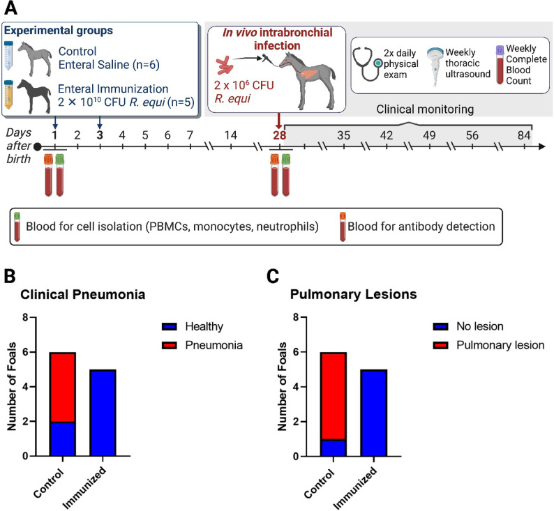
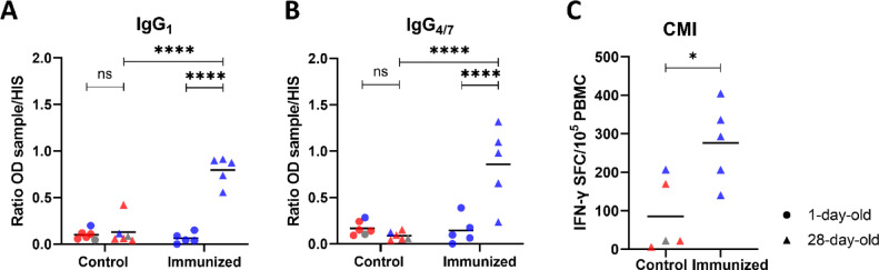
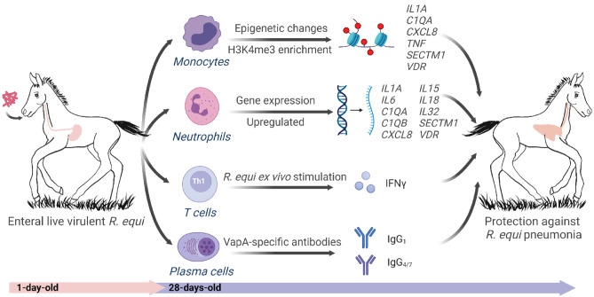
Using a horse foal model, we show that enteral immunization of newborn foals with Rhodococcus equi overcomes neonatal vaccination challenges by reprogramming innate immune responses, inducing R. equi-specific adaptive humoral and cell-mediated immune responses and protecting foals against experimental pneumonia challenge. Foals were immunized twice via gavage of R. equi (immunized group) or saline (control group) at ages 1 and 3 days. At age 28 days, all foals were challenged intrabronchially with R. equi. Post-challenge, all 5 immunized foals remained healthy, whereas 67% (4/6) of control foals developed clinical pneumonia. Immunized foals exhibit changes in the epigenetic profile of blood monocytes, > 1,000 differentially-expressed genes in neutrophils, higher concentrations of R. equi-specific IgG1 and IgG4/7, and a higher number of IFN-γ producing lymphocytes in response to R. equi stimulation indicating T helper type 1 response compared to control foals. Together, our data indicate that early life exposure to R. equi in the gastrointestinal tract can modulate innate immune responses, generate specific antibodies and cell-mediated immunity, and protect against pneumonia.

Keywords: Rhodococcus equi; Horse; Infection; Trained Immunity; Vaccine.


Conflict of interest statement

Declarations. Competing interests: The authors declare no competing interests.

Figures

Fig. 1
Fig. 1
Enteral immunization of newborn foals protected against R. equi intrabronchial challenge at age 28 days. (A) At ages 1 and 3 days, foals were gavaged with either 2 × 1010 CFU of R. equi (ATCC 33701+; immunized group) or saline (control group). At age 28 days, all foals were challenged intrabronchially with 2 × 106 CFU of R. equi (strain EIDL-5331). Blood for cell isolation was collected at ages 1 and 28 days. (B) All 5 foals in the immunized group remained healthy after intrabronchial challenge with R. equi, in contrast to foals in the control group in which 67% (4/6) developed clinical pneumonia (P = 0.0454). (C) Pulmonary lesions were detected by thoracic ultrasonography in 83% (5/6) in the control group, while no lesion developed in immunized foals (P = 0.013).
Fig. 2
Fig. 2
Enteral immunization with R. equi altered neutrophil gene expression. (A) Volcano plot representation of RNA-seq results showing differential expression analysis of genes in neutrophils of foals at ages 1 and 28 days between controls and immunized foals (dots represent difference on gene expression; ● = non-significant, formula image= upregulated, formula image= downregulated). (B) Heatmap of RNA-seq results of neutrophil from 28-day-old foals including all genes with fold-change higher than 11 between immunized and control groups (royal blue = healthy; red = pneumonic; gray = subclinical pneumonia).
Fig. 3
Fig. 3
Enteral immunization induced H3K4me3 enrichment in the promoter regions of monocytes. Monocytes from immunized foals had an increase of H3K4me3 enrichment in the promoter regions at age 28 days. At ages 1 and 28 days, we isolated blood monocytes from foals and immunoprecipitated chromatin extracts with monoclonal antibody (mAb) recognizing the trimethylated form of histone 3 lysine 4 (H3K4me3). We purified DNA and performed qPCR using primers recognizing the region proximal to the transcriptional start sites of genes encoding IL1A, IL1B, IL1RN, IL32, C1QA, CCL8, CXCL8, CXCL10, RPL30, SECTM1, TNF, and VDR. Data are shown as % of input. Different symbols represent timepoints (● = 1-day-old and ▲ = 28-day-old). Blue symbols represent monocytes isolated from foals that remained healthy after intrabronchial infection, gray symbol the foal that developed subclinical pneumonia, and red symbols foals that had clinical pneumonia. The lines connect timepoints from the same animal. Statistical difference is represented by ** for P < 0.01, * for P < 0.05, # for 0.1 > P > 0.05, and ns for not significant.
Fig. 4
Fig. 4
Enteral immunization increased H3K4me3:H3K27me3 ratio in the promoter regions of monocytes. We isolated blood monocytes from foals at ages 1 and 28 days. We immunoprecipitated chromatin extracts with mAbs recognizing the trimethylated forms of histone 3 lysine 4 (H3K4me3) and lysine 27 (H3K27me3). We performed qPCR using purified DNA and primers amplifying the regulatory regions of genes encoding IL1A, IL1B, IL1RN, IL32, C1QA, CCL8, CXCL8, CXCL10, RPL30, SECTM1, TNF, and VDR. Data show the ratio of % of input of H3K4me3 divided by % of input of H3K27me3. Different symbols represent different timepoints (● = 1-day-old and ▲ = 28-day-old). Blue symbols represent monocytes isolated from foals that remained healthy after intrabronchial infection, gray symbol the foal that developed subclinical pneumonia, and red symbols foals that had clinical pneumonia. The lines connect timepoints from the same animal. Statistical difference is represented by *** for P < 0.001, ** for P < 0.01, * for P < 0.05, # for 0.1 > P > 0.05, and ns for not significant.
Fig. 5
Fig. 5
Neutrophils from foals exhibit a bivalent chromatin signature at genes encoding IL32, C1QA, CCL8, SECTM1, and VDR. We isolated blood neutrophils from all foals at ages 1 and 28 days. We immunoprecipitated chromatin extracts with mAbs recognizing the trimethylated forms of histone 3 lysine 4 (H3K4me3) and lysine 27 (H3K27me3), and the normal H3 histone (H3), normal IgG (mock control). We performed qPCR using primers specific for the region proximal to the transcriptional start site of genes encoding IL1A, IL1B, IL1RN, IL32, C1QA, CCL8, CXCL8, CXCL10, RPL30, SECTM1, TNF, and VDR. Bivalent domains were identified as those, having enrichment for both H3K4me3 and H3K27me3. Different symbols represent different timepoints (● = 1-day-old and ▲ = 28-day-old). Blue symbols represent neutrophils isolated from foals that remained healthy after intrabronchial infection, gray symbol the foal that developed subclinical pneumonia, and red symbols foals that had clinical pneumonia. Different letters (a, b, c, and d) represent statistical difference (P < 0.05).
Fig. 6
Fig. 6
Enteral immunization induced R. equi-specific cell-mediated immunity (CMI) and humoral immunity. (A and B) Anti-VapA IgG1 (B) and IgG4/7 (C) activities were measured in foal serum at ages 1 and 28 days by ELISA. The ratio of the optical density (OD) for each sample was calculated by dividing the sample OD minus blank by the positive control R. equi-hyper immune serum (HIS) minus blank. Both VapA-specific IgG1 and IgG4/7 subisotypes were increased at age 28 days in immunized foals, but not in control foals. C) Peripheral blood mononuclear cells (PBMCs) isolated from foals at age 28 days that received enteral R. equi had increased (p = 0.0168) IFN-γ spot-forming cells (SFC) following stimulation with R. equi lysate, suggesting induction of higher R. equi-specific immune responses compared to control foals. Different symbols represent different timepoints (● = 1-day-old and ▲ = 28-day-old). Blue symbols represent foals that remained healthy after intrabronchial infection, gray symbol the foal that developed subclinical pneumonia, and red symbols foals that had clinical pneumonia. Statistical difference is represented by * for P < 0.05, **** for P < 0.00001, and ns for not significant.
Fig. 7
Fig. 7
Summary of our results. Foals immunized enterally with live virulent R. equi (ATCC 33701+) at ages 1 and 3 days developed enhanced innate and adaptive immune responses at age 28 days. Innate cells of immunized foals had several important changes by age 28 days, including a modified epigenetic profile in monocytes and more than 1,000 differentially-expressed genes (DEG) in neutrophils. Adaptive immune responses to immunization included R. equi-specific IFN-γ producing cells among the PBMCs and the generation of VapA-specific IgG1 and IgG4/7 antibodies. In summary, immunized foals had enhanced innate and adaptive immune responses and were protected foals against intrabronchial challenge with R. equi.

References

    1. Mohr, E. & Siegrist, C. A. Vaccination in early life: Standing up to the challenges. Curr. Opin. Immunol.41, 1–8. 10.1016/j.coi.2016.04.004 (2016). - PubMed
    1. Flaminio, M. J. et al. The effect of CpG-ODN on antigen presenting cells of the foal. J. Immune Based Ther. Vaccines5, 1. 10.1186/1476-8518-5-1 (2007). - PMC - PubMed
    1. Flaminio, M. J., Nydam, D. V., Marquis, H., Matychak, M. B. & Giguere, S. Foal monocyte-derived dendritic cells become activated upon Rhodococcus equi infection. Clin. Vaccine Immunol.: CVI. 16, 176–183. 10.1128/CVI.00336-08 (2009). - PMC - PubMed
    1. Pargass, I. S. et al. The influence of age and Rhodococcus equi infection on CD1 expression by equine antigen presenting cells. Vet. Immunol. Immunopathol.130, 197–209. 10.1016/j.vetimm.2009.02.007 (2009). - PubMed
    1. Lopez, B. S. et al. The effect of age on foal monocyte-derived dendritic cell (MoDC) maturation and function after exposure to killed bacteria. Vet. Immunol. Immunopathol.210, 38–45. 10.1016/j.vetimm.2018.11.020 (2019). - PubMed

MeSH terms

LinkOut - more resources