. 2025 May;13(10):e70383.
doi: 10.14814/phy2.70383.

Regulation of TFEB in human placental Cytotrophoblasts and Syncytiotrophoblasts

Affiliations

Regulation of TFEB in human placental Cytotrophoblasts and Syncytiotrophoblasts

A Mathew et al. Physiol Rep. 2025 May.

Abstract

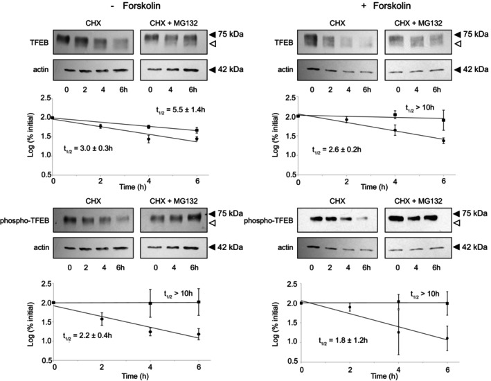
While cellular proteins exist in a dynamic state maintained by the balance of synthesis and degradation, there is a paucity of information on these processes in placental trophoblasts, including within cytotrophoblasts which differentiate into multi-nucleate syncytiotrophoblasts. TFEB, a transcription factor with a myriad of cellular activities, is one of the most abundant genes expressed in syncytiotrophoblasts compared to cytotrophoblasts. TFEB is localized to the nucleus of human BeWo differentiated syncytiotrophoblasts and to the cytoplasm of the undifferentiated cytotrophoblasts. Within both the cytotrophoblasts and syncytiotrophoblasts, TFEB exists in subcellular compartments as both phosphorylated and unphosphorylated forms and translocates between cytoplasm and nucleus upon amino acid starvation/refeeding. Endogenous TFEB and endogenous phospho-TFEB are both rapidly (t1/2 ~ 2-3 h) degraded via the ubiquitin proteasome system in cytotrophoblasts and in syncytiotrophoblasts. These results suggest dynamic regulatory processes during trophoblast development/differentiation.

Keywords: cytotrophoblast; degradation; placental; syncytiotrophoblast; ubiquitin.


Conflict of interest statement

The authors declare no conflicts of interest.

Figures

FIGURE 1
FIGURE 1
Triple‐label Immunofluorescence Localization of BeWo Cytotrophoblasts (left) and Syncytiotrophoblasts (right). BeWo cells were treated with vehicle control (left) or with forskolin (right) and after 72 h cells were fixed and immunostained. Nuclei were stained with DAPI (blue), cell junctions were stained with anti‐E cadherin (red) and TFEB was stained with anti‐TFEB (green). Inset in left panel is of naturally occurring syncytia. Bars in lower right represent 10 μm.
FIGURE 2
FIGURE 2
Quantification of TFEB Localization in BeWo Cytotrophoblasts and Syncytotrophoblasts. Triple immunostained cells as seen in Figure 1 were scored for TFEB expression as predominantly cytoplasmic (C), predominantly nuclear (N) or present in both cytoplasm and nucleus (C/N) in single cells (a, b) or within syncytia (c, d) in cells treated with vehicle alone (a, c) or with forskolin (b, d). In cells treated with vehicle alone, greater than 99% of cells were single cells, whereas in cells treated with forskolin 58% of nuclei were in syncytia and 42% were in single cells. 5329 cells were scored from 3 independent experiments (panel a: 3785; b: 629; c: 49; d: 866). Data are displayed as mean ± SEM of the 3 experiments. Statistical comparison of C versus N: A, b, d: p < 0.001; c: p < 0.02.
FIGURE 3
FIGURE 3
TFEB Localization in Cell Fractions of BeWo Cytotrophoblasts and Syncytiotrophoblasts. BeWo cells were treated with vehicle alone or forskolin for 72 h and then harvested following which whole cell extract (WCE), post‐nuclear supernatant extract (PNS, cytoplasm) and nuclear extract (NE) were prepared and analyzed via Western blot with anti‐TFEB antibody. Blots were then reprobed with anti‐phospho TFEB antibodies as indicated. Controls of anti‐lamin B1 and anti‐alpha tubulin are shown. The 75 kDa marker is indicated by closed triangle. The open triangle denotes lower molecular weight (unphosphorylated) TFEB.
FIGURE 4
FIGURE 4
TFEB Localization Following Amino Acid Starvation and Refeeding in BeWo Cytotrophoblasts and Synctiotrophoblasts. Following treatment ± forskolin, cells were fed with complete media for 2 h, amino acid starved in EBSS for 1 h, and then refed for 1 h with complete media, as indicated. Thereafter, cells were harvested, fractionated, run on gels, and blots thereafter probed with anti‐TFEB as in Figure 3. Controls and markers are as in Figure 3. Data from individual experiments are displayed as circles and the mean as a bar.
FIGURE 5
FIGURE 5
TFEB Localization Following Torin Treatment in BeWo Cytotrophoblasts and Synctiotrophoblasts. Following treatment ± forskolin, cells were incubated without or with 250 nM Torin, as indicated. Thereafter cells were harvested, fractionated, run on gels, and blots thereafter probed with anti‐TFEB as in Figure 3. Controls and markers are as in Figure 3. Data from individual experiments are displayed as circles and the mean as a bar.
FIGURE 6
FIGURE 6
Degradation of Endogenous TFEB and Phospho‐TFEB in BeWo Cytotrophoblasts and Syncytiotrophoblasts. Following 72 h treatment with vehicle alone (cytotrophoblasts) or 100 μM forskolin (syncytiotrophoblasts), cells were incubated with 100 μg/mL cycloheximide (CHX) in the absence or presence of 20 μM MG132. At 0, 2, 4 and 6 h (CHX) and 0, 4 and 6 h (CHX + MG132) cells were harvested and analyzed via Western blot with anti‐TFEB antibody and subsequently reprobed with anti‐phospho‐TFEB as described in Methods. The TFEB and phospho‐TFEB were quantified as described in Methods and displayed in the Figure, CHX alone (●), CHX + MG132 (■). Half‐lives were calculated as indicated (mean ± SEM of 4 independent experiments). Upper panels are representative Western blots with anti‐TFEB, anti‐phospho‐TFEB and anti‐Actin, as control. Statistical comparison of TFEB and of phospho‐TFEB t 1/2 (−forskolin versus + forskolin) are not significant (p > 0.05). Statistical comparison of TFEB and of phospho‐TFEB (− forskolin versus + forskolin) at 2, 4 and 6 h are each not significant (p > 0.05).

Similar articles

References

    1. Azar, C. , Valentine, M. , Trausch‐Azar, J. , Druley, T. , Nelson, D. M. , & Schwartz, A. L. (2018). RNA‐Seq identifies genes whose proteins are transformative in the differentiation of cytotrophoblast to syncytiotrophoblast, in human primary villous and BeWo trophoblasts. Scientific Reports, 8(1), 5142. 10.1038/S41598-018-23379-2 - DOI - PMC - PubMed
    1. Azar, C. , Valentine, M. C. , Trausch‐Azar, J. , Rois, L. , Mahjoub, M. , Nelson, D. M. , & Schwartz, A. L. (2021). RNA‐Seq identifies genes whose proteins are upregulated during syncytia development in murine C2C12 myoblasts and human BeWo trophoblasts. Physiological Reports, 9(1), e14671. 10.14814/phy2.14671 - DOI - PMC - PubMed
    1. Banani, S. F. , Lee, H. O. , Hyman, A. A. , & Rosen, M. K. (2017). Biomolecular condensates: Organizers of cellular biochemistry. Nature Reviews Molecular Cell Biology, 18(5), 285–298. 10.1038/nrm.2017.7 - DOI - PMC - PubMed
    1. Breitschopf, K. , Bengal, E. , Ziv, T. , Admon, A. , & Ciechanover, A. (1998). A novel site for ubiquitination: The N‐terminal residue, and not internal lysines of MyoD, is essential for conjugation and degradation of the protein. EMBO Journal, 17(20), 5964–5973. 10.1093/emboj/17.20.5964 - DOI - PMC - PubMed
    1. Cesana, M. , Tufano, G. , Panariello, F. , Zampelli, N. , Soldati, C. , Mutarelli, M. , Montefusco, S. , Grieco, G. , Sepe, L. V. , Rossi, B. , Nusco, E. , Rossignoli, G. , Panebianco, G. , Merciai, F. , Salviati, E. , Sommella, E. M. , Campiglia, P. , Martello, G. , Cacchiarelli, D. , … Ballabio, A. (2024). TFEB controls syncytiotrophoblast formation and hormone production in placenta. Cell Death and Differentiation, 31(11), 1439–1451. 10.1038/s41418-024-01337-y - DOI - PMC - PubMed

MeSH terms

Substances

LinkOut - more resources