Review
. 2025 May 9:15:1565513.
doi: 10.3389/fcimb.2025.1565513. eCollection 2025.

Primary cell culture systems to investigate host-pathogen interactions in bacterial respiratory tract infections of livestock

Affiliations
Review

Primary cell culture systems to investigate host-pathogen interactions in bacterial respiratory tract infections of livestock

Yenehiwot Berhanu Weldearegay et al. Front Cell Infect Microbiol. .

Abstract

Respiratory infections of livestock represent a major health issue for the animals and cause high economic losses for the farmers. Still, little is known about the intricate interactions between host cells and the many different pathogens that cause respiratory diseases, leaving a substantial knowledge gap to be filled in order to develop effective therapies. Immortalized cell lines and two-dimensional cultures of primary respiratory epithelial cells do not reflect the complex architecture and functionality of the respiratory tract tissues. Thus, it is essential to develop and apply appropriate primary cell culture systems to study respiratory diseases. In human research, the use of complex cell culture systems, such as air-liquid interface (ALI) cultures, organoids and lung-on-chip, has proceeded significantly during the last years, whereas in veterinary research, these models are only rarely used. Nevertheless, there are several three-dimensional, primary cell culture systems available to study respiratory infections of livestock. Here, we give an overview on models that are currently used in this field: nasal mucosa explants, tracheal organ cultures, ALI cultures, and precision-cut lung slices. All these models align with the 3R principle, as they can replace animal experiments to some extent and the tissue material for these culture systems can be obtained from abattoirs or veterinary research facilities. We aim to encourage other researchers to use these versatile cell culture systems to drive investigations of respiratory tract infections of livestock forward. Finally, these models are not limited to infection research, but can also be applied in other research fields and can be transferred to other animal species than livestock.

Keywords: air-liquid interface cultures; livestock diseases; nasal mucosa explants; organotypic models; precision-cut lung slices; respiratory tract infections; tracheal organ cultures.


Conflict of interest statement

The authors declare that the research was conducted in the absence of any commercial or financial relationships that could be construed as a potential conflict of interest.

Figures
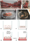
Figure 1
Figure 1
Schematic overview of preparation and culturing of NMEs. As an example, a pig snout is shown in (A, B). To obtain NMEs, the interior of the nasal cavity is exposed (A) and the mucosal layer is stripped from the cartilage (B, C). The tissue is then cut into appropriately sized pieces (NMEs) (D) and inoculated by submerging the NME in medium containing pathogen (E). Following incubation and washing, the NME is placed on a scaffold (shown here as a transwell culture plate insert) and cultured at the air-liquid interphase (F). Alternatively, a polarized setup can be employed. Prior to inoculation, the NME can be embedded in agarose to occlude the basolateral and side surfaces of the NME, thus allowing infection to occur only from the apical side (G). Following incubation and washing, the embedded NME is cultured at the air-liquid interphase in fresh medium (H). Drawings in panels G and H have been prepared taking inspiration from Frydas et al (Frydas et al., 2013). Photos provided by L. Brogaard, Department of Biotechnology and Biomedicine, Section for Medical Biotechnology, Technical University of Denmark, Kgs. Lyngby, Denmark. Created by L. Brogaard in BioRender. https://BioRender.com/dwhk64n.
Figure 2
Figure 2
Preparation of chicken TOC. (A) Preparation should be done under a magnification glass in a petri dish with pre-warmed medium and on filter paper. (B) After removal of the trachea from the embryo or post hatch bird, connective tissue is stripped off (black arrow indicates direction of stripping). (C) Cutting of the trachea into 0.8–1 mm evenly thick rings with a sharp blade manually or with a tissue chopper. (D) Placement of the ring in pre-warmed medium (arrow indicates TOC) and incubation in an overhead rotator at 37°C. (E) Microscopical evaluation of the tracheal ring after incubation for ciliary activity using an inverted microscope (arrow indicates cilia and a clear lumen of the ring without mucus accumulation). (F) Histological section of the TOC showing goblet cells (filled arrowhead) and epithelial cells with cilia (open arrowhead). Photos provided by N. Rüger and S. Rautenschlein, Clinic for Poultry, University of Veterinary Medicine Hannover.
Figure 3
Figure 3
Preparation of ALI cultures from porcine trachea/bronchi. (A) Lungs from slaughtered animals from the abattoir are transported on ice to the laboratory and the trachea and/or the main bronchi are separated from the connective lung tissue. (B) The trachea and/or the main bronchi are incubated in digestion buffer at 4°C for up to three days. (C) Epithelial cells are scraped off from the luminal surface using a scalpel blade and transferred to DMEM supplemented with 10% FBS. After filtration and washing, cells are incubated in non-coated plastic cell culture dishes at 37°C for 1–2 h to reduce fibroblast contamination. (D) Non-adherent epithelial cells are then transferred to a collagen I-coated cell culture flask and incubated at 37°C until cells are confluent. (E) Epithelial cells are detached from the cell culture flask and seeded on collagen IV-coated polycarbonate membranes with a pore size of 0.4 µm and (F) incubated under submerged conditions for four days. (G) Medium from the apical compartment is removed and (H) the epithelial cells differentiate under ALI conditions within 3–4 weeks. Photos provided by (D) Schaaf, Institute for Microbiology, University of Veterinary Medicine Hannover, Germany. Created by D. Schaaf in BioRender. https://BioRender.com/b62a774.
Figure 4
Figure 4
Fully differentiated primary porcine bronchial epithelial cells (PBEC) under ALI conditions. (A) Hematoxylin-eosin staining of PBEC (D. Schaaf, Institute for Microbiology, University of Veterinary Medicine Hannover, Germany). (B) Immunofluorescence staining of PBEC under ALI conditions. Visualization of cilia (β-tubulin, red), tight junctions (β-catenin, green), and nuclei (DAPI, blue) (D. Schaaf, Institute for Microbiology, University of Veterinary Medicine Hannover, Germany). (C) SEM of PBEC (M. Rohde, Helmholtz Center for Infection Research, Braunschweig, Germany). Bars represent 10 µm.
Figure 5
Figure 5
Preparation of porcine PCLS. (A) Lungs from slaughtered animals from the abattoir are transported on ice to the laboratory. (B) The cranial lobes of apparently healthy lungs are dissected and filled with 37°C low melting-point agarose through a cannula introduced in the bronchus. Filled lung lobes are kept on ice until the agarose becomes solid. (C) By using a tissue coring tool, cylindrical pieces of lung tissue with a bronchioles in the middle are punched out and cut into approximately 300 µm thin tissue slices by using a tissue slicer or vibratome (here: Krumdieck tissue slicer, model MD 4000-01; TSE Systems, Chesterfield, MO, USA). (D) The tissue slices are transferred to cell culture medium (e.g., RPMI) supplemented with antibiotics and antimycotics and bubbled with a normoxic gas mixture at 37°C to remove the agarose from the airway lumen. (E) PCLS can be maintained for up to two weeks in cell culture medium (e.g., RPMI) supplemented with antibiotics and antimycotics in, e.g., a 24-well plate. Photos provided by D. Schaaf, Institute for Microbiology, University of Veterinary Medicine Hannover, Germany.
Figure 6
Figure 6
Visualization of PCLS. (A) Hematoxylin-eosin staining of porcine PCLS; bar represents 50 µm (R. Spriewald, Institute for Microbiology, University of Veterinary Medicine Hannover, Germany). (B) Immunofluorescence staining of porcine PCLS infected with Bordetella bronchiseptica. Bacteria are shown in green, cilia (β-tubulin) in red, and nuclei (DAPI) in blue; bar represents 20 µm (D. Schaaf, Institute for Microbiology, University of Veterinary Medicine Hannover, Germany). (C) SEM of porcine PCLS; bar represents 5 µm (M. Rohde, Helmholtz Center for Infection Research, Braunschweig, Germany).

References

    1. Abd El Rahman S., Winter C., El-Kenawy A., Neumann U., Herrler G. (2010). Differential sensitivity of well-differentiated avian respiratory epithelial cells to infection by different strains of infectious bronchitis virus. J. Virol 84, 8949–8952. doi: 10.1128/JVI.00463-10 - DOI - PMC - PubMed
    1. Abdul-Wahab O. M., Ross G., Bradbury J. M. (1996). Pathogenicity and cytadherence of Mycoplasma imitans in chicken and duck embryo tracheal organ cultures. Infect. Immun. 64, 563–568. doi: 10.1128/iai.64.2.563-568.1996 - DOI - PMC - PubMed
    1. Abraham G., Zizzadoro C., Kacza J., Ellenberger C., Abs V., Franke J., et al. . (2011). Growth and differentiation of primary and passaged equine bronchial epithelial cells under conventional and air-liquid-interface culture conditions. BMC Vet Res. 7, 26. doi: 10.1186/1746-6148-7-26 - DOI - PMC - PubMed
    1. Abs V., Bonicelli J., Kacza J., Zizzadoro C., Abraham G. (2019). Equine bronchial fibroblasts enhance proliferation and differentiation of primary equine bronchial epithelial cells co-cultured under air-liquid interface. PLoS One 14, e0225025. doi: 10.1371/journal.pone.0225025 - DOI - PMC - PubMed
    1. Abugisisa L., Royse E. X., Kemp M. W., Jobe A. H., Hillman N. H. (2023). Preterm ovine respiratory epithelial cell responses to mechanical ventilation, lipopolysaccharide, and interleukin-13. Am. J. Physiol. Lung Cell Mol. Physiol. 324, L815–L824. doi: 10.1152/ajplung.00355.2022 - DOI - PMC - PubMed

MeSH terms

LinkOut - more resources