Review
. 2025 May 9:13:1592391.
doi: 10.3389/fbioe.2025.1592391. eCollection 2025.

Illuminating extracellular nanovesicles through the spectroscopic lens: a mini review of cutting-edge insights and emerging applications

Affiliations
Review

Illuminating extracellular nanovesicles through the spectroscopic lens: a mini review of cutting-edge insights and emerging applications

Debarati Bhowmik et al. Front Bioeng Biotechnol. .

Abstract

Extracellular vesicles (EVs) are cell-derived particles that facilitate intercellular communication by carrying bioactive molecules like proteins and RNA, impacting both health and disease. Herein, the EVs' significance in physiological and pathological processes is reviewed, emphasising their potential as biomarkers for diseases including for instance, cancer, neurodegenerative disorders and cardiovascular conditions. The principles and applications of Raman spectroscopy (RS) - a powerful tool offering detailed molecular insights into EVs, are further examined. The non-destructive nature of this spectroscopic technique renders it invaluable for studying the molecular composition, purity and concentration of EVs. When EVs are isolated from accessible biofluids such as blood, urine or saliva, the overall process remains minimally invasive, enhancing its clinical applicability. The review highlights Raman spectroscopy's role in identifying disease-related EVs, distinguishing subpopulations and enhancing our understanding of EVs in disease mechanisms and therapeutic applications.

Keywords: Raman imaging; Raman spectroscopy; SERS (surface enhanced Raman scattering); disease diagnostics; extracellular vesicles.


Conflict of interest statement

The authors declare that the research was conducted in the absence of any commercial or financial relationships that could be construed as a potential conflict of interest.

Figures

FIGURE 1
FIGURE 1
Schematic illustrating the importance of EVs in various diseases and conditions, including cancer (Sandfeld-Paulsen B et al.,2016), neurological (Arifin et al., 2022) and neurodegenerative disorders (Valle-Tamayo et al., 2022), autoimmune diseases, cardiovascular diseases (Chong et al., 2019), COPD (Gomez et al., 2022), liver (Richter et al., 2021) and kidney diseases (Estefanía et al., 2023) and diabetes (Hu et al., 2020). Created with Biorender (Bhowmik, 2025).
FIGURE 2
FIGURE 2
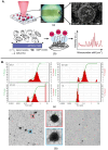
(A) SERS substrate irradiated by laser light initiates (a) Raman scattering. (b) The substrate facilitates the transport of EVs near AgNP clusters on the silicate scaffold (Zheng et al., 2022). (B) Characterization of EVs from citrate plasma samples: (a) Particle size distribution by dynamic light scattering. (b) TEM of EV120-enriched fraction, with enlarged views showing typical EV structures (Osei et al., 2021). All figures are reprinted with permission.
FIGURE 3
FIGURE 3
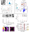
(A) The EVs released from Pseudomonas aeruginosa PAO1 were characterized using several techniques. (a) Transmission electron microscopy images showed the morphology of these EVs, with a scale bar of 100 nm. (b) The particle size distribution was analysed by nanoflow cytometry, revealing sizes ranging from 68 to 155 nm using standard-sized particles as references. (c) Raman spectroscopy provided the spectral profile of the EVs, with the mean and standard deviation represented by a solid line and light shading, respectively, with different colours indicating various substance types (Qin et al., 2023). (B) Deep learning-enabled Raman spectroscopic identification of pathogen-derived EVs and their biogenesis (Qin et al., 2022). (C). Positively charged particle-based EV detection. (a) TEM images of DMAP-AuNPs-coated EVs at different ratios, scale bars 100 nm. (b) SERS measurements of DMAP-AuNP coated EVs, with DMAP and EV peaks indicated. (c) EM field enhancement with a zoom-in of a single DMAP-AuNP on a vesicle surface. (d) Relation between EV coverage ratio and EFEF before and after Ag shell formation. (e) SERS characterization of EVs with Ag shell-AuNPs or DMAP-AuNPs, showing various coverage percentages (Shin et al., 2020). All figures are reprinted with permission.

References

    1. Abels E. R., Breakefield X. O. (2016). Introduction to extracellular vesicles: biogenesis, RNA cargo selection, content, release, and uptake. Cell. Mol. Neurobiol. 36, 301–312. 10.1007/s10571-016-0366-z - DOI - PMC - PubMed
    1. Arifin D. R., Witwer K. W., Bulte J. W. M. (2022). Non‐Invasive imaging of extracellular vesicles: Quo vaditis in vivo?. Journal of Extracellular Vesicles, 11 (7) 12241. 10.1002/jev2.12241 - DOI - PMC - PubMed
    1. Baker R. E., Mahmud A. S., Miller I. F., Rajeev M., Rasambainarivo F., Rice B. L., et al. (2022). Infectious disease in an era of global change. Nat. Rev. Microbiol. 20 (4), 193–205. 10.1038/s41579-021-00639-z - DOI - PMC - PubMed
    1. Banwell C. N. (1972). Fundamentals of molecular spectroscopy.
    1. Beekman P., Enciso-Martinez A., Hoon S. R., Pujari S. P., Aufrid T. M. L., Zuilhof H., et al. (2019). Immuno-capture of extracellular vesicles for individual multi-modal characterization using AFM, SEM and Raman spectroscopy. Lab a Chip 19 (15), 2526–2536. 10.1039/c9lc00081j - DOI - PubMed

LinkOut - more resources