

A Case of Gastric Atypical Lipomatous Tumor/Well-Differentiated Liposarcoma With Endoscopic Morphological Changes
- PMID: 40416586
- PMCID: PMC12098964
- DOI: 10.1002/deo2.70146
A Case of Gastric Atypical Lipomatous Tumor/Well-Differentiated Liposarcoma With Endoscopic Morphological Changes
Abstract
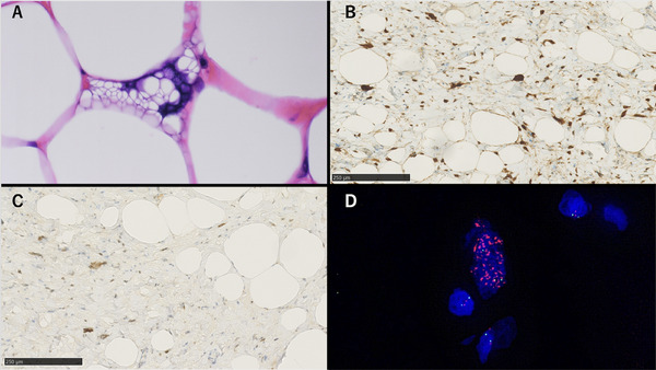
Atypical lipomatous tumor/well-differentiated liposarcoma is a locally aggressive mesenchymal neoplasm composed of adipocytes and stromal cells. Gastric cases are exceedingly rare, and their malignant potential remains unclear. We report a case of a woman in her 60s who was found to have multiple submucosal tumor-like lesions of the stomach. Over time, the tumors increased in size, requiring a laparoscopic partial gastrectomy. Histological examination revealed a tumor composed of both fatty tissue and fibrous stroma with nuclear atypia. Immunohistochemistry showed positivity for CDK4 and MDM2, and fluorescence in situ hybridization confirmed MDM2 amplification, leading to a diagnosis of atypical lipomatous tumor/well-differentiated liposarcoma. This case presented an unusual gastric manifestation, with multiple submucosal tumor-like lesions on endoscopy and exhibiting progressive morphological changes over several years.
Keywords: CDK4; MDM2; atypical lipomatous tumor; stomach; well‐differentiated liposarcoma.
© 2025 The Author(s). DEN Open published by John Wiley & Sons Australia, Ltd on behalf of Japan Gastroenterological Endoscopy Society.
Conflict of interest statement
The authors declare no conflicts of interest.
Figures


References
-
- Marta S., World Health Organization Classification of Tumours of Soft Tissue (IARC Press, 2019)
-
- Fletcher C. D. M. and Hogendoom P. C. W., “Pathology and Genetics of Tumours of Soft Tissue and Bone,” in World Health Organization Classification of Tumours, eds. C. D. M. Fletcher, K. K. Unni, and F. Mertens (IARC Press, 2013), 10–238.
-
- Saegusa H., Simizu T., Ochi Y., et al., “Primary Gastric Liposarcoma, Report of a Case and Review of the Literature,” Gastrointestinal Endoscopy 44 (2002): 1168–1174.
LinkOut - more resources
Full Text Sources
Research Materials