

Progress to Date on Cranial Electromagnetic Field Stimulation to Modulate Brain Activity
- PMID: 40416912
- PMCID: PMC12101138
- DOI: 10.7759/cureus.84653
Progress to Date on Cranial Electromagnetic Field Stimulation to Modulate Brain Activity
Abstract
Background: The electromagnetic field (EMF) of the brain can be modulated through EMF stimulation. The authors investigate whether longer duration of continuous EMF stimulation using a novel method to identify and provide feedback and adjustment of EMF recording would translate into sustained improvement in EMF patterns, such as higher amplitude with correlating improvement in clinical symptoms or deficits.
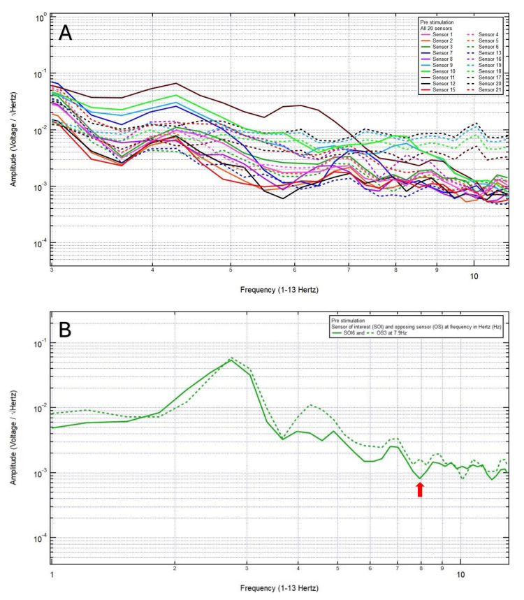
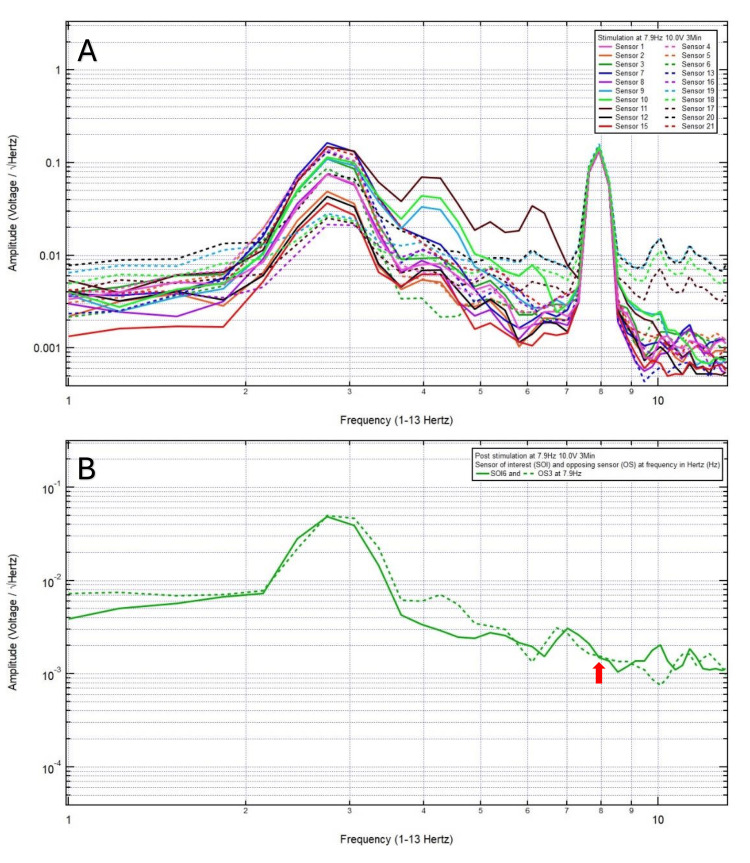
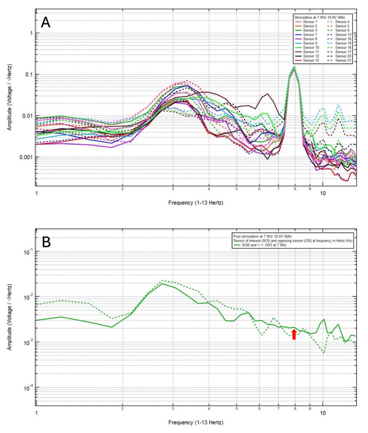
Methods: From January 2025 to February 2025, a prospective study enrolled patients greater than 18 years old diagnosed with atraumatic and traumatic brain injury who underwent EMF stimulation within 24 hours of presentation. EMF data were collected using DAQami software (Dataq Instruments, Akron, Ohio) and analyzed using fast Fourier transformation (FFT) with Igor Pro 8 software (Wavemetrics Inc., Lake Oswego, Oregon). Based on each patient's clinical presentations and/or radiographic findings, localization of brain injuries, frequency selection, and optimal voltage stimulation were determined in real-time followed by delivery of incremental increase in duration of stimulation from 3, 5, 8, and 10 minutes until improvement in clinical symptoms and/or neurological deficits and sustained EMF change was achieved.
Results: Ten patients were included in this study, with a mean age of 47.1 years. Mechanisms of injury included spontaneous hypertensive intracranial hemorrhage (1 patient) and head trauma after motor vehicle collision, dirt bike accident, and ground-level fall (9 patients). Radiographic findings included spontaneous basal ganglia hemorrhage (1 patient), isolated traumatic subdural hematoma (1 patient), traumatic subarachnoid hemorrhage (1 patient), and no intracranial abnormalities (7 patients). Clinical resolution of their neurological symptoms or remaining asymptomatic was achieved in five patients after three minutes of continuous EMF stimulation, two patients after five minutes of continuous EMF stimulation, and one patient after 10 minutes of continuous EMF stimulation (Table 1). Patient 8 declined to continue with the study after three minutes of continuous EMF stimulation, and patient 9 declined to continue with the study after five minutes of continuous EMF stimulation.
Conclusions: This study reveals the progress made to date utilizing a novel technology of EMF measurement at a distance, in real-time, using the non-invasive, lightweight portable helmet, and continuous feedback. The range of brain EMF can be stimulated at the optimal frequency and voltage with or without longer duration of stimulation in a precise and prescribed manner to produce sustained genetic and neuronal changes to improve, recover, and enhance the brain function in a sample of patients with atraumatic and traumatic brain injury and improve or resolve their neurological symptoms or deficits. It illustrates the necessity of real-time evaluation and adjustment of brain EMF for EMF stimulation. It further indicates the efficacy of tailored and precise EMF stimulation to the specific patient, the specific area of abnormality, and for a specific pathology studied. The range of unique EMF corresponds to macroscopic and microscopic functions, the vast majority of which have yet to be qualified and quantified, and for which most brain diseases have yet to be studied.
Keywords: duration of treatment; electromagnetic field frequency; electromagnetic field stimulation; head trauma; voltage.
Copyright © 2025, Wang et al.
Conflict of interest statement
Human subjects: Consent for treatment and open access publication was obtained or waived by all participants in this study. Arrowhead Regional Medical Center issued approval #23-58. PROTOCOL: Transcranial Electromagnetic Field Stimulation for Modulation of Brain Activity in Patients with Neurological Disorders. The information provided was reviewed and approved by the Institutional Review Board on May 16, 2024. Animal subjects: All authors have confirmed that this study did not involve animal subjects or tissue. Conflicts of interest: In compliance with the ICMJE uniform disclosure form, all authors declare the following: Payment/services info: All authors have declared that no financial support was received from any organization for the submitted work. Financial relationships: All authors have declared that they have no financial relationships at present or within the previous three years with any organizations that might have an interest in the submitted work. Other relationships: All authors have declared that there are no other relationships or activities that could appear to have influenced the submitted work.
Figures
Similar articles
-
Optimal Voltage for Cranial Electromagnetic Field Stimulation to Modulate Brain Activity.Cureus. 2025 Apr 10;17(4):e82011. doi: 10.7759/cureus.82011. eCollection 2025 Apr. Cureus. 2025. PMID: 40351961 Free PMC article.
-
Localization of Brain Injuries Using Cranial Electromagnetic Fields.Cureus. 2025 Mar 13;17(3):e80518. doi: 10.7759/cureus.80518. eCollection 2025 Mar. Cureus. 2025. PMID: 40225463 Free PMC article.
-
Optimal Frequency for Cranial Electromagnetic Field Stimulation.Cureus. 2025 Mar 29;17(3):e81436. doi: 10.7759/cureus.81436. eCollection 2025 Mar. Cureus. 2025. PMID: 40303536 Free PMC article.
-
The effects of radiofrequency electromagnetic field exposure on biomarkers of oxidative stress in vivo and in vitro: A systematic review of experimental studies.Environ Int. 2024 Dec;194:108940. doi: 10.1016/j.envint.2024.108940. Epub 2024 Aug 14. Environ Int. 2024. PMID: 39566441
-
Neuropathology of Mild Traumatic Brain Injury: Correlation to Neurocognitive and Neurobehavioral Findings.In: Kobeissy FH, editor. Brain Neurotrauma: Molecular, Neuropsychological, and Rehabilitation Aspects. Boca Raton (FL): CRC Press/Taylor & Francis; 2015. Chapter 31. In: Kobeissy FH, editor. Brain Neurotrauma: Molecular, Neuropsychological, and Rehabilitation Aspects. Boca Raton (FL): CRC Press/Taylor & Francis; 2015. Chapter 31. PMID: 26269912 Free Books & Documents. Review.
References
-
- Effect of pulsed electromagnetic field (PEMF) on infarct size and inflammation after cerebral ischemia in mice. Pena-Philippides JC, Yang Y, Bragina O, Hagberg S, Nemoto E, Roitbak T. Transl Stroke Res. 2014;5:491–500. - PubMed
-
- Pulsed electromagnetic field and relief of hypoxia-induced neuronal cell death: The signaling pathway. Gessi S, Merighi S, Bencivenni S, et al. J Cell Physiol. 2019;234:15089–15097. - PubMed
-
- Pulsed electromagnetic field exposure reduces hypoxia and inflammation damage in neuron-like and microglial cells. Vincenzi F, Ravani A, Pasquini S, et al. J Cell Physiol. 2017;232:1200–1208. - PubMed
-
- Effect of pulsed electromagnetic field exposure on adenosine receptors in rat brain. Varani K, Vincenzi F, Targa M, et al. Bioelectromagnetics. 2012;33:279–287. - PubMed
-
- Signaling pathways involved in anti-inflammatory effects of pulsed electromagnetic field in microglial cells. Merighi S, Gessi S, Bencivenni S, et al. Cytokine. 2020;125:154777. - PubMed
LinkOut - more resources
Full Text Sources