. 2025 May 22;7(12):3867-3880.
doi: 10.1039/d4na01072h. eCollection 2025 Jun 10.

Synergistic effect of gold nanorods coated with type I collagen and LED irradiation on wound healing in human skin fibroblast cells

Affiliations

Synergistic effect of gold nanorods coated with type I collagen and LED irradiation on wound healing in human skin fibroblast cells

Sasiprapa Poomrattanangoon et al. Nanoscale Adv. .

Abstract

Delayed wound healing poses a significant risk to human health, especially when wounds are infected by pathogens. Therefore, the development of effective methods for accelerating wound healing is important. This study investigated the synergistic effect of gold nanorods (GNRs) coated with type I collagen (GNRs@C) combined with light-emitting diode (LED) irradiation on wound healing in scratched human skin fibroblast (HSF) cells. Scratched HSF cells were treated with 3 μg mL-1 GNRs@C, followed by LED irradiation. This combined treatment significantly enhanced cell proliferation, increasing from the control cell base line (scratched HSF cells without any treatment) to 104.08 ± 2.96% and 107.82 ± 3.25% after 24 and 48 h of incubation, respectively. GNRs@C demonstrated superior cellular uptake compared to uncoated GNRs. Notably, complete closure of scratched HSF cells was observed in scratched HSF cells treated with GNRs@C with LED irradiation and then incubated for 40 h. Additionally, the treatment significantly reduced interleukin-6 (IL-6) and tumor necrosis factor-alpha (TNF-α) levels, while upregulating key growth factors, including vascular endothelial growth factor (VEGF) and basic fibroblast growth factor (bFGF). These findings demonstrate the wound healing potential of GNRs@C combined with LED irradiation.


Conflict of interest statement

There are no conflicts to declare.

Figures

Fig. 1
Fig. 1. Light absorption spectra of (a) GNRs and GNRs@PSS (b) GNRs@C.
Fig. 2
Fig. 2. TEM images of (a) GNRs, (b) GNRs@PSS and (c) GNRs@C and (d) zeta potentials of GNRs, GNRs@PSS, and GNRs@C.
Fig. 3
Fig. 3. FTIR spectra of GNRs@PSS, GNRs@C, and type I collagen.
Fig. 4
Fig. 4. Gel electrophoresis of type I collagen, GNRs@C, and GNRs@PSS.
Fig. 5
Fig. 5. Viability of HSF cells after treating with GNRs, GNRs@PSS, and GNRs@C at concentrations of 3, 5, 10, 15, and 25 μg mL−1 for 24 h. *Significant difference in cell viability in comparison with control cells (untreated cells) and cells treated with GNRs@PSS, and GNRs@C at P < 0.05, n ≥ 4.
Fig. 6
Fig. 6. Viability of HSF cells treated with (a) GNRs@PSS and (b) GNRs@C (concentrations of 0, 3, 5, 10, 15, and 25 μg mL−1) under LED irradiation and non-LED irradiation and then incubated for 24 h. *Significant difference in cell viability when HSF cells were treated with 10, 15, and 25 μg mL−1 GNRs@PSS plus LED irradiation, compared with control cells (untreated) and control cells treated with LED irradiation at P < 0.05, n ≥ 4.
Fig. 7
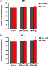
Fig. 7. Cell proliferations of HSF cells treated with GNRs@PSS and GNRs@C (3 μg mL−1) under LED irradiation and non LED irradiation and then incubated for (a) 24 h and (b) 48 h. * denotes significant difference in cell proliferation compared with scratched HSF cells with/without LED irradiation (P < 0.05; n ≥ 5).
Fig. 8
Fig. 8. Images of scratched HSF cells under different conditions, immunofluorescently labelled for Ki-67 (green fluorescence in the cell nuclei). (a) Scratched HSF cells without any treatment, (b) treated with GNRs@PSS, (c) treated with GNRs@C, (d) treated with LED irradiation, (e) treated with GNRs@PSS and LED irradiation, and (f) treated with GNRs@C and LED irradiation.
Fig. 9
Fig. 9. Gold contents in scratched HSF cells incubated with 3 μg mL−1 of GNRs@PSS and GNRs@C for 5 h and then exposed to LED irradiation for 5 min, followed by 24 and 48 h of incubation. * Denotes a significant difference in cellular uptake of scratched HSF cells compared with control and scratched HSF cells treated with GNRs@PSS for the same incubation period (P < 0.05; n ≥ 3).
Fig. 10
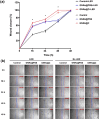
Fig. 10. (a) Percentages of wound closure in scratched HSF cells treated with 3 μg mL−1 GNRs@PSS and GNRs@C with/without LED irradiation for 0, 16, 24, 40, and 48 h.*Significant difference in wound closure compared with the control sample (untreated HSF cells). (P < 0.05; n ≥ 3). (b) Images of scratched HSF cell migration of scratched HSF cells treated with 3 μg mL−1 GNRs@PSS and GNRs@C with/without LED irradiation 0, 16, 24, 40, and 48 h.
Fig. 11
Fig. 11. Levels of (a) IL-6, (b) TNF-α, (c) VEGF, and (d) bFGF in scratched HSF cells treated with GNRs@PSS and GNRs@C with/without irradiation at 48 h post-wounding. In (a), * denotes a significant difference compared to scratched HSF cells with/without LED irradiation and ** denotes a significant difference compared to scratched HSF cells treated with GNRs@PSS plus LED irradiation. In (b), # denotes a significant difference compared to scratched HSF cells with/without LED irradiation. In (d), * denotes a significant difference in scratched HSF cells with GNRs@C with/without LED irradiation, ** denotes a significant difference compared to scratched HSF cells treated with GNRs@C with/without LED irradiation, # denotes a significant difference compared to control scratched HSF cells with/without LED irradiation (P < 0.05; n ≥ 3).

References

    1. Chen H. Shao L. Li Q. Wang J. Chem. Soc. Rev. 2013;42:2679–2724. - PubMed
    1. Pissuwan D. Valenzuela S. M. Cortie M. B. Biotechnol. Genet. Eng. Rev. 2008;25:93–112. doi: 10.5661/bger-25-93. - DOI - PubMed
    1. Salah D. Moghanm F. S. Arshad M. Alanazi A. A. Latif S. El-Gammal M. I. Shimaa E. M. Elsayed S. Diagnostics. 2021;11:1196. - PMC - PubMed
    1. Wan J. Wang J.-H. Liu T. Xie Z. Yu X.-F. Li W. Sci. Rep. 2015;5:1–16. - PMC - PubMed
    1. Chaves M. E. d. A. Araújo A. R. d. Piancastelli A. C. C. Pinotti M. An. Bras. Dermatol. 2014;89:616–623. doi: 10.1590/abd1806-4841.20142519. - DOI - PMC - PubMed

LinkOut - more resources