Review
. 2025 May 22;13(23):11499-11514.
doi: 10.1039/d4tc05420b. eCollection 2025 Jun 12.

Molecular spectroscopies with semiconductor metasurfaces: towards dual optical/chemical SERS

Affiliations
Review

Molecular spectroscopies with semiconductor metasurfaces: towards dual optical/chemical SERS

Alexander Berestennikov et al. J Mater Chem C Mater. .

Abstract

Surface-enhanced Raman spectroscopy (SERS) has emerged as a powerful technique for the ultra-sensitive detection of molecules and has been widely applied in many fields, ranging from biomedical diagnostics and environmental monitoring to trace-level detection of chemical and biological analytes. While traditional metallic SERS substrates rely predominantly on electromagnetic field enhancement, emerging semiconductor SERS materials have attracted growing interest because they offer the additional advantage of simultaneous chemical and electromagnetic enhancements. Here, we review some of the recent advancements in the design and optimization of semiconductor SERS substrates, with a focus on their dual enhancement mechanisms. We also discuss the transition from nanoparticle-based platforms to more advanced nanoresonator-based SERS metasurfaces, highlighting their superior sensing performance.


Conflict of interest statement

There are no conflicts to declare.

Figures

Fig. 1
Fig. 1. Advances in fabrication of metal and semiconductor SERS substrates. The progression illustrates the transition from simple colloidal particles to complex metasurfaces with precise nanoscale patterns, enabling enhanced Raman signal detection and broad application potential. (1995) Adapted with permission from ref. . Copyright 1995, AAAS. (2005) Adapted with permission from ref. . Copyright 2005, American Chemical Society. (2006) Adapted with permission from ref. . Copyright 2006, the Royal Society of Chemistry. (2009) Adapted with permission from ref. . Copyright 2009, American Chemical Society. (2006) Adapted with permission from ref. . Copyright 2006, the Royal Society of Chemistry. (2007) Adapted with permission from ref. . Copyright 2007, AIP Publishing. (2015) Adapted with permission from ref. . Copyright 2015, Springer Nature. (2018) Adapted with permission from ref. . Copyright 2018, American Chemical Society. (2024) Adapted with permission from ref. . Copyright 2024, John Wiley and Sons.
Fig. 2
Fig. 2. SERS enhancement mechanisms: (a) electromagnetic enhancement illustrated by LSPRs in metals and Mie resonances in semiconductors. (b) Chemical enhancement illustrating CT interactions between the metal or semiconductor energy bands and the molecular energy levels (HOMO and LUMO).
Fig. 3
Fig. 3. Strategies for enhancing CT mechanisms in semiconductor-based SERS substrates: (a) creation of oxygen vacancies, where vacancy states facilitate PICT between the CB and the molecular HOMO/LUMO levels. (b) Material doping illustrated by doping of ZnO NPs with Ga atoms, which adjusts the energy band alignment to optimize CT and enhance SERS activity. (a) (2019) Adapted with permission from ref. . Copyright 2019, John Wiley and Sons. (b) (2019) Adapted with permission from ref. . Copyright 2019, Frontiers Media SA.
Fig. 4
Fig. 4. Overview of advanced semiconductor-based metasurfaces for SERS applications: (a) scattering cross-sections of silicon nanospheres demonstrating multipole resonances (ED: electric dipole, MD: magnetic dipole, EQ: electric quadrupole, and MQ: magnetic quadrupole) and their contributions to SERS enhancement. (b) Bound states in the continuum (BIC) in optical systems, showing protected and unprotected channels for enhanced light–matter interactions. (c) SEM image of the gallium nitride L3 photonic crystal cavity, illustrating their potential for optical confinement and field enhancement. (d) Schematic representation of a label-free aptamer sensor based on silicon microring resonators, highlighting its functional application in SERS-based molecular detection. (a) (2020) Adapted with permission from ref. Copyright 2020, De Gruyter. (b) (2016) Adapted with permission from ref. . Copyright 2016, Springer Nature. (2017) Adapted with permission from ref. . Copyright 2017, American Chemical Society. (c) (2014) Adapted with permission from ref. . Copyright 2014, AIP Publishing. (d) (2013) Adapted with permission from ref. . Copyright 2013, Elsevier.
Fig. 5
Fig. 5. Examples of randomly grown semiconductor-based substrates for SERS applications: (a) SEM image of nanospheres with an inset showing the electric near-field distribution, demonstrating the “hot spots” due to Mie resonances. (b) SEM image of amorphous ZnO NPs. (c) Cross-sectional SEM image of Ge nanostructures, with the inset showing XRD patterns confirming their crystalline structure. (d) SEM images of the as-grown CuO nanowires. (a) (2019) Adapted with permission from ref. . Copyright 2019, John Wiley and Sons. (b) (2017) Adapted with permission from ref. . Copyright 2017, John Wiley and Sons. (c) (2011) Adapted with permission from ref. . Copyright 2011, American Chemical Society. (d) (2012) Adapted with permission from ref. . Copyright 2012, Elsevier.
Fig. 6
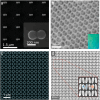
Fig. 6. Ordered nanostructured platforms for enhanced SERS applications: (a) SEM image of the Si dimer nanoantennas. (b) SEM image of a plasmon-free TiO2 photonic microarray, showing its periodic structure designed for enhanced light–matter coupling. (c) SEM image of the Si3N4 BIC photonic crystal. (d) Metasurface pattern featuring elliptical nanostructures, designed to exploit BICs for enhanced light–matter interactions. (a) (2018) Adapted with permission from ref. . Copyright 2018, American Chemical Society. (b) (2014) Adapted with permission from ref. . Copyright 2014, American Chemical Society. (c) (2018) Adapted with permission from ref. . Copyright 2018, American Chemical Society. (d) (2024) Adapted with permission from ref. . Copyright 2024, John Wiley and Sons.

Similar articles

References

    1. Vo-Dinh T. Sens. Actuators, B. 1995;29:183–189. doi: 10.1016/0925-4005(95)01681-3. - DOI
    1. Bantz K. C. Meyer A. F. Wittenberg N. J. Im H. Kurtuluş Ö. Lee S. H. Lindquist N. C. Oh S.-H. Haynes C. L. Phys. Chem. Chem. Phys. 2011;13:11551–11567. doi: 10.1039/C0CP01841D. - DOI - PMC - PubMed
    1. Ngo H. T. Wang H.-N. Fales A. M. Vo-Dinh T. Anal. Bioanal. Chem. 2016;408:1773–1781. doi: 10.1007/s00216-015-9121-4. - DOI - PubMed
    1. Muhammad M. Huang Q. Talanta. 2021;227:122188. doi: 10.1016/j.talanta.2021.122188. - DOI - PubMed
    1. Zhao H. Chin L. K. Shi Y. Liu P. Y. Zhang Y. Cai H. Yap E. P. H. Ser W. Liu A.-Q. Sens. Actuators, B. 2021;331:129428. doi: 10.1016/j.snb.2020.129428. - DOI

LinkOut - more resources