Comment
. 2025 Feb;32(1):181-187.
doi: 10.21315/mjms-01-2025-077. Epub 2025 Feb 28.

Delineating Neuroanatomical Structures for the Measurement of Temporal Horn Dilatation

Affiliations
Comment

Delineating Neuroanatomical Structures for the Measurement of Temporal Horn Dilatation

Devalagan Muthalagan et al. Malays J Med Sci. 2025 Feb.
No abstract available


Conflict of interest statement

Conflict of Interest: None.

Figures

Figure 1
Figure 1
The measurement taken for transverse width of temporal horn Note: Adapted from Frisoni et al. (3); Parallel lines are drawn tangentially to the tip of the temporal horn where the width is maximum (arrows), at the level whereby the right and left temporal horns can be appreciated in their full length
Figure 2
Figure 2
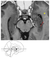
Head of hippocampus, uncal apex, temporal (inferior) horn of the lateral ventricle, amygdala, body of hippocampus, cornu Ammonis and collateral eminence (ce) Note: Adapted from Duvernoy et al. (Figure 7.26, pp. 212) (4); 1, 2 = head of hippocampus; 3 = uncal apex; 4 = temporal (inferior) horn of the lateral ventricle; 5 = amygdala; black star = body of hippocampus; red arrow = cornu Ammonis; yellow star = collateral eminence (ce)
Figure 3
Figure 3
The collateral eminence, 4 bordering the lateral aspect of the hippocampus at the level of the upper mesencephalon, which constitutes the floor of the temporal horn. Note: Adapted from Duvernoy et al. (Figure 4.20, pp 63) (4)
Figure 4
Figure 4
Ten contiguous 2-mm-thick CT axial slices
Figure 5
Figure 5
The second method, whereby (B) shows an axial slice for which ventricular measurements for temporal horns of the lateral ventricles are undertaken; which is typically best seen at the level of the petrous apex Note: Adapted from Greenberg (Figure 24.4, pp. 432) (5)
Figure 6
Figure 6
Head of hippocampus, uncal apex, temporal (inferior) horn of the lateral ventricle, amygdala, body of hippocampus and amygdalohippocampal transition area (AHi) Note: Adapted from Duvernoy et al. (Figure 7.26, pp. 212) (4); 1, 2 = head of hippocampus; 3 = uncal apex; 4 = temporal (inferior) horn of the lateral ventricle; 5 = amygdala; black star = body of hippocampus; yellow arrow = amygdalohippocampal transition area (AHi)
Figure 7
Figure 7
The anterior extent of the temporal horn can be depicted by the amygdalohippocampal transition area (AHi) (blue arrow), which delineates the amygdala (A) from the hippocampus, labelled as CA (cornu Ammonis) Note: Adapted from Schaltenbrand and Wahren (6)
Figure 8
Figure 8
CT images of the similar 3 years old child with grade 4 medulloblastoma as in the case in Figure 4
Figure 9
Figure 9
MRI Brain of the similar 3 years old child with grade 4 medulloblastoma as in the case in Figure 4

Comment on

References

    1. Zakaria Z, Van Rostenberghe H, Ramli N, Suhaimi MS, Hazlan SNH, Abdullah JM. The key aspects of neonatal and infant neurological examination: the Ballard score, the infant’s head with hydrocephalus and assessment in a clinical setting. Malays J Med Sci. 2023;30(4):193–206. doi: 10.21315/mjms2023.30.4.16. - DOI - PMC - PubMed
    1. Missori P, Paolini S, Peschillo S, Mancarella C, Scafa AK, Rastelli E, et al. Temporal horn enlargements predict secondary hydrocephalus diagnosis earlier than evans’ index. Tomography. 2022;8(3):1429–1436. doi: 10.3390/tomography8030115. - DOI - PMC - PubMed
    1. Frisoni GB, Geroldi C, Beltramello A, Bianchetti A, Binetti G, Bordiga G, et al. Radial width of the temporal horn: a sensitive measure in Alzheimer disease. AJNR Am J Neuroradiol. 2002;23(1):35–47. - PMC - PubMed
    1. Duvernoy H, Cattin F, Risold P. The human hippocampus: functional anatomy, vascularization and serial sections with MRI. 4th ed. Heidelberg: Springer; 2013. - DOI
    1. Greenberg MS. Greenberg’s handbook of neurosurgery. 10th ed. New York: Thieme; 2023.

LinkOut - more resources