. 2025 May 9:16:1503961.
doi: 10.3389/fphar.2025.1503961. eCollection 2025.

Sanqi oral solution alleviates podocyte apoptosis in experimental membranous nephropathy by mediating EMT through the ERK/CK2-α/β-catenin pathway

Affiliations

Sanqi oral solution alleviates podocyte apoptosis in experimental membranous nephropathy by mediating EMT through the ERK/CK2-α/β-catenin pathway

Xiaowan Wang et al. Front Pharmacol. .

Abstract

Introduction: Sanqi oral solution (SQ) is a Chinese medicine that has been used well to treat idiopathic membranous nephropathy (IMN). It has been demonstrated to mitigate IMN proteinuria by inhibiting podocyte apoptosis. however, the precise mechanism has not been fully elucidated.

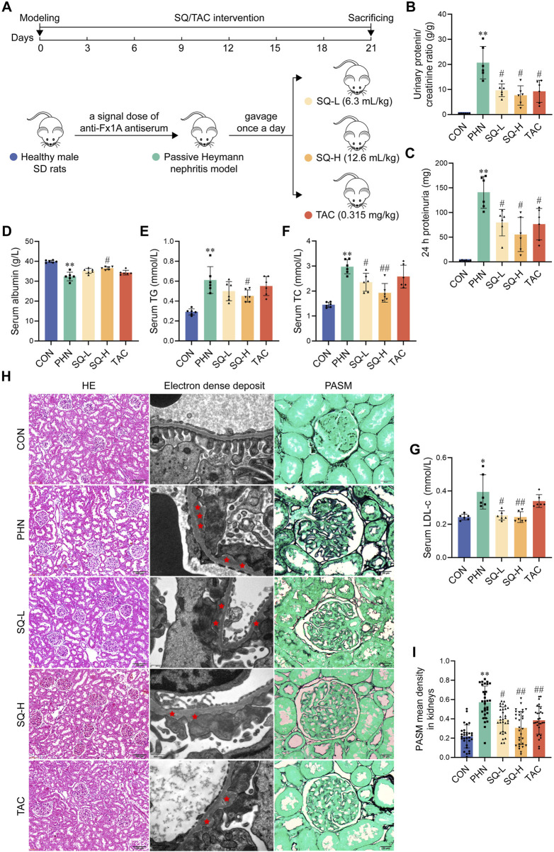
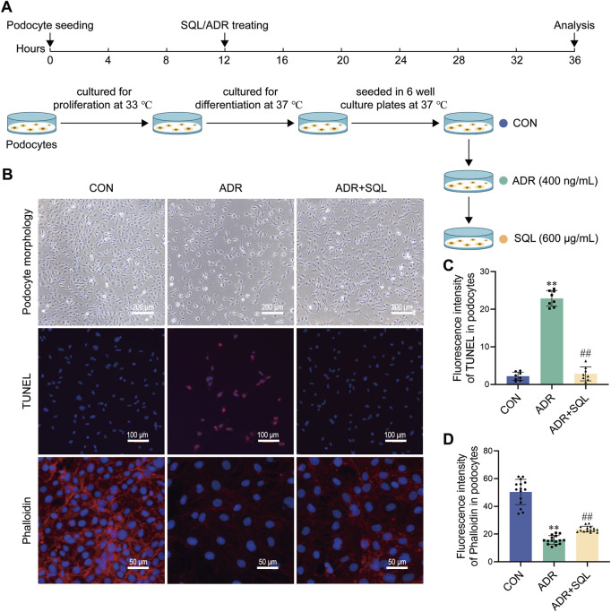
Methods: A passive Heymann nephropathy (PHN) rat model was used to mimic the in vivo disease characteristics of IMN. The PHN rats were intragastrically administered SQ (12.6/6.3 mL/kg) or tacrolimus (0.315 mg/kg) for 21 days. SQ was applied to ADR-induced podocytes in vitro. The effects of SQ on IMN and its underlying mechanisms were determined by measuring biochemical indices, pathomorphological characteristics, membrane attack complex (MAC), cell morphology, and protein levels.

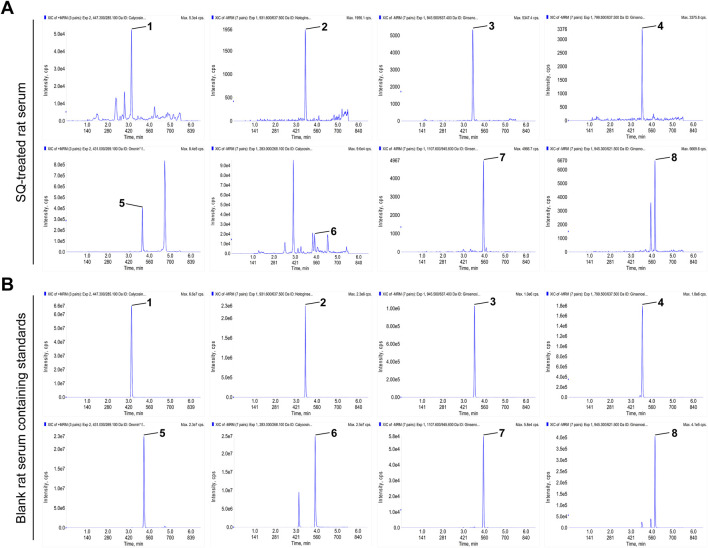
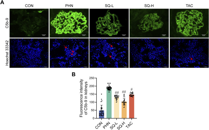
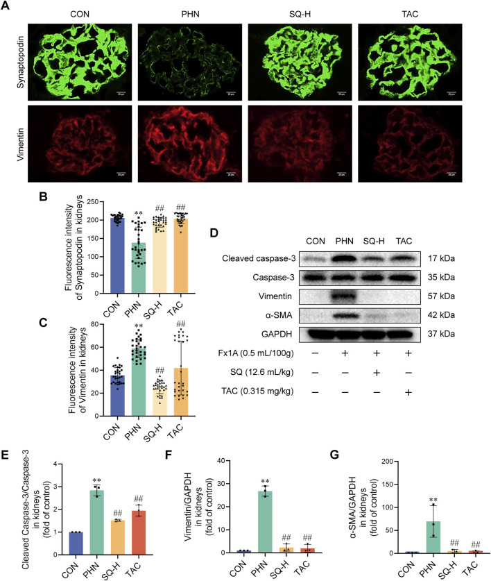
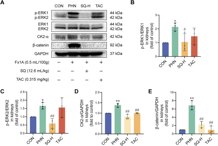
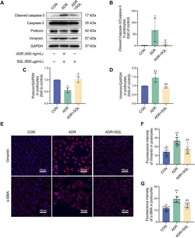
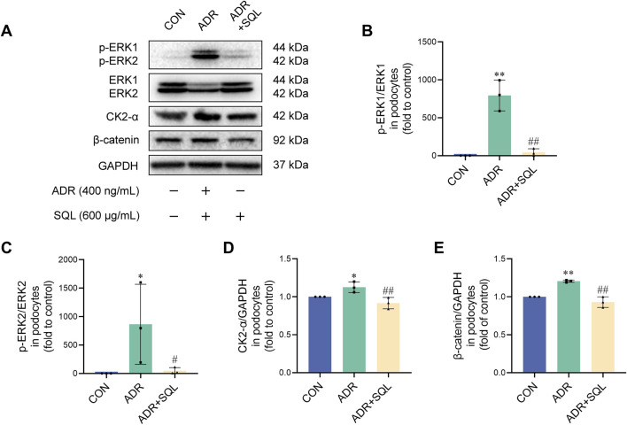
Results: The SQ ingredients found in rat serum underscored their successful absorption in rats. In PHN rats, SQ induced a significant reduction in proteinuria, MAC, C5b-9, and glomerular basement membrane thickness, along with a drop in apoptotic podocytes. Similarly, SQ exerted a protective effect against ADR-induced podocyte injury by inhibiting apoptosis. Furthermore, inhibition of the ERK/CK2-α/β-catenin pathway-mediated epithelial-to-mesenchymal transition (EMT) was found to be involved in the anti-apoptotic effect of SQ in PHN rats and podocytes, marked by the reduction in vimentin and α-SMA and the induction of Synaptopodin and Podocin protein levels.

Conclusion: Inhibition of EMT via the ERK/CK2-α/β-catenin pathway may be the main mechanism by which SQ suppresses podocyte apoptosis in IMN.

Keywords: apoptosis; epithelialmesenchymal transition; passive heymann nephropathy; sanqi oral solution; the ERK/CK2-α/β-catenin pathway.


Conflict of interest statement

The authors declare that the research was conducted in the absence of any commercial or financial relationships that could be construed as a potential conflict of interest.

Figures

FIGURE 1
FIGURE 1
The main active ingredients of SQ were absorbed by rats. (A, B) MRM chromatograms of the main active ingredients of SQ in SQ-treated rat serum and blank rat serum-containing standards. (1) caylcosin-7-O-β-D-glucopyranoside, (2) notoginsenoside R1, (3) ginsenoside Re, (4) ginsenoside Rg1, (5) ononin, (6) calycosin, (7) ginsenoside Rb1, and (8) ginsenoside Rd.
FIGURE 2
FIGURE 2
SQ relieved albuminuria, hyperlipidemia, and glomerular histopathological injury in PHN rats. (A) Diagram of the animal experiments. (B, C) Proteinuria was detected in rats from all groups, and the urinary protein/creatinine ratio and 24 h proteinuria were calculated (n = 6). (D–G) Serum albumin, TG, TC, and LDL-c levels were measured in the peripheral blood of rats in all the groups (n = 6). (H) HE staining (magnification ×200) and Transmission Electron Microscopy (magnification ×30000) showed overall histomorphological changes and microscopic electron dense deposits in the kidneys of all groups, and PASM staining (magnification ×1000) indicated the thickness of GBM in the glomeruli of all groups (n = 6). (I) The thickness of the GBM was semi-quantified (n = 6). Data are presented as the mean ± SD from independent groups. *p < 0.05 vs. the CON group. **p < 0.01 vs. the CON group. #p < 0.05 vs. the PHN group. ##p < 0.01 vs. the PHN group.
FIGURE 3
FIGURE 3
SQ reduced glomerular apoptotic podocytes in PHN rats. (A, B) C5b-9 assembly was visualized by immunofluorescence staining (magnification ×1000) and semi-quantified using the ImageJ software. A Hoechst 33342 staining (magnification ×400) revealed apoptotic podocytes. Data are presented as the mean ± SD from independent groups. **p < 0.01 vs. the CON group. #p < 0.05 vs. the PHN group. ##p < 0.01 vs. the PHN group.
FIGURE 4
FIGURE 4
SQ reduced apoptotic podocytes by inhibiting EMT in PHN rats. (A–C) Synaptopodin (magnification ×1000) and Vimentin (magnification ×1000) were visualized by immunofluorescence staining and semi-quantified using the ImageJ software. (D–G) Protein levels of cleaved caspase-3, Caspase-3, vimentin, and α-SMA in the kidneys of all groups were detected by Western blotting. Data are presented as the mean ± SD from independent groups. **p < 0.01 vs. the CON group. ##p < 0.01 vs. the PHN group.
FIGURE 5
FIGURE 5
The ERK/CK2-α/β-catenin pathway mediated the effects of SQ on EMT in PHN rats. (A–E) Protein expression of p-ERK1, ERK1, p-ERK2, ERK2, CK2-α, and β-catenin was evaluated in the kidneys of all groups by Western blotting. Data are presented as the mean ± SD from independent groups. *p < 0.05 vs. the CON group. **p < 0.01 vs. the CON group. #p < 0.05 vs. the PHN group. ##p < 0.01 vs. the PHN group.
FIGURE 6
FIGURE 6
SQ alleviated ADR-induced podocyte injury. (A) Diagram of the cell experiments. (B–D) Morphological changes (magnification ×100) in podocytes were recorded using a light microscope, the nuclear DNA fragmentation of the podocyte was visualized by TUNEL staining (magnification ×200), and the podocyte cytoskeleton (magnification ×400) was visualized by immunofluorescence staining. Semi-quantified using the ImageJ software. Data are presented as the mean ± SD from independent groups. **p < 0.01 vs. the CON group. ##p < 0.01 vs. the PHN group.
FIGURE 7
FIGURE 7
SQ reduced apoptosis in ADR-injured podocytes by suppressing EMT. (A–D) Cleaved caspase-3, Caspase-3, podocin, and vimentin protein levels in podocytes were measured by Western blotting. (E–G) Vimentin (magnification ×200) and α-SMA (magnification ×200) in podocytes were visualized (E) by immunofluorescence staining and semi-quantified using ImageJ software. Data are presented as the mean ± SD from independent groups. *p < 0.05 vs. the CON group. **p < 0.01 vs. the CON group. #p < 0.05 vs. the PHN group. ##p < 0.01 vs. the PHN group.
FIGURE 8
FIGURE 8
SQ inhibited the ERK/CK2-α/β-catenin pathway in ADR-injured podocytes. (A–E) Protein expression of p-ERK1, ERK1, p-ERK2, ERK2, CK2-α, and β-catenin in podocytes was measured by Western blotting. Data are presented as the mean ± SD from independent groups. *p < 0.05 vs. the CON group. **p < 0.01 vs. the CON group. #p < 0.05 vs the PHN group. ##p < 0.01 vs. the PHN group.

Similar articles

References

    1. Cai A., Meng Y., Zhou H., Cai H., Shao X., Wang Q., et al. (2024). Podocyte pathogenic bone morphogenetic protein-2 pathway and immune cell behaviors in primary membranous nephropathy. Adv. Sci. Weinheim, Baden-Wurttemberg, Ger. 11, e2404151. 10.1002/advs.202404151 - DOI - PMC - PubMed
    1. Chen S. M., Wang J., Wang J. Y., Jia X. Y., Xuan Z. H., Cheng Z. L., et al. (2023). Wnt/β-catenin signaling pathway promotes abnormal activation of fibroblast-like synoviocytes and angiogenesis in rheumatoid arthritis and the intervention of Er Miao San. Phytomedicine 120, 155064. 10.1016/j.phymed.2023.155064 - DOI - PubMed
    1. Dai C. S., Stolz D. B., Kiss L. P., Monga S. P., Holzman L. B., Liu Y. H. (2009). Wnt/beta-catenin signaling promotes podocyte dysfunction and albuminuria. J. Am. Soc. Nephrol. 20 (9), 1997–2008. 10.1681/ASN.2009010019 - DOI - PMC - PubMed
    1. Dai X. G., Feng J. X., Chen J. (2013). Syndrome differentiation and treatment of chronic kidney disease treated with traditional Chinese medicine. Chin. J. Integr. Tradit. West. Nephrol. 14 (10), 933–934.
    1. Dan Hu Q., Wang H.-L., Liu J., He T., Tan R.-Z., Zhang Q., et al. (2023). Btg2 promotes focal segmental glomerulosclerosis via smad3-dependent podocyte-mesenchymal transition. Adv. Sci. Weinheim, Baden-Wurttemberg, Ger. 10 (32), e2304360. 10.1002/advs.202304360 - DOI - PMC - PubMed

LinkOut - more resources