

Glioblastoma Mimicking Autoimmune Encephalitis
- PMID: 40417244
- PMCID: PMC12102076
- DOI: 10.1177/19418744251343174
Glioblastoma Mimicking Autoimmune Encephalitis
Abstract
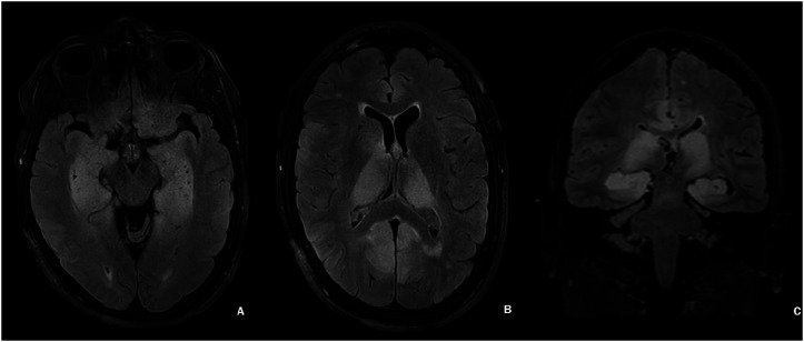
A 66-year-old female patient presented with progressive short-term memory loss over a period of three months and mild gait imbalance. MRI of the brain demonstrated symmetric expansile T2 FLAIR hyperintensities within the bilateral mesial temporal lobes, thalami, and cingulate gyri (Figure 1). Due to the symmetry of the signal changes in the bilateral cerebral hemispheres and especially the mesial temporal lobes, an autoimmune encephalitis was strongly favored on imaging. Glioblastoma was a consideration on the initial scan; however, it was thought to be much less likely, and the patient received immunosuppression with plasmapheresis and IV steroids. At that time, it was even presumed that the patient improved mildly with plasmapheresis. The patient was discharged on PO steroids; however, a few weeks later the patient presented in status epilepticus. On repeat MRI brain, findings were not significantly changed, and the diagnosis of an autoimmune process was again favored on imaging. The patient received plasmapheresis and IV steroids. However, on the second admission the patient's neurologic function was markedly below baseline per the family's report. The patient had received an extensive autoimmune, infectious and metabolic work up, including testing for Creutzfeldt-Jacob disease, with all the tests coming back negative. Therefore, a brain biopsy was performed to understand the underlying pathology, IDH wild-type glioblastoma. On MRI, the expansile signal changes were bilateral and multifocal, affecting more than three cerebral lobes. This is a case of gliomatosis cerebri, which was misdiagnosed as autoimmune encephalitis due to the symmetry of cerebral involvement.
Keywords: autoimmune encephalitis; clinical specialty; epilepsy; glioblastoma; gliomatosis cerebri; neuroradiology; seizures.
© The Author(s) 2025.
Conflict of interest statement
The author(s) declared no potential conflicts of interest with respect to the research, authorship, and/or publication of this article.
Figures
References
LinkOut - more resources
Full Text Sources