. 2025 May 13;16(24):11110-11122.
doi: 10.1039/d5sc01863c. eCollection 2025 Jun 18.

Evolutionary insights into the selectivity of sterol oxidising cytochrome P450 enzymes based on ancestral sequence reconstruction

Affiliations

Evolutionary insights into the selectivity of sterol oxidising cytochrome P450 enzymes based on ancestral sequence reconstruction

Daniel Z Doherty et al. Chem Sci. .

Abstract

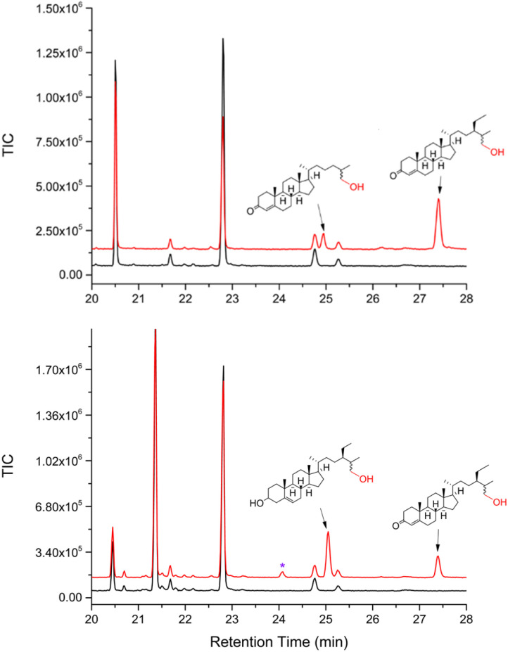
The cytochrome P450 (CYP) enzyme CYP125A1 is a crucial enzyme for the long-term survival and pathogenicity of Mycobacterium tuberculosis. CYP125 genes are found not only in pathogenic mycobacteria but are also widely dispersed within the Actinobacteria phylum, with many species possessing multiple copies of CYP125 encoding genes. Their primary function is the catalytic hydroxylation of the terminal methyl group of cholesterol and phytosterols. We have previously shown that CYP125 enzymes from distinct mycobacteria have substrate selectivity preferences for animal versus plant steroid oxidation. An evolutionary understanding of this selectivity is not known. Here, we use Ancestral Sequence Reconstruction (ASR), to support the hypothesis that some CYP125 enzymes evolved in a manner reflective of their adaptation to a pathogenic niche. We constructed a maximum-likelihood, most-recent common ancestor of the CYP125 clade (CYP125MRCA). We were then able to produce and characterise this enzyme both functionally and structurally. We found that CYP125MRCA was able to catalyse the terminal hydroxylation of cholesterol, phytosterols, and vitamin D3 (cholecalciferol); the latter was hydroxylated at both C-25 and C-26. This is the first example to date of vitamin D3 oxidation by a CYP125 enzyme, thereby demonstrating an increased substrate range of CYP125MRCA relative to its characterised extant relatives. The X-ray crystal structures of CYP125MRCA bound with sitosterol and vitamin D3 were determined, providing important insight into the changes that enable the expanded substrate range.


Conflict of interest statement

There are no conflicts to declare.

Figures

Scheme 1
Scheme 1. Oxidation of the cholesterol side-chain by CYP125 enzymes.
Fig. 1
Fig. 1. Ancestral Sequence Reconstruction of CYP125MRCA. Only a portion of the constructed phylogenetic tree is shown for clarity (see Fig. S1 for full tree). CYP125A1 was used as the search sequence (shown in magenta). The ancestrally reconstructed CYP125MRCA node is highlighted in blue.
Fig. 2
Fig. 2. CYP125MRCA can bind and oxidise both animal and phytosterols. (a) Terminal methyl oxidation of both animal and phytosterols in their 3-keto or 3-hydroxy forms by CYP125MRCA. (b) Bar-chart demonstrating the binding constants (Kd) of various animal and phytosterols to CYP125MRCA. * Indicates that the substrate bound but was not oxidised. (c) GC chromatogram of the in vitro oxidation of cholest-4-en-3-one (left) and sitosterol (right) by CYP125MRCA using a reconstituted NADPH and spinach ferredoxin/ferredoxin reductase electron transfer system. * Indicates substrate/product peaks arising from a campesterol impurity in the sitosterol sample. The enzymatic reactions are shown in red and the no P450 substrate control reactions are shown in black. (d) Crystal structure of CYP125MRCA in complex with sitosterol. Left: Feature-enhanced map depicting the electron density (σ = 1.0, carve radius = 2.0) of the sitosterol substrate within the CYP125MRCA active-site (sitosterol in green, protein in yellow). Right: The CYP125MRCA structure overlaid with cholest-4-en-3-one (magenta) bound CYP125A1 (blue, PDB ID: 2X5W). Residues shown are within 5 Å of the substrates.
Fig. 3
Fig. 3. CYP125MRCA is capable of oxidising vitamin D3 (a) Proposed oxidation of vitamin D3 (cholecalciferol) and isomer by CYP125MRCA. The blue arrows indicate the pathway in which isomerism occurs before hydroxylation by CYP125MRCA, while the magenta arrows indicate the opposite pathway, in which isomerism occurs after hydroxylation. While isotachysterol is shown here as the alternative isomer leading to hydroxylation, due to acidification of the sample prior to GC-MS, hydroxylation of the other isomers cannot be ruled out. (b) The shift of the Soret band of CYP125MRCA in the UV-Vis region that is induced by the binding of vitamin D3 in the active-site. (c) GC chromatogram of the in vitro oxidation of vitamin D3 by CYP125MRCA using a reconstituted NADPH and spinach ferredoxin/ferredoxin reductase electron transfer system. * Indicates the two substrate peaks. * (blue) are products arising from hydroxylation and * (green) is a partially underivatised hydroxylation product. The enzymatic reaction is shown in red and the no P450 substrate control reaction in black. Left: Crystal structure of CYP125MRCA in complex with vitamin D3 (vitamin D3 in orange, protein in purple). The feature-enhanced map depicting the electron density of the substrate (σ = 1.0, carve radius = 2.0) within the CYP125MRCA active-site Right: The CYP125MRCA-vitamin D3-bound structure overlaid with the CYP125MRCA sitosterol-bound structure, with the sitosterol-bound structure residues shown in yellow and the sitosterol ligand in green. Residues shown are within 5 Å of the substrates.
Fig. 4
Fig. 4. GC-MS chromatograms of the competitive oxidations of 1 : 1 mixtures of cholest-4-en-3-one versus stigmast-4-en-3-one (top), and sitosterol versus stigmast-4-en-3-one (bottom). The substrate control is shown in black and the enzyme catalysed oxidation reaction in red. * indicates a product peak arising from a campesterol impurity in the sitosterol stock. The top chromatogram shows a greater amount of the plant sterol stigmast-4-en-3-one 26-hydroxylated product than that of the animal sterol cholest-4-en-3-one (ratio of 3.2 : 1 versus a ratio of 1.5 : 1 for an equimolar mixture of the substrates). The bottom chromatogram demonstrates a slightly greater amount of hydroxylated product for sitosterol over the 3-keto form stigmast-4-en-3-one (a ratio of 1.9 : 1 versus a ratio of 1.2 : 1 for an equimolar mixture of the substrates).

References

    1. Ouellet H. Guan S. Johnston J. B. Chow E. D. Kells P. M. Burlingame A. L. Cox J. S. Podust L. M. de Montellano P. R. Mol. Microbiol. 2010;77:730–742. doi: 10.1111/j.1365-2958.2010.07243.x. - DOI - PMC - PubMed
    1. Rosloniec K. Z. Wilbrink M. H. Capyk J. K. Mohn W. W. Ostendorf M. van der Geize R. Dijkhuizen L. Eltis L. D. Mol. Microbiol. 2009;74:1031–1043. doi: 10.1111/j.1365-2958.2009.06915.x. - DOI - PMC - PubMed
    1. McLean K. J. Lafite P. Levy C. Cheesman M. R. Mast N. Pikuleva I. A. Leys D. Munro A. W. J. Biol. Chem. 2009;284:35524–35533. doi: 10.1074/jbc.M109.032706. - DOI - PMC - PubMed
    1. McLeod M. P. Warren R. L. Hsiao W. W. Araki N. Myhre M. Fernandes C. Miyazawa D. Wong W. Lillquist A. L. Wang D. Dosanjh M. Hara H. Petrescu A. Morin R. D. Yang G. Stott J. M. Schein J. E. Shin H. Smailus D. Siddiqui A. S. Marra M. A. Jones S. J. Holt R. Brinkman F. S. Miyauchi K. Fukuda M. Davies J. E. Mohn W. W. Eltis L. D. Proc. Natl. Acad. Sci. U. S. A. 2006;103:15582–15587. doi: 10.1073/pnas.0607048103. - DOI - PMC - PubMed
    1. Haussmann U. Wolters D. A. Franzel B. Eltis L. D. Poetsch A. J. Proteome Res. 2013;12:1188–1198. doi: 10.1021/pr300816n. - DOI - PubMed

LinkOut - more resources