Review
. 2024 Oct 17;6(4):332-339.
doi: 10.24875/RECIC.M24000476. eCollection 2024 Oct-Dec.

[The role of implant projection in optimizing transcatheter aortic valve implantation]

[Article in Spanish]
Affiliations
Review

[The role of implant projection in optimizing transcatheter aortic valve implantation]

[Article in Spanish]
Rut Álvarez-Velasco et al. REC Interv Cardiol. .

Abstract

Severe aortic stenosis is the most frequent valve condition requiring surgery, and its incidence is increasing yearly. Transcatheter aortic valve implantation (TAVI) is the first-line treatment for patients at all levels of surgical risk. Nevertheless, modifications to the procedure often appear to improve clinical outcomes. A major concern after TAVI is the higher rate of permanent pacemaker implantation (PPMI) compared with surgical valve replacement. Optimal implantation depth is crucial to reduce the burden of PPMI without causing serious complications such as valve embolization. The classic implantation technique, where the 3 cusps are aligned in the same plane, has been modified to a cusp overlap projection by isolating the noncoronary cusp and superimposing the left and right cusps. This simple modification provides optimal visualization during deployment and helps to achieve the desired implant depth to reduce conduction disturbances and PPMI. Another limitation after TAVI is coronary reaccess due to the frame of the transcatheter valve obstructing the coronary ostia. Commissural alignment of the prostheses with the native valve may facilitate selective cannulation of the coronary arteries after this procedure. This review will discuss the techniques and supporting evidence for these modifications to the deployment and implant projection methods, and how they can improve TAVI outcomes.

Keywords: Commissural alignment; Cusp overlap projection; Transcatheter aortic valve replacement.


Conflict of interest statement

CONFLICTO DE INTERESES: Ninguno.

Figures
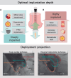
Figure 1
Figure 1. A: achieving optimal implantation depth is fundamental to improving outcomes. A deeply implanted valve may impair mitral valve function, produce paravalvular leaks, and interact with the conduction system, increasing permanent pacemaker rates. In contrast, high valve implantation may produce coronary obstruction, valve embolization, and paravalvular leak. B: cusp overlap projection where the left and right cusp overlap on the right side of the screen, isolating the noncoronary cusp, has been shown to optimize implantation depth. CAU, caudal; CRA, cranial; PPM, permanent pacemaker; RAO, right anterior oblique.
Figure 2
Figure 2. Central illustration. Minor modifications during deployment can achieve commissural alignment (left side of the panel), facilitate coronary access in future procedures, reduce the burden of coronary occlusion in redo-TAVI, and improve valve hemodynamics. A cusp overlap projection (right side of the panel) can improve the implanter’s view to help optimize implantation depth, reduce conduction disturbances and permanent pacemaker rates, and subsequently improve outcomes and length of stay. ECG, electrocardiogram; PPM, permanent pacemaker; TAVI, transcatheter aortic valve implantation.
Figure 3
Figure 3. Step-by-step tutorial for commissural alignment in self-expanding valves. A: Evolut FX valve. First, the flush port is positioned at 3 o’clock. The hat marker band must be placed toward the outer curvature when advancing the valve in the descending aorta. Second, during deployment, the hat marker faces the NCC. Finally, 2 of the radiopaque markers of the Evolut FX valve should be viewed on the left side of the screen and the other marker on the right. B: Acurate Neo2 valve. First, insert the valve with the flush port positioned to 6 o’clock. Second, torque the delivery catheter counterclockwise. Finally, during valve deployment, 2 radiopaque posts should be viewed in the major curve of the aorta and 1 on the other side. Using the classic implantation technique, 1 post should be viewed in the middle of the aortic annulus and the other 2 on each side. CIT, classic implantation technique; COP, cusp overlapping projection.
Figure 4
Figure 4. Major predictors of impaired coronary reaccess. THV, transcatheter heart valve.
Figura 1
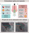
Figura 1. A: lograr una profundidad óptima de implante es esencial para mejorar los resultados. Una válvula implantada profundamente podría afectar la función de la válvula mitral, provocar fugas periprotésicas llegando, incluso, a interactuar con el sistema de conducción aumentando las tasas de implante de marcapasos permanente. Por otro lado, un implante alto de la válvula podría provocar obstrucciones coronarias, embolizaciones de la válvula y fugas periprotésicas. B: se ha demostrado que la proyección de superposición de cúspides, donde las cúspides izquierda y derecha se superponen en el lado derecho de la pantalla, aislando la cúspide no coronaria, sirve para optimizar la profundidad de implante. CAU: caudal; CRA: craneal; MP: marcapasos permanente; OAD: oblicuo anterior derecho.
Figura 2
Figura 2. Figura central. Pequeñas modificaciones durante el implante pueden conseguir el alineamiento comisural (lado izquierdo del panel), facilitar el acceso coronario en futuras intervenciones, reducir el riesgo de oclusiones coronarias en el redo-TAVI y mejorar la hemodinámica valvular. Una proyección de superposición de cúspides (lado derecho del panel) mejora la visión del implantador para ayudar a optimizar la profundidad de implante, reduce los trastornos de la conducción y las tasas de implante de marcapasos permanente y, en última instancia, mejora tanto los resultados como la duración de la hospitalización. ECG: electrocardiograma; IMPP: implante de marcapasos permanente; TAVI: implante percutáneo de válvula aórtica.
Figura 3
Figura 3. Tutorial paso a paso para alineamientos comisurales en válvulas autoexpandibles. A: válvula Evolut FX. En primer lugar, se coloca el puerto de lavado en la posición de las 3 en punto. La banda de marcaje en forma de sombrero se debe colocar mirando a la curvatura externa mientras se avanza la válvula por la aorta descendente. Después, durante la fase de liberación de la válvula, la banda debe mirar a la CNC. Por último, 2 de los marcadores radiopacos de la válvula Evolut FX deben verse en el lado izquierdo de la pantalla y el tercero en el lado derecho. B: válvula Acurate Neo2. En primer lugar, insertar la válvula con el puerto de lavado en la posición de las 6 en punto. Después, girar el catéter de liberación en sentido contrario al de las ajugas del reloj. Por último, durante la fase de despliegue de la válvula, deben verse 2 postes radiopacos en la curva mayor de la aorta y el tercero en el otro lado. Empleando la técnica clásica de implante, 1 poste debe visualizarse en el centro del anillo aórtico y los otros 2 a cada lado. TIC: técnica de implante clásica; TSC: técnica de superposición de cúspides.
Figura 4
Figura 4. Principales predictores de un acceso coronario deteriorado. THV: prótesis valvular cardiaca percutánea.

References

    1. Ferrer-Sistach E, Teis A, Bayés-Genís A, Delgado V. Multimodality imaging in aortic stenosis:new diagnostic and therapeutic frontiers. Rev Esp Cardiol. 2023;76:40-46. - PubMed
    2. Ferrer-Sistach E, Teis A, Bayés-Genís A, Delgado V. Multimodality imaging in aortic stenosis:new diagnostic and therapeutic frontiers. Rev Esp Cardiol. 2023;76:40–46. - PubMed
    1. D'Arcy JL, Coffey S, Loudon MA, et al. Large-scale community echocardiographic screening reveals a major burden of undiagnosed valvular heart disease in older people:the OxVALVE Population Cohort Study. Eur Heart J. 2016;37:3515-3522. - PMC - PubMed
    2. D'Arcy JL, Coffey S, Loudon MA, et al. Large-scale community echocardiographic screening reveals a major burden of undiagnosed valvular heart disease in older people:the OxVALVE Population Cohort Study. Eur Heart J. 2016;37:3515–3522. - PMC - PubMed
    1. Nguyen V, Willner N, Eltchaninoff H, et al. Trends in aortic valve replacement for aortic stenosis:a French nationwide study. Eur Heart J. 2022;43:666-679. - PubMed
    2. Nguyen V, Willner N, Eltchaninoff H, et al. Trends in aortic valve replacement for aortic stenosis:a French nationwide study. Eur Heart J. 2022;43:666–679. - PubMed
    1. Vahanian A, Beyersdorf F, Praz F, et al. 2021 ESC/EACTS Guidelines for the management of valvular heart disease. Eur Heart J. 2022;43:561-632. - PubMed
    2. Vahanian A, Beyersdorf F, Praz F, et al. 2021 ESC/EACTS Guidelines for the management of valvular heart disease. Eur Heart J. 2022;43:561–632. - PubMed
    1. Pinar E, García de Lara J, Hurtado J, et al. Cost-effectiveness analysis of the SAPIEN 3 transcatheter aortic valve implant in patients with symptomatic severe aortic stenosis. Rev Esp Cardiol. 2022;75:325-333. - PubMed
    2. Pinar E, García de Lara J, Hurtado J, et al. Cost-effectiveness analysis of the SAPIEN 3 transcatheter aortic valve implant in patients with symptomatic severe aortic stenosis. Rev Esp Cardiol. 2022;75:325–333. - PubMed

LinkOut - more resources