Review
. 2025 May 9:19:1570596.
doi: 10.3389/fncel.2025.1570596. eCollection 2025.

Mitochondria: the hidden engines of traumatic brain injury-driven neurodegeneration

Affiliations
Review

Mitochondria: the hidden engines of traumatic brain injury-driven neurodegeneration

Olusola A Olatona et al. Front Cell Neurosci. .

Abstract

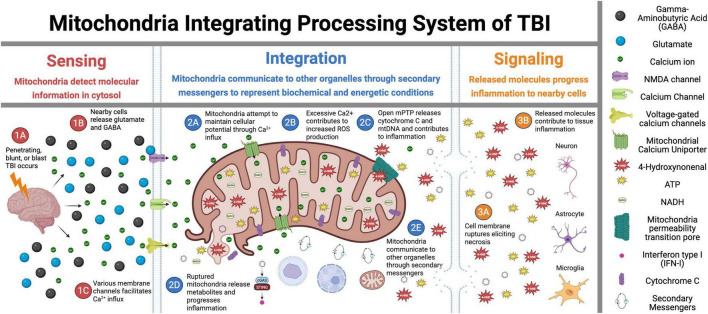
Mitochondria play a critical role in brain energy metabolism, cellular signaling, and homeostasis, making their dysfunction a key driver of secondary injury progression in traumatic brain injury (TBI). This review explores the relationship between mitochondrial bioenergetics, metabolism, oxidative stress, and neuroinflammation in the post-TBI brain. Mitochondrial dysfunction disrupts adenosine triphosphate (ATP) production, exacerbates calcium dysregulation, and generates reactive oxygen species, triggering a cascade of neuronal damage and neurodegenerative processes. Moreover, damaged mitochondria release damage-associated molecular patterns (DAMPs) such as mitochondrial DNA (mtDNA), Cytochrome C, and ATP, triggering inflammatory pathways that amplify tissue injury. We discuss the metabolic shifts that occur post-TBI, including the transition from oxidative phosphorylation to glycolysis and the consequences of metabolic inflexibility. Potential therapeutic interventions targeting mitochondrial dynamics, bioenergetic support, and inflammation modulation are explored, highlighting emerging strategies such as mitochondrial-targeted antioxidants, metabolic substrate supplementation, and pharmacological regulators of mitochondrial permeability transition pores. Understanding these mechanisms is crucial for developing novel therapeutic approaches to mitigate neurodegeneration and enhance recovery following brain trauma.

Keywords: bioenergetics; brain injury; metabolism; mitochondria; neurodegeneration.


Conflict of interest statement

The authors declare that the research was conducted in the absence of any commercial or financial relationships that could be construed as a potential conflict of interest.

Figures

FIGURE 1
FIGURE 1
Mitochondrial signal transduction in traumatic brain injury (TBI). This figure illustrates the sequential role of mitochondria in TBI across three critical phases: Sensing, Integration, and Signaling. In the Sensing phase (1A–1C), the initial trauma triggers glutamate release and activation of membrane ion channels, initiating the early cellular responses. During the Integration phase (2A–2E), mitochondria interpret injury signals through calcium influx, reactive oxygen species (ROS) production, and mitochondria permeability pore (mPTP) opening while also interacting with other cellular components. The Signaling phase (3A–3E) demonstrates how these integrated responses lead to mitochondria membrane rupture, necrosis, and inflammatory propagation to surrounding tissues. Overall, this figure depicts the central role of mitochondria as information processors that detect initial injury signals, integrate biochemical responses, and ultimately determine cellular fate in the progressive pathophysiology of TBI. Figure created in Biorender.com.
FIGURE 2
FIGURE 2
Metabolic pathways and bioenergetics. (A) Glycolysis, (B) The Tricarboxylic acid cycle (TCA) cycle and (C) Fatty Acid β-Oxidation are metabolically interconnected, with key intermediates serving as inputs for downstream pathways. In glycolysis (A), glucose is converted into pyruvate through 10 enzymatically controlled steps, producing adenosine triphosphate (ATP) and reduced nicotinamide adenine dinucleotide (NADH). Pyruvate subsequently enters the TCA cycle (B) as acetyl-CoA, driving the production of NADH and FADH2 (reduced flavin adenine dinucleotide) for oxidative phosphorylation. Fatty acid β-oxidation (C) degrades fatty acids into acetyl-CoA, further fueling the TCA cycle and linking lipid metabolism to energy production. Together, these pathways sustain cellular bioenergetics by integrating carbohydrate and lipid metabolism. Figure created in Biorender.com.
FIGURE 3
FIGURE 3
Comparison of metabolism and bioenergetics pre- and post-injury. (A) Pre-injury: Under normal conditions, cellular metabolism is well-regulated, with neurons relying on oxidative phosphorylation (OXPHOS) for adenosine triphosphate (ATP) production, while glial cells (microglia and astrocytes) preferentially utilize glycolysis. Key metabolic pathways - including glycolysis, the tricarboxylic acid (TCA) cycle, and OXPHOS – operate efficiently to maintain cellular homeostasis and energy balance. (B) Post-injury: Following injury, metabolic activity shifts toward glycolysis as the dominant energy source, enabling rapid ATP production under stress conditions. This metabolic reprogramming results in glucose depletion, intracellular acidosis, increased reactive oxygen species (ROS) and nitric oxide synthase levels, and overall hypo-metabolism. Green and red arrows indicate upregulation or downregulation of specific pathways following injury. Figure created in Biorender.com.
FIGURE 4
FIGURE 4
Glutamate-glutamine cycle and its disruption following acute traumatic brain injury (TBI). (A) Pre-TBI: Under normal physiological conditions, glutamate released from presynaptic neurons is efficiently taken up by astrocytes via Excitatory Amino Acids Transporters (EAATs). Within astrocytes, glutamate is either converted to glutamine – subsequently shuttled back to neurons for reuse - or metabolized into α-ketoglutarate (α-KG), which enters the tricarboxylic acid (TCA) cycle to support astrocytic energy metabolism and maintain cellular homeostasis. (B) Post-TBI: Following TBI, mechanical forces trigger excessive glutamate release and impair astrocytic uptake via EAATs. This dysregulation leads to extracellular glutamate accumulation, resulting in excitotoxicity, mitochondrial dysfunction, and progressive neuronal injury. Figure created in Biorender.com.
FIGURE 5
FIGURE 5
Cellular and mitochondrial ionic dysregulation in response to acute brain injury. (A) Pre-traumatic brain injury (TBI): Under normal physiological conditions, ionic homeostasis is maintained at the synapse. Glutamate is released from the presynaptic neuron and binds to N-methyl-D-Aspartate receptor (NMDA) and α- Amino-3-hydroxy-5-methyl-4-isoxazolepropionic acid (AMPA) receptors on the post-synaptic membrane, facilitating the controlled influx of calcium (Ca2+) and Sodium (Na+). Mitochondria play a key role in maintaining ionic balance and supporting neuronal function through efficient energy production. (B) Post-TBI: TBI disrupts this homeostatic balance, causing excessive glutamate release and overactivation of NMDA and AMPA receptors. This results in uncontrolled Na+ influx, pathological Ca2+ accumulation, mitochondrial dysfunction, ROS production, and initiation of apoptotic signaling pathways. These disturbances contribute to excitotoxicity, oxidative stress, and progressive neuronal injury. Figure created in Biorender.com.
FIGURE 6
FIGURE 6
Inflammatory cascade following traumatic brain injury (TBI). Following TBI, damaged neurons release mitochondrial damage-associated molecular patterns (DAMPs) – including adenosine triphosphate (ATP), Cytochrome C, and mitochondrial DNA (mtDNA) - into the extracellular space. These DAMPs activate surrounding glial cells (both microglia and astrocytes) through distinct signaling pathways. ATP binds to purinergic receptors (e.g., P2X7) on microglia, promoting NLRP3 inflammasome formation and cytokine release. Cytochrome C activates TLR4, triggering apoptosome assembly and caspase activation, leading to apoptosis and neuroinflammation. mtDNA stimulates the cGAS-STING pathway in microglia, resulting in IRF3 phosphorylation and IFN-1 production. Additionally, mtDNA engages TLR9 receptors in endosomes, activating nuclear factor kappa B (NF-κB) pathway in both microglia and astrocytes, driving pro-inflammatory cytokine release (IL-1β, IL-6, TNF-α). Together, these processes amplify neuroinflammation through a positive feedback loop, perpetuating mitochondrial dysfunction and sustaining the inflammatory response in injured brain tissue. cGAMP, cyclic guanosine monophosphate–adenosine monophosphate; cGAS, cyclic GMP-AMP synthase; IFN-1, type I interferons; IL, interleukin; IRF3, interferon regulatory factor 3; LPS, lipopolysaccharide; MYD88, myeloid differentiation primary response 88; NLRP3, nucleotide-binding oligomerization domain-like receptor family pyrin domain containing 3; P2x7, purinoreceptor 7; ROS, reactive oxygen species; STING, stimulator of interferon genes; TBK1, TANK-binding kinase 1; TLR, toll-like receptor; TNF-α, tumor necrosis factor alpha; TRAF6, TNF receptor-associated factor 6. Figure created in Biorender.com.

References

    1. Abdullah M., Zeng R., Margerum C., Papadopoli D., Monnin C., Punter K., et al. (2022). Mitochondrial hyperfusion via metabolic sensing of regulatory amino acids. Cell Rep. 40:111198. 10.1016/j.celrep.2022.111198 - DOI - PubMed
    1. Ahluwalia M., Kumar M., Ahluwalia P., Rahimi S., Vender J., Raju R., et al. (2021). Rescuing mitochondria in traumatic brain injury and intracerebral hemorrhages - A potential therapeutic approach. Neurochem. Int. 150:105192. 10.1016/j.neuint.2021.105192 - DOI - PMC - PubMed
    1. Alirezaei M., Kemball C., Flynn C., Wood M., Whitton J., Kiosses W. (2010). Short-term fasting induces profound neuronal autophagy. Autophagy 6 702–710. 10.4161/auto.6.6.12376 - DOI - PMC - PubMed
    1. Amorini A., Lazzarino G., Di Pietro V., Signoretti S., Lazzarino G., Belli A., et al. (2017). Severity of experimental traumatic brain injury modulates changes in concentrations of cerebral free amino acids. J. Cell Mol. Med. 21 530–542. 10.1111/jcmm.12998 - DOI - PMC - PubMed
    1. Anderson R., Barger J., Edwards M., Braun K., O’Connor C., Prolla T., et al. (2008). Dynamic regulation of PGC-1alpha localization and turnover implicates mitochondrial adaptation in calorie restriction and the stress response. Aging Cell. 7 101–111. 10.1111/j.1474-9726.2007.00357.x - DOI - PMC - PubMed

LinkOut - more resources