. 2025 Jun 3;14(11):e040687.
doi: 10.1161/JAHA.124.040687. Epub 2025 May 26.

Interleukin-1 Signaling on Vascular Smooth Muscle Cells Accelerates Atherosclerosis in a Murine Model of Kawasaki Disease

Affiliations

Interleukin-1 Signaling on Vascular Smooth Muscle Cells Accelerates Atherosclerosis in a Murine Model of Kawasaki Disease

Shuang Chen et al. J Am Heart Assoc. .

Abstract

Background: We previously showed that Lactobacillus casei cell wall extract-induced Kawasaki disease (KD) vasculitis significantly accelerates atherosclerosis in hypercholesterolemic mice on high-fat diet. Here, we investigated the contribution of IL-1 (interleukin-1) signaling on vascular smooth muscle cells in this model.

Methods: Tamoxifen-inducible vascular smooth muscle cell-specific IL-1 receptor (Il1r1) knockout (SMCΔ/Δ) mice and Il1r1SMCWT/WT [wildtype/wildtype] littermate controls, all on ApoE-/- background, were injected with either PBS or Lactobacillus casei cell wall extract. Two weeks later, mice were fed a tamoxifen diet for 2 weeks to induce Il1r1 deletion on vascular smooth muscle cells before being exposed to 8 weeks of Western diet to promote atherosclerosis.

Results: KD vasculitis led to a significant acceleration of atherosclerosis. Il1r1SMCΔ/Δ mice had significantly diminished atherosclerotic plaque size, macrophage infiltration, and necrotic core formation in the aortic root, as well as diminished lipid accumulation in the aorta en face compared with control mice, despite similar serum cholesterol levels. Il1r1SMCΔ/Δ mice also had significantly diminished expression of endothelial adhesion molecules VCAM-1 (vascular cell adhesion molecule 1) and ICAM-1 (intercellular adhesion molecule 1) in the lesion area, as well as reduced serum MCP-1 (monocyte chemotaxis protein-1) levels compared with Il1r1SMCWT/WT control mice. Monocyte and macrophage recruitment was significantly reduced in the Il1r1SMCΔ/Δ group compared with the Il1r1SMCWT/WT group.

Conclusions: Our results suggest an important pathophysiologic link between IL-1 signaling on vascular smooth muscle cells and subsequent acceleration of atherosclerosis in hypercholesterolemic mice following KD vasculitis. Thus, further studies are warranted to investigate the role of IL-1 signaling not only in acute KD but also in the subsequent vascular remodeling and long-term complications of KD vasculitis, including accelerated atherosclerosis.

Keywords: Kawasaki disease; atherosclerosis; interleukin‐1; vascular smooth muscle cell; vasculitis.


Conflict of interest statement

None.

Figures
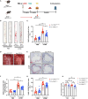
Figure 1
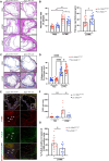
Figure 1. Deletion of Il1r1 on VSMCs reduces vasculitis‐induced acceleration of atherosclerosis.
A, Schematic for LCWE‐ induced KD mouse model and tamoxifen‐inducible VSMC‐specific IL‐1 receptor (Il1r1) knockout mice (Il1r1SMCΔ/Δ) to study acceleration of atherosclerosis. B, Aorta en face lesion coverage representative and (C), quantification of Oil Red O staining from tamoxifen‐induced l1r1SMCΔ/Δ and Il1r1SMCWT/WT control mice injected with either PBS or LCWE. D, Picture of aortic arch in LCWE injected Il1r1SMCΔ/Δ mice and Il1r1 SMC WT/WT mice. E, Representative of Oil Red O staining and (F) Quantification of lesion area in aortic sinus and (G) Lipid content in lesion area from tamoxifen‐treated l1r1SMCΔ/Δ and Il1r1SMCWT/WT control mice injected with either PBS or LCWE. Data represented as mean value±SEM. n=14–20/group. H, Total serum cholesterol level. *P≤0.05; **P≤0.01; ***P≤0.001. IL‐1 indicates interleukin‐1; KD, Kawasaki disease; LCWE, Lactobacillus casei cell wall extract; TAM, tamoxifen diet; VSMC, vascular smooth muscle cell; WD, Western diet; and WT, wildtype.
Figure 2
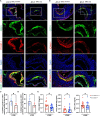
Figure 2. Il1r1 deletion on VSMCs reduces infiltration of macrophages and activation of Caspase‐1 in vasculitisassociated acceleration of atherosclerosis.
A, Representative images and (B) Quantification of hematoxylin and eosin staining for necrotic core and fibrous cap from tamoxifen‐induced l1r1SMC Δ/Δ and Il1r1SMCWT/WT control mice injected with either PBS or LCWE. C, Representative images and (D) Quantification of Masson staining for collagen content from tamoxifen‐induced l1r1SMC Δ/Δ and Il1r1SMCWT/WT control mice injected with either PBS or LCWE. Scale bars: 500 μm. E, Immunofluorescent staining for F4/80 (red) and caspase‐1 activity (FLICA, green) in atherosclerotic lesions from Tamoxifen‐induced l1r1SMC Δ/Δ and Il1r1SMCWT/WT control mice injected with either PBS or LCWE. Nuclei were stained with DAPI. The arrows show the positive cells. F, Quantification of F4/80 positive cells in aortic sinus lesions. G, Quantification of active caspase‐1 positive and F4/80 positive cells in lesion. Data represented as mean value±SEM. n=14–20/group. Significance was determined using 2‐way ANOVA with Bonferroni's posttest (B, D, F) and unpaired Student's t test (G). *P≤0.05; **P≤0.01; ***P≤0.001. LCWE indicates Lactobacillus casei cell wall extract; VSMC, vascular smooth muscle cell; and WT, wildtype.
Figure 3
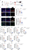
Figure 3. Reduced expression of endothelial cell adhesion molecules in CD31 positive cells and reduced serum levels of CCL2 in LCWE‐injected Il1r1SMCΔ/Δ atherogenic mice.
Immunofluorescent staining for (A) CD31 (green), VCAM‐1 (red); (B) CD31 (green), ICAM‐1 (red); (C) Quantification of VCAM staining in atherosclerotic lesions from Il1r1SMCΔ/Δ and Il1r1SMCWT/WT control mice injected with LCWE; (D) Quantification of ICAM staining in aortic sinus lesions from Il1r1SMCΔ/Δ and Il1r1SMCWT/WT control mice injected with LCWE (n=6/group). Nuclei were stained with DAPI. Scale bars: 200 μm. E, Serums were collected from Il1r1SMCΔ/Δ and Il1r1SMCWT/WT mice injected with LCWE. CCL2, CCL5, and CXCL1 levels were measured by ELISA (n=16–20/group). Data represented as mean value±SEM. Significance was determined using unpaired Student's t test. *P≤0.05; **P≤0.01; ***P≤0.001. CCL2 indicates monocyte chemotaxis protein‐1; CCL5, chemokine (C‐C motif) ligand 5; CXCL1, C‐X‐C motif chemokine ligand 1; ICAM‐1, intercellular adhesion molecule‐1; LCWE, Lactobacillus casei cell wall extract; SMC, smooth muscle cell; VCAM‐1, vascular cell adhesion protein‐1; and WT, wildtype.
Figure 4
Figure 4. Il1r1 deletion on VSMCs reduces recruitment of macrophages in vasculitis associated‐atherosclerotic aortic sinus and the tissue of aorta following adaptive transfer of CD45.1 BM cells.
A, Schematic for LCWE‐induced KD mouse model and tracing of CD45.2 and CD45.1 monocytes/macrophages from tamoxifen‐induced Il1r1SMCΔ/Δ and Il1r1SMCWT/WT control mice injected with LCWE. B, Immunofluorescent staining for macrophages (F4/80, red) and CD45.1 (green) in atherosclerotic sinus lesions from Il1r1SMCΔ/Δ and Il1r1SMCWT/WT control mice injected with LCWE. Nuclei were stained with DAPI. Scale bars: 200 μm. C, Quantification of CD45.1 positive macrophages in aortic sinus lesion (n=8–9). D, Immunofluorescent staining for macrophages (F4/80, red) and CD45.2 (green) in atherosclerotic sinus lesions from Il1r1SMCΔ/Δ and Il1r1SMCWT/WT control mice injected with LCWE. Nuclei were stained with DAPI. Scale bars: 200 μm (n=8–9). E, Quantification of CD45.2 positive macrophages in aortic sinus lesion (n=8–9). F, Live CD11b+ cells were reduced in Il1r1SMCΔ/Δ compared with Il1r1SMCWT/WT mice (n=4/group). GI, Both CD45.1 and CD45.2 CD11b+ macrophages and monocytes in the aorta were significantly less abundant in the Il1r1SMCΔ/Δ group compared with Il1r1SMCWT/WT group (n=4/group). Data represented as mean value±SEM. *P≤0.05; **P≤0.01. VSMCs: vascular smooth muscle cells. BM indicates bone marrow; KD, Kawasaki disease; LCWE, Lactobacillus casei cell wall extract; MFI, mean fluorescence intensity; MHCII, major histocompatibility complex class II; TAM, tamoxifen diet; VSMC, vascular smooth muscle cell; WD, Western diet; and WT, wildtype.
Figure 5
Figure 5. MCP‐1 and CXCL1 levels from isolated VSMCs in response to rIl‐1β stimulation.
Supernatants were collected from isolated VSMCs from Il1r1SMCΔ/Δ and Il1r1SMCWT/WT and treated with PBS or rIl‐1β (5 nmol/L/mL) for 24 hours. MCP‐1 (A) and CXCL1 (B) levels were measured by ELISA (n=6/group). Data represented as mean value±SEM. Significance was determined using 1‐way ANOVA with Bonferroni's posttest. ****P≤0.0001. CXCL1 indicates C‐X‐C motif chemokine ligand 1; MCP‐1, monocyte chemotaxis protein‐1; VSMCs, vascular smooth muscle cells; and WT, wildtype.

References

    1. Newburger JW, Takahashi M, Gerber MA, Gewitz MH, Tani LY, Burns JC, Shulman ST, Bolger AF, Ferrieri P, Baltimore RS, et al. Diagnosis, treatment, and long‐term management of Kawasaki disease: a statement for health professionals from the committee on rheumatic fever, endocarditis and Kawasaki disease, Council on Cardiovascular Disease in the Young, American Heart Association. Circulation. 2004;110:2747–2771. doi: 10.1161/01.CIR.0000145143.19711.78 - DOI - PubMed
    1. Sosa T, Brower L, Divanovic A. Diagnosis and management of Kawasaki disease. JAMA Pediatr. 2019;173:278–279. doi: 10.1001/jamapediatrics.2018.3307 - DOI - PubMed
    1. McCrindle BW, Rowley AH, Newburger JW, Burns JC, Bolger AF, Gewitz M, Baker AL, Jackson MA, Takahashi M, Shah PB, et al. Diagnosis, treatment, and long‐term management of Kawasaki disease: a scientific statement for health professionals from the American Heart Association. Circulation. 2017;135:e927–e999. doi: 10.1161/CIR.0000000000000484 - DOI - PubMed
    1. Histopathological aspects of cardiovascular lesions in Kawasaki disease. Int J Rheum Dis. 2018;21:2131–2135. doi: 10.1111/1756-185X.13207 - DOI - PubMed
    1. Orenstein JM, Shulman ST, Fox LM, Baker SC, Takahashi M, Bhatti TR, Russo PA, Mierau GW, de Chadarévian JP, Perlman EJ, et al. Three linked vasculopathic processes characterize Kawasaki disease: a light and transmission electron microscopic study. PLoS One. 2012;7:e38998. doi: 10.1371/journal.pone.0038998 - DOI - PMC - PubMed

MeSH terms

Substances

LinkOut - more resources