. 2025 Jun 3;14(11):e040214.
doi: 10.1161/JAHA.124.040214. Epub 2025 May 26.

Initiating Empagliflozin and Sacubitril/Valsartan Early After Acute Myocardial Infarction: Mechanistic Study

Affiliations

Initiating Empagliflozin and Sacubitril/Valsartan Early After Acute Myocardial Infarction: Mechanistic Study

Daina Martínez-Falguera et al. J Am Heart Assoc. .

Abstract

Background: Empagliflozin and sacubitril/valsartan are established in heart failure treatment, but their effects after myocardial infarction (MI) are less clear. This study evaluated early empagliflozin initiation, with or without sacubitril/valsartan, on post-MI inflammation, oxidative stress, metabolism, fibrosis, cardiac function, and ventricular tachycardia (VT) risk in a pig model.

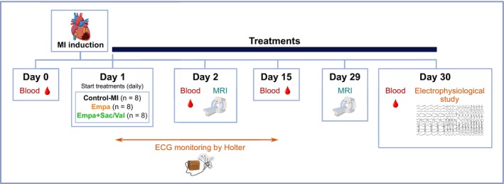
Methods: A total of 24 of 30 pigs survived the MI procedure and were subsequently randomized to receive beta-blocker treatment alone (control-MI), beta-blocker+empagliflozin, or beta-blocker+empagliflozin+sacubitril/valsartan. Immune response, metabolic profile, and cardiac function were monitored. At 30 days after MI, programmed electrical stimulation and high-density mapping were performed and VT inducibility was assessed. Tissue samples were collected for cardiac inflammation, oxidative stress, and metabolic analyses.

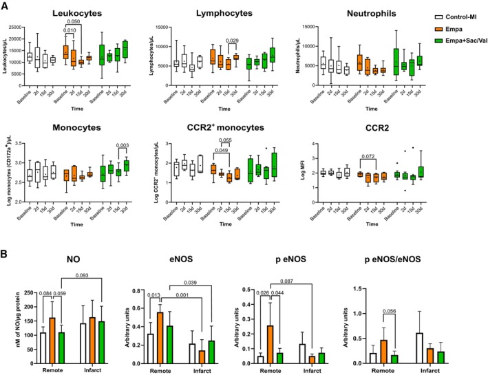
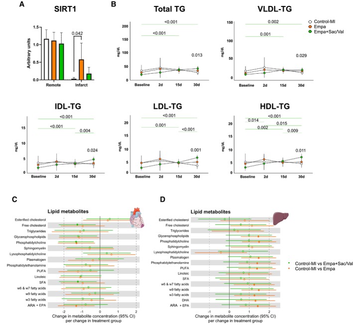
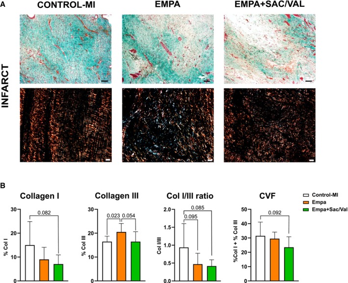
Results: Empagliflozin reduced circulating leukocytes at 2 and 15 days after MI (P=0.010 and P=0.050, respectively) and decreased C-C chemokine receptor 2+ monocytes at 15 days (P=0.049). Nitric oxide bioavailability increased in remote myocardium (P=0.059), along with cardioprotective liver lipids and collagen III in the myocardial scar (P=0.023). No effect on cardiac function or VT inducibility was observed at 30 days. With empagliflozin+sacubitril/valsartan, scar collagen I decreased (P=0.082), left ventricular compliance improved (P=0.029), electrophysiological remodeling improved (reduced border-zone corridors [P=0.006] and deceleration zones [P=0.008]), and VT inducibility decreased (P=0.025).

Conclusions: In this pig model of nonreperfused MI treated with beta-blocker, early initiation of empagliflozin reduced inflammation, improved nitric oxide bioavailability, increased protective liver lipids, and modified scar composition without affecting cardiac function or VT risk. With empagliflozin+sacubitril/valsartan treatment, scar collagen I and VT inducibility declined and left ventricular remodeling was enhanced.

Keywords: empagliflozin; fibrosis; inflammation; myocardial infarction; sacubitril/valsartan; ventricular arrhythmia.


Conflict of interest statement

This work was partially funded by Boehringer Ingelheim through an unrestricted grant with Germans Trias i Pujol Health Science Research Institute. Boehringer Ingelheim was given the opportunity to review the manuscript for medical and scientific accuracy as well as intellectual property considerations in relation to potential mention of Boehringer Ingelheim substances.

Dr Delgado has received speaker fees from Abbott Vascular, Edwards Lifesciences, GE Healthcare, Medtronic, Novartis, JenaValve, and Philips and consulting fees from Edwards Lifesciences, MSD, and Novo Nordisk. Dr Bayes‐Genis has lectured and/or participated in ad boards for Abbott, AstraZeneca, Bayer, Boehringer Ingelheim, Medtronic, Novartis, Novo Nordisk, Roche Diagnostics, and Vifor. Dr Bisbal has received speaker fees from Abbott, Biosense Webster, and Biotronik and consulting fees from Abbott. Dr Gálvez‐Montón is a cofounder and the CSO of NIMBLE Diagnostics. Dr Ferrer‐Curriu has received fees from Boehringer Ingelheim. Núria Amigó is a stock owner of Biosfer Teslab and has a patent on the method for lipoprotein profiling described in the present article.

Figures

Figure 1
Figure 1. Chronogram and workflow of the animal study.
Empa indicates empagliflozin; Empa+Sac/Val, empagliflozin+sacubitril/valsartan; MI, myocardial infarction; and MRI magnetic resonance imaging.
Figure 2
Figure 2. Systemic immune response and NO analysis.
A, Histograms representing total number of leukocytes, lymphocytes, neutrophils, monocytes, CCR2+ monocytes, and expression of CCR2 (mean fluorescence intensity), at baseline and at 2, 15, and 30 days of follow‐up of animals from the control‐MI (n=8), empagliflozin (n=8), and empagliflozin+sacubitril/valsartan (n=8) groups. Statistical differences according to the modified Tukey post hoc test from repeated‐measures ANOVA. B, Histograms showing NO bioavailability (control‐MI, n=7; empagliflozin, n=8; empagliflozin+sacubitril/valsartan, n=8) and quantification of eNOS (control‐MI, n=5; empagliflozin, n=7; empagliflozin+sacubitril/valsartan, n=7) and phosphorylated eNOS (control‐MI, n=4, empagliflozin, n=4; empagliflozin+sacubitril/valsartan, n=4) in remote and infarct zones of animals from the groups. Statistical differences according to Tukey post hoc test from ANOVA or paired t test, as appropriate. CCR2 indicates C‐C chemokine receptor 2; Empa, empagliflozin; Empa+Sac/Val, empagliflozin+sacubitril/valsartan; eNOS, endothelial nitric oxide synthase; MI, myocardial infarction; NO, nitric oxide; and p eNOS, phosphorylated endothelial nitric oxide synthase.
Figure 3
Figure 3. Metabolomic analysis.
A, Sirtuin 1 quantification (control‐MI, n=5; empagliflozin, n=7; empagliflozin+sacubitril/valsartan, n=7) in remote and infarct zones. Statistical differences according to Tukey post hoc test from ANOVA; (B) lipoprotein profile of animals at baseline and at 2, 15, and 30 days after MI from the control‐MI (n=8), empagliflozin (n=8), and empagliflozin+sacubitril/valsartan (n=8) groups. Statistical differences according to modified Tukey post hoc test from repeated‐measures ANOVA. C and D, Association analyses of odds ratios from comparing control‐MI vs empagliflozin groups and control‐MI vs empagliflozin+sacubitril/valsartan groups, in the infarct myocardium and liver tissue, respectively. Control‐MI (n=6), empagliflozin (n=6), and empagliflozin+sacubitril/valsartan (n=6). Variables with a filled circle indicate P<0.05. ARA+EPA indicates arachidonic acid+eicosapentaenoic acid; DHA, docosahexaenoic acid; Empa, empagliflozin; Empa+Sac/Val, empagliflozin+sacubitril/valsartan; HDL, high‐density lipoprotein; IDL, intermediate‐density lipoprotein; LDL, low‐density lipoprotein; PUFA, polyunsaturated fatty acids; and SFA, saturated fatty acids; TG, triglycerides; and VLDL, very low‐density lipoprotein.
Figure 4
Figure 4. Fibrosis analysis.
A, Top: Microphotographs of Masson's trichrome staining from the infarct core; collagen, light green; muscle, dark pink; cytoplasm, pink; nuclei, black (scale bar=100 μm). Bottom: Polarized light microscopy images of Picrosirius Red staining in infarct core samples, showing collagen fibrils type I (red/yellow) and type III (green) (scale bar=25 μm). B, Percentage of collagen I and collagen III, collagen I/ collagen III ratio, and collagen volume fraction in the infarct core; control‐MI (n=8), empagliflozin (n=8), and empagliflozin+sacubitril/valsartan (n=8). Statistical differences according to Welch t test or t test, as appropriate. Col indicates collagen; CVF, collagen volume fraction; Empa, empagliflozin; Empa+Sac/Val, empagliflozin+sacubitril/valsartan; and MI, myocardial infarction.

Similar articles

References

    1. Lippi G, Sanchis‐Gomar F. Global epidemiology and future trends of heart failure. AME Med J. 2020;5:1–6. doi: 10.21037/amj.2020.03.03 - DOI
    1. Gerber Y, Weston SA, Enriquez‐Sarano M, Berardi C, Chamberlain AM, Manemann SM, Jiang R, Dunlay SM, Roger VL. Mortality associated with heart failure after myocardial infarction: a contemporary community perspective. Circ Heart Fail. 2016;9:e002460. doi: 10.1161/CIRCHEARTFAILURE.115.002460 - DOI - PMC - PubMed
    1. Zhang Q, Wang L, Wang S, Cheng H, Xu L, Pei G, Wang Y, Fu C, Jiang Y, He C, et al. Signaling pathways and targeted therapy for myocardial infarction. Signal Transduct Target Ther. 2022;7:78. doi: 10.1038/s41392-022-00925-z - DOI - PMC - PubMed
    1. Azevedo PS, Polegato BF, Minicucci MF, Paiva SAR, Zornoff LAM. Cardiac remodeling: concepts, clinical impact, pathophysiological mechanisms and pharmacologic treatment. Arq Bras Cardiol. 2016;106:62–69. doi: 10.5935/abc.20160005 - DOI - PMC - PubMed
    1. Bauersachs J. Heart failure drug treatment: the fantastic four. Eur Heart J. 2021;42:681–683. doi: 10.1093/eurheartj/ehaa1012 - DOI - PMC - PubMed

MeSH terms

LinkOut - more resources