. 2025 Aug;24(8):e70115.
doi: 10.1111/acel.70115. Epub 2025 May 26.

Accumulation of GSK-3β in Interneurons Impairs Adult Hippocampal Neurogenesis by Inhibiting GABAergic Transmission

Affiliations

Accumulation of GSK-3β in Interneurons Impairs Adult Hippocampal Neurogenesis by Inhibiting GABAergic Transmission

Fei Liu et al. Aging Cell. 2025 Aug.

Abstract

The activation of glycogen synthase kinase 3β (GSK-3β) and the deterioration of spatial memory represent prominent pathological and clinical manifestations of Alzheimer's disease (AD). Nevertheless, the precise intrinsic mechanisms linking these pathological features remain poorly elucidated. In this study, we identified significant upregulation of GSK-3β activity in inhibitory interneurons within the hippocampal dentate gyrus (DG) of 3×Tg-AD mice. Subsequent investigations demonstrated that targeted overexpression of GSK-3β in these interneurons triggered aberrant activation of neural stem cells (NSCs), culminating in apoptotic cell death and consequent deficits in adult hippocampal neurogenesis (AHN). Utilizing in vivo fiber-optic recording techniques, we further established that GSK-3β overexpression in DG inhibitory interneurons elicited hyperactivation of excitatory neurons, thereby disrupting the excitation-inhibition (E/I) balance within the DG circuitry. Notably, these pathological alterations were ameliorated through chemogenetic suppression of excitatory neuronal activity. Mechanistically, we determined that impaired GABAergic transmission, characterized by reduced GABA release in the DG region, underlies these observed effects. Pharmacological intervention with GABA receptor agonists effectively rescued AHN impairment and attenuated spatial cognitive deficits. Collectively, these findings demonstrate that GSK-3β overexpression in GABAergic interneurons compromises AHN and promotes NSC apoptosis via disruption of GABAergic signaling, while pharmacological potentiation of GABAergic transmission exerts neuroprotective effects. This study elucidates a previously unrecognized mechanism contributing to AHN impairment in AD and identifies a promising therapeutic target for pro-neurogenic strategies.

Keywords: GABA transmission; adult hippocampal neurogenesis; glycogen synthase kinase‐3β; interneuron.


Conflict of interest statement

The authors declare no conflicts of interest.

Figures
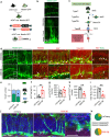
FIGURE 1
FIGURE 1
Interneuron‐specific overexpression of GSK‐3β induced AHN deficits. (a) GSK‐3β‐tyr216 aggregated in interneurons within the DG of 3×tg mice compared to vehicle 129 mice. (b) Cells labeled for GSK‐3β‐tyr216 co‐express GAD67. Arrowheads indicate co‐labeled cells. Scale bars were 50 μm (black) or 20 μm (white). (c) Experimental scheme: AAV‐EF1α‐DIO‐GSK‐3β‐mCherry or AAV‐EF1α‐DIO‐mCherry was stereotaxically infused in 2‐month‐old vGAT‐cre mice for specific overexpression of GSK‐3β in interneurons for 4 weeks. Some mice were randomly selected for sacrificing 3 days following a continuous intraperitoneal injection of BrdU. (d) Representative images showing the expression pattern of AAV and ROV in vGAT‐cre mice. The image on the right is an enlarged representation of the square area depicted in the image on the left. Scale bars are indicated in the figure. (e) AAV‐labeled cells co‐express GAD. Arrowheads indicate co‐labeled cells. Scale bar is 20 μm. (f) Representative images showing AHN. Scale bars were 50 μm (white) or 10 μm (red). (g–k) Overexpression of GSK‐3β in GABAergic interneurons reduced the number of BrdU‐ and DCX‐immunoreactive cells (e, f), as well as a decline in the dendritic length (g), complexity (h) and spine density (i) among ROV‐GFP‐labeled newborn neurons. Unpaired t‐tests and two‐way ANOVA, *p < 0.05, **p < 0.01. n = 6–7 mice (dots) or 17–19 neurons.
FIGURE 2
FIGURE 2
Interneuron‐specific overexpression of GSK‐3β activated NSCs and increased NSCs apoptosis. (a) Experimental scheme: vGAT‐cre: Nestin‐GFP mice were generated by crossbreeding vGAT‐cre with Nestin‐GFP strains. AAV was stereotaxically infused to selectively overexpress GSK‐3β in GABAergic interneurons. (b) A representative image showing the typical morphology of quiescent NSCs. Scale bar, 20 μm. (c) A cartoon illustrating the various stages of differentiation from NSCs to newborn neurons. (d) Representative images showing the NSCs in vGAT‐Cre: Nestin‐GFP mice. (e–i) Overexpression of GSK‐3β in GABAergic interneurons activated NSCs: Morphology of GFP‐labeled Nestin+ cells was altered with an increased proportion of type II cells (e). The number of GFP positive cells co‐labeled with MCM2 increased (f), whereas the count of GFP positive cells co‐labeled with Sox2 remained unchanged (g). Conversely the number of GFP positive cells co‐labeled with NeuroD1 decreased (h). Meanwhile, the number of cells co‐labeled with Ki67 and BrdU decreased (i). (j, k) GSK‐3β overexpression in GABAergic interneurons led to enhanced NSCs apoptosis and increased proportion of GFP positive cells co‐labeled with cleaved casepase3 (j), as illustrated in the representative image (k). Unpaired t‐tests and two‐way ANOVA, *p < 0.05, **p < 0.01. n = 4 mice (dots).
FIGURE 3
FIGURE 3
Interneuron‐specific overexpression of GSK‐3β induced network hyperactivation. (a) Experimental procedure of AAVs injection and in vivo optical fiber recording. (b) The representative image showed the pattern of virus infection. Arrows indicate the position of optic fiber. (c, d) Specific GSK‐3β overexpression enhanced calcium response in DG excitatory neurons, as evidenced by an increased number of calcium response (d, left) and a greater area under the ΔF/F curve (AUC) (d, right). Representative ΔF/F signals are presented in (c). (e) Experimental procedure for AAVs injection and in vivo optical fiber recording. (f, g) Administration of hM4Di + CNO for a month resulted in a decreased in both the number of calcium response (f, left) and the area under the ΔF/F curve (AUC) (f, right). Representative ΔF/F signals are shown in (g). (h) Representative images depicting AHN. Scale bars were 50 μm (white) or 10 μm (red). (i–l) Chemogenetic inhibition partially rescued the AHN deficits induced by interneuron GSK‐3β overexpression. Dendritic complexity (i) and dendritic spine density (j) showed recovery in ROV‐labeled newborn neurons; this recovery was observed within BrdU‐ and DCX‐labeled cell numbers (k, l). (m) The increased proportion of type II cells showed a similar recovery. Unpaired t‐tests and two‐way ANOVA, *p < 0.05, **p < 0.01. n = 5 mice (dots) or 13–17 neurons.
FIGURE 4
FIGURE 4
Interneuron‐specific overexpression of GSK‐3β attenuated GABAergic transmission. (a) Experimental procedure for AAVs injection and in vivo optic fiber recording. (b) The representative image showed the pattern of virus infection. (c–e) Interneuron GSK‐3β overexpression attenuated GABA responses (take 5% ΔF/F as threshold) in DG, compared to the mCherry control group. Representative ΔF/F signals of iGABASnFR are shown in (c). Unpaired t‐tests, n = 6 mice in each group, *p < 0.05. (f) Representative images showing AHN. Scale bars were 50 μm (white) or 10 μm (red). (g–k) Gaboxadol rescued AHN deficits caused by GSK‐3β overexpression, as evidenced by the recovery of dendritic complexity (g), dendritic length (h) and dendritic spine density (i) in ROV‐labeled newborn neurons, along with recovery in the number of BrdU (j) and DCX (k) labeled cells. (l) The number of cells co‐labeled with GFP and cleaved‐casepase3 was reduced. (m) The morphology of the GFP‐labeled Nestin+ cells showed improvement. The proportion of type II IPCs was restored. Unpaired t‐tests and two‐way ANOVA, *p < 0.05, **p < 0.01, ***p < 0.001. n = 4–6 mice (dots) or 6–9 neurons.
FIGURE 5
FIGURE 5
GABA receptor agonist reverses GSK‐3β overexpression induced learning and memory deficits. (a) Experimental procedure: VGAT‐cre mice (2 months old) received AAV virus injections and THIP treatment for 5 weeks. (b) Behavioral training and testing protocol for contextual learning and pattern separation. (c) Mice effectively distinguished environment A from environment B (*p < 0.05, unpaired t‐test; n = 6–8/group). (d) Alternating training in environment A and environment C. (e) Mice gradually improved discrimination between the two environments, with increasing preference scores (*p < 0.05, unpaired t‐test; n = 6–8/group). (f) GSK‐3β‐overexpressing mice showed slower learning in distinguishing environment A from environment C. By the third block, controls reduced freezing in environment C, while overexpression mice showed no difference. THIP restored discrimination ability. (g) Freezing times in environment A and environment C during the third block (*p < 0.05, **p < 0.01; repeated measures ANOVA, Tukey's test, or paired t‐test; n = 6–8/group).

References

    1. Andang, M. , Hjerling‐Leffler J., Moliner A., et al. 2008. “Histone H2AX‐Dependent GABAA Receptor Regulation of Stem Cell Proliferation.” Nature 451, no. 7177: 460–464. 10.1038/nature06488. - DOI - PubMed
    1. Bao, H. , Asrican B., Li W., et al. 2017. “Long‐Range GABAergic Inputs Regulate Neural Stem Cell Quiescence and Control Adult Hippocampal Neurogenesis.” Cell Stem Cell 21, no. 5: 604–617. 10.1016/j.stem.2017.10.003. - DOI - PMC - PubMed
    1. Barbara, S. , Friederike K., Ping W. L., Monika K., Helmut K., and Gerd K.. 2006. “Type‐2 Cells as Link Between Glial and Neuronal Lineage in Adult Hippocampal Neurogenesis.” Glia 54, no. 8: 805–814. 10.1002/glia.20407. - DOI - PubMed
    1. Boldrini, M. , Fulmore C. A., Tartt A. N., et al. 2018. “Human Hippocampal Neurogenesis Persists Throughout Aging.” Cell Stem Cell 22, no. 4: 589–599. 10.1016/j.stem.2018.03.015. - DOI - PMC - PubMed
    1. Braak, H. , Braak E., and Bohl J.. 1993. “Staging of Alzheimer‐Related Cortical Destruction.” European Neurology 33, no. 6: 403–408. 10.1159/000116984. - DOI - PubMed

Substances

LinkOut - more resources