

Epstein-Barr virus-associated smooth muscle tumor partially occluding the superior sagittal sinus: illustrative case
- PMID: 40418886
- PMCID: PMC12105596
- DOI: 10.3171/CASE24759
Epstein-Barr virus-associated smooth muscle tumor partially occluding the superior sagittal sinus: illustrative case
Abstract
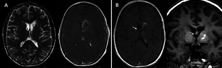
Background: Venous sinus-occlusive mass lesions are infrequent and commonly include meningiomas, with Epstein-Barr virus-associated smooth muscle tumor (EBV-SMT) being much less common.
Observations: The authors present a case report of a venous sinus-occlusive EBV-SMT in an adolescent immunosuppressed male after liver transplantation.
Lessons: The case highlights the importance of considering blood flow dynamics and the use of advanced imaging modalities including incident dark field microcirculation microscopy and indocyanine green video angiography when managing venous sinus-occlusive mass lesions. To the best of the authors' knowledge, this is the first report of a primary intracranial venous sinus EBV-SMT occurring in an HIV-negative patient after liver transplantation, and it illustrates management dilemmas of a sinus-occlusive tumor at a sensitive region of the superior sagittal sinus. https://thejns.org/doi/10.3171/CASE24759.
Keywords: Epstein-Barr virus; superior sagittal sinus; tumor.
Figures


References
-
- Hussein K Rath B Ludewig B Kreipe H Jonigk D.. Clinico-pathological characteristics of different types of immunodeficiency-associated smooth muscle tumours. Eur J Cancer. 2014;50(14):2417-2424. - PubMed
-
- Jonigk D, Laenger F, Maegel L.Molecular and clinicopathological analysis of Epstein-Barr virus-associated posttransplant smooth muscle tumors. Am J Transplant. 2012;12(7):1908-1917. - PubMed
-
- Kaphan E, Eusebio A, Witjas T.Primary leiomyosarcoma of the cavernous sinus associated with Epstein-Barr virus in a kidney graft. Article in French. Rev Neurol (Paris). 2003;159(11):1055-1059. - PubMed
-
- Kazmi SAJ Aizenberg MR Harper JL McComb RD.. Multifocal histologically malignant Epstein-Barr virus-associated smooth muscle tumor in a pediatric transplant patient with an indolent course. Int J Surg Pathol. 2014;22(2):186-189. - PubMed
-
- Boudjemaa S Boman F Guigonis V Boccon-Gibod L.. Brain involvement in multicentric Epstein-Barr virus-associated smooth muscle tumours in a child after kidney transplantation. Virchows Arch. 2004;444(4):387-391. - PubMed
LinkOut - more resources
Full Text Sources
