. 2025 May 26;15(1):18368.
doi: 10.1038/s41598-025-02380-6.

Growth inhibition of Saccharomyces cerevisiae by SUMO-specific nanobodies

Affiliations

Growth inhibition of Saccharomyces cerevisiae by SUMO-specific nanobodies

Pavana Suresh et al. Sci Rep. .

Abstract

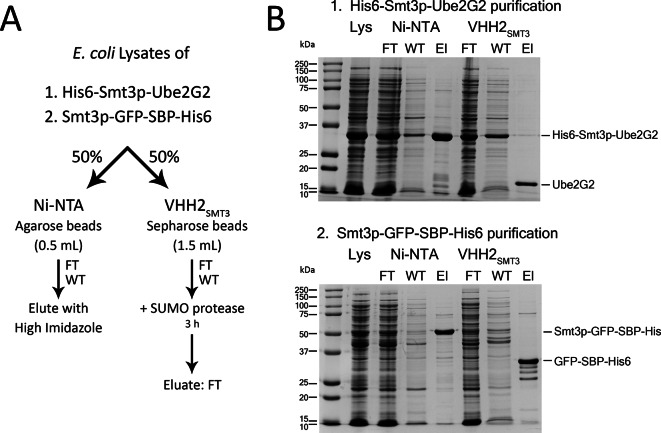
Four nanobodies (VHH1-4SMT3) that target the yeast SUMO protein Smt3p were isolated and characterized. VHH1-4SMT3 bind to Smt3p and Smt3p-tagged proteins with high affinity (Kd: low nM). NMR analysis shows that the four nanobodies all bind near the C-terminus of Smt3p, partially overlapping with the binding site for the SUMO protease Ulp1p. Binding of Smt3p-specific nanobodies impairs Ulp1-mediated cleavage of Smt3p-tagged proteins, with VHH1SMT3 showing complete inhibition. The use of immobilized VHH2SMT3 enabled efficient purification of Smt3p-tagged proteins, while VHH1SMT3 can be used for immunoblotting and detects both Smt3p-tagged and free Smt3p. When expressed in yeast, VHH1SMT3 causes significant growth defects, particularly when targeted to the nucleus or fused with GFP, indicative of interference with essential SUMOylation-dependent processes.


Conflict of interest statement

Declarations. Competing interests: The authors declare no competing interests. Ethics approval: The animal study was approved by IACUC University of Massachusetts Amherst. The study was conducted in accordance with the local legislation and institutional requirements. All methods reported are in accordance with ARRIVE guidelines.

Figures

Fig. 1
Fig. 1
Nanobodies specific for yeast SUMO (Smt3p) protein and their binding characteristics. (A) Anti-Smt3p nanobody sequences with their complementarity determining regions (CDRs) indicated, based on IMGT database. The sequence identity for CDR3 in VHH2-4SMT3 suggests a common progenitor, with accumulation of several somatic mutations in its descendants. Using the VHH2SMT3 sequence as the basis for comparison, these somatic mutations are color-coded blue in VHHs 2-4SMT3. (B) Binding of nanobodies to His6- Smt3p as determined by size exclusion chromatography (SEC). Profiles of His6-Smt3p (Blue), VHHs (Green), and an equimolar mixture of Smt3p:VHH (red) are shown. The fractions corresponding to the VHH:Smt3p complex (red peak) were analyzed by SDS PAGE and Native PAGE, as indicated.
Fig. 2
Fig. 2
Smt3p tagged proteins are recognized by Smt3p-specific VHHs in native PAGE and immunoblot. (A) Left; Native PAGE shows that all four VHHs bind to Smt3p and Smt3p tagged proteins when added in a twofold molar excess over Smt3p. Smt3p-tagged proteins show reduced electrophoretic mobility in the presence of twofold molar excess of VHHs, concomitant with the disappearance of free Smt3p. Right; SDS PAGE gels of the corresponding samples. (B) SDS PAGE of purified Smt3p and Smt3p-tagged proteins used for immunoblots are shown on the left. Immunoblots are displayed on the right as indicated. Immunoblots were performed with VHHs as the primary probes, followed by anti-HA monoclonal antibody to detect HA-tagged VHHs. (C) Only VHH1SMT3 is capable of detecting free Smt3p and its Smt3p-SBP-His6 tagged version by immunoblot. (D) VHH1SMT3 detects free Smt3p in mammalian whole cell lysates spiked with purified Smt3p.
Fig. 3
Fig. 3
Epitope mapping by 15N NMR shows that all VHHs bind near the C terminus of Smt3p. (AC) 1H 15N-TROSY-HSQC spectrum of 15N-labeled Smt3p in the presence of excess VHH1SMT3 (red) (A), VHH2SMT3 (blue) (B), and VHH4SMT3 (pink) (C). The spectra are overlaid on those of Smt3p in the absence of added VHH (black). Individual Smt3p residues that show estimated lower limit for combined chemical shift changes in parts per million (ppm) upon VHH binding are shown in the middle panels. Residues with chemical shift changes with ppm > 0.1 are indicated in yellow, and those with ppm > 0.2 are indicated in red. Residues with > 0.1 ppm, yellow and red, mapped onto a surface rendering of Smt3p (PDB:1EUV) are shown on the right. (D) Comparison of 1H 15N-TROSY-HSQC spectrum (left) and estimated lower limit of combined chemical shift changes of Smt3p residues (right) in the presence of VHH2SMT3 (blue) overlaid on the spectrum obtained in the presence of VHH4SMT3 (pink) highlights the similarity of the presumptive contact residues. (E) Overlay of the spectra collected in the presence of VHH1SMT3 (red) and VHH2SMT3 (blue) showcases the differences in residues affected by their respective VHH binding. The inclusion of VHH1SMT3 shows stronger perturbation towards the C-terminus of Smt3p consistent with a distinct mode of binding. Note that the Kd values for VHH1SMT3 and VHH2SMT3 are similar (Table 1).
Fig. 4
Fig. 4
VHH1SMT3 and VHH2SMT3 inhibit cleavage of Smt3p-tagged proteins by Ulp1p. (A,B) X-ray crystal structure (PDB: 1EUV) of SUMO protease (Ulp1p) in complex with Smt3p. Residues perturbed upon binding of VHH1SMT3 (A) and VHH2SMT3 (B) as determined by NMR are indicated in the color code of Fig. 3. (CE) Cleavage of Smt3p-tagged proteins by Ulp1p in the presence and absence of a twofold molar excess of VHH over Smt3p at the indicated time points for: Smt3p-SBP-His6 (C), His6- Smt3p-Ube2G2 (D), and Smt3p-GFP-SBP-His6 (E). Quantification of cleavage, based on 3 independent experiments, is shown on the left. A representative SDS PAGE gel of the reaction is shown on the right. Major protein species are identified to the right. (F) Overlay of plots shown in (CE) highlight the distinction in amount of cleaved product for the indicated substrates.
Fig. 5
Fig. 5
VHH2SMT3 can be conjugated to a solid support for retrieval of Smt3p-tagged proteins from bacterial extracts. (A) Workflow showing purification done using a Ni–NTA agarose column and VHH2SMT3 conjugated Sepharose beads. (B) SDS PAGE gel showing different stages of purification of His6-Smt3p-Ube2G2 (top) and Smt3p-GFP-SBP-His6 (bottom). Lys: Lysate, FT: Flow through, WT: Wash through, El: Elution.
Fig. 6
Fig. 6
Smt3p VHHs do not cross react with human SUMO proteins but retrieve Smt3p and Smt3pCTD-tagged GFP from HEK 293T transfectants. (A) Multiple sequence alignment of yeast SUMO (SMT3) and human SUMO variants 1–5 (hSUMO1-5). (B) Native PAGE of VHHs added to human SUMO1 in a threefold molar excess over SUMO1. The migration of hSUMO1 is not affected upon inclusion of the nanobodies. The absence of an additional species that could correspond to a complex indicates the absence of binding. The corresponding SDS PAGE gel is shown below the native PAGE gel. (C) Native PAGE of hSUMO2 showing absence of binding towards Smt3p VHHs similar to (B). (D) VHH1SMT3 fails to recognize human SUMO1 and human SUMO2 in immunoblot. (E) Expression of Smt3p-GFP or GFP tagged with the C terminal region of Smt3p (Smt3pCTD-GFP) in HEK 293T transfectants. SDS PAGE gel of whole cell lysate is shown on the top and the corresponding immunoblots performed with anti-GFP monoclonal antibody and with anti-β actin monoclonal antibody are shown below. (F) Anti-GFP and anti-HA tag immunoblots after retrieval by VHH1SMT3 and VHH2SMT3 of Smt3p tagged GFP from HEK 293T cells. Anti-HA tag indicates the nanobody used for retrieval.
Fig. 7
Fig. 7
Cytosolically expressed VHH1SMT3 and VHH2SMT3 compromise yeast growth. (A) Smt3p overexpressed under the control of the CUP1 promoter in EBY100 yeast is readily detected by VHH1SMT3. Left: whole cell lysate of EBY100 yeast transformed with His7-Smt3p plasmid, as indicated. Shown is an immunoblot of overexpressed His7- Smt3p upon induction with 100 µM CuSO4 detected by anti-His antibody (middle) and VHH1SMT3 (right). (B) Anti-HA tag immunoblot to detect expression of VHH1SMT3, VHH2SMT3 and VHH1SMT3 equipped with a C terminal nuclear localization tag (NLS), induced in response to varying concentrations (0, 100, 300 µM) of CuSO4. (C) Anti-HA tag immunoblot of GFP- and NLS-tagged versions of VHH1SMT3 in yeast. (D) Yeast spot dilution growth assay with the VHH constructs shown in (B,C) on CuSO4-containing (0, 100, 300 µM) yeast synthetic media agar plates. The four constructs showing a significant growth defect with increasing concentration of CuSO4 are shown on the right. (E) Fluorescence microscopy showing EBY100 yeast cells expressing VHH1SMT3-GFP tagged versions with and without an NLS. Images for phase-contrast (left), the GFP channel (middle) and an overlay of both (right) are shown. Notice increased nuclear localization of VHH1SMT3 with an NLS tag, regardless of whether the NLS is located at the N or C terminus.

References

    1. Meluh, P. B. & Koshland, D. Evidence that the MIF2 gene of Saccharomyces cerevisiae encodes a centromere protein with homology to the mammalian centromere protein CENP-C. Mol. Biol. Cell.6(7), 793–807. 10.1091/mbc.6.7.793 (1995). - PMC - PubMed
    1. Celen, A. B. & Sahin, U. Sumoylation on its 25th anniversary: Mechanisms, pathology, and emerging concepts. FEBS J.287(15), 3110–3140. 10.1111/febs.15319 (2020). - PubMed
    1. Vertegaal, A. C. O. Signalling mechanisms and cellular functions of SUMO. Nat. Rev. Mol. Cell Biol.23(11), 715–731. 10.1038/s41580-022-00500-y (2022). - PubMed
    1. Jentsch, S. & Psakhye, I. Control of nuclear activities by substrate-selective and protein-group SUMOylation. Annu. Rev. Genet.47, 167–186. 10.1146/annurev-genet-111212-133453 (2013). - PubMed
    1. Psakhye, I. & Jentsch, S. Protein group modification and synergy in the SUMO pathway as exemplified in DNA repair. Cell151(4), 807–820. 10.1016/j.cell.2012.10.021 (2012). - PubMed

MeSH terms

LinkOut - more resources