

Burst of gyrification in the human brain after birth
- PMID: 40419689
- PMCID: PMC12106832
- DOI: 10.1038/s42003-025-08155-z
Burst of gyrification in the human brain after birth
Abstract
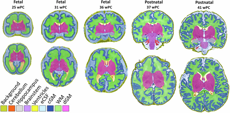
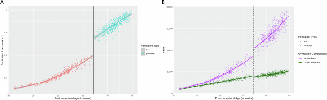
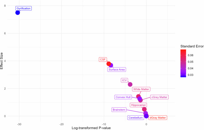
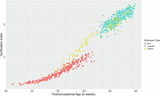
Gyrification, the intricate folding of the brain's cortex, begins mid-gestation and surges dramatically throughout the perinatal period. Yet, a critical factor has been largely overlooked in neurodevelopmental research: the profound impact of birth on brain structure. Leveraging the largest known perinatal MRI dataset-819 sessions spanning 21 to 45 postconceptional weeks-we reveal a burst in gyrification immediately following birth (~37 weeks post-conception), amounting to half the entire gyrification expansion occurring during the fetal period. Using state-of-the-art, homogenized imaging processing tools across varied acquisition protocols, and applying a regression discontinuity design approach that is novel to neuroimaging, we provide the first evidence of a sudden, birth-triggered shift in cortical development. Investigation of additional cortical features confirms that this effect is uniquely confined to gyrification. This finding sheds light onto the understanding of early brain development, suggesting that the neurobiological consequences of birth may hold significant behavioral and physiological relevance.
© 2025. The Author(s).
Conflict of interest statement
Competing interests: The authors declare no competing interests.
Figures
Similar articles
-
Aberrant gyrification contributes to the link between gestational age and adult IQ after premature birth.Brain. 2019 May 1;142(5):1255-1269. doi: 10.1093/brain/awz071. Brain. 2019. PMID: 31032850
-
The dynamics of cortical folding waves and prematurity-related deviations revealed by spatial and spectral analysis of gyrification.Neuroimage. 2019 Jan 15;185:934-946. doi: 10.1016/j.neuroimage.2018.03.005. Epub 2018 Mar 6. Neuroimage. 2019. PMID: 29522888
-
A gyrification analysis approach based on Laplace Beltrami eigenfunction level sets.Neuroimage. 2021 Apr 1;229:117751. doi: 10.1016/j.neuroimage.2021.117751. Epub 2021 Jan 15. Neuroimage. 2021. PMID: 33460799 Free PMC article.
-
Cortical Gyrification Morphology in Individuals with ASD and ADHD across the Lifespan: A Systematic Review and Meta-Analysis.Cereb Cortex. 2021 Mar 31;31(5):2653-2669. doi: 10.1093/cercor/bhaa381. Cereb Cortex. 2021. PMID: 33386405 Free PMC article.
-
Development of the cerebral cortex and the effect of the intrauterine environment.J Physiol. 2018 Dec;596(23):5665-5674. doi: 10.1113/JP277151. Epub 2018 Nov 2. J Physiol. 2018. PMID: 30325048 Free PMC article. Review.
References
-
- Helfer, R. E. The perinatal period, a window of opportunity for enhancing parent-infant communication: An approach to prevention. Child Abuse Negl.11, 565–579 (1987). - PubMed
-
- World Health Organization. The WHO Application of ICD-10 to Deaths during the Perinatal Period: ICD-PM (World Health Organization, Geneva, 2016).
-
- Kostović, I., Sedmak, G. & Judaš, M. Neural histology and neurogenesis of the human fetal and infant brain. NeuroImage188, 743–773 (2019). - PubMed
-
- Girard, N. & Huisman, T. A. G. M. Fetal magnetic resonance imaging of the central nervous system. in Pediatric Neuroradiology: Brain (eds Tortori-Donati, P. & Rossi, A.) 1219–1253. 10.1007/3-540-26398-5_27 (Springer, 2005).
MeSH terms
Grants and funding
LinkOut - more resources
Full Text Sources
