. 2025 Aug;6(8):1350-1369.
doi: 10.1038/s43018-025-00975-6. Epub 2025 May 26.

Skeletal muscle endothelial dysfunction through the activin A-PGC1α axis drives progression of cancer cachexia

Affiliations

Skeletal muscle endothelial dysfunction through the activin A-PGC1α axis drives progression of cancer cachexia

Young-Mee Kim et al. Nat Cancer. 2025 Aug.

Abstract

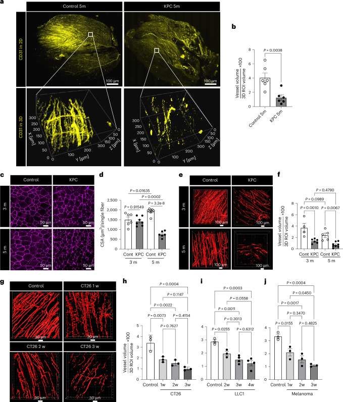
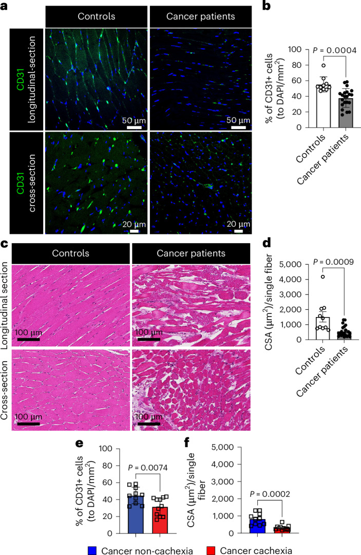
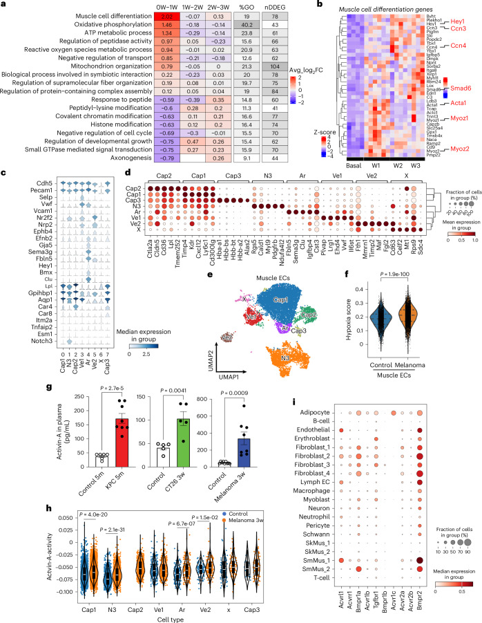
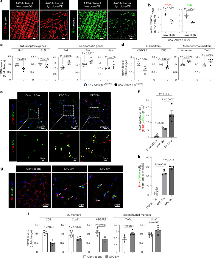
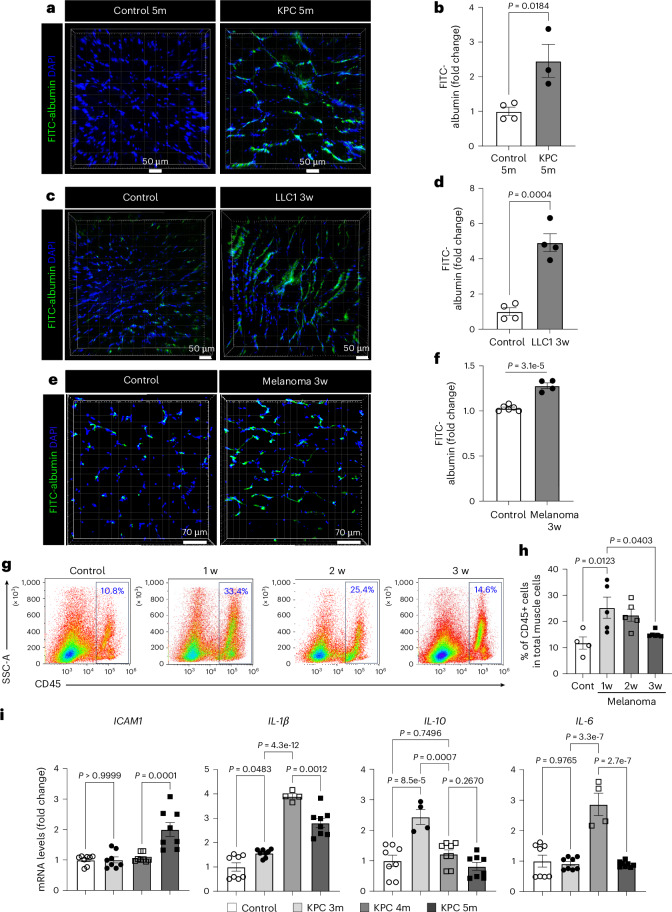
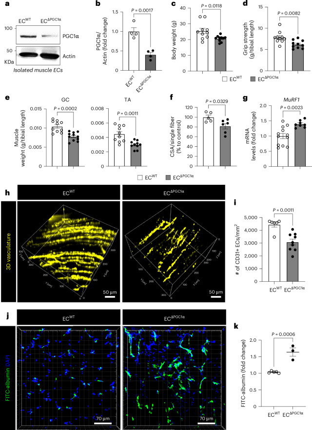
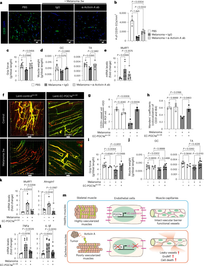
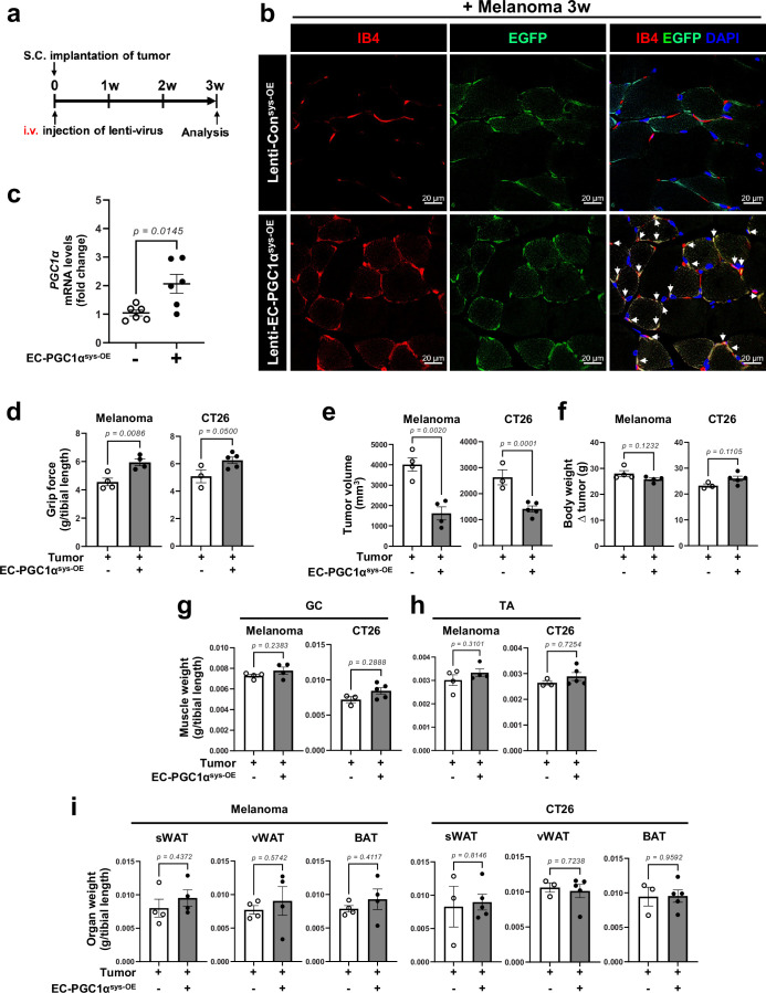
Cachexia is the wasting of skeletal muscle in cancer and is a major complication that impacts a person's quality of life. We hypothesized that cachexia is mediated by dysfunction of the vascular system, which is essential for maintaining perfusion and tempering inappropriate immune responses. Using transparent tissue topography, we discovered that loss of muscle vascular density precedes muscle wasting in multiple complementary tumor models, including pancreatic adenocarcinoma, colon carcinoma, lung adenocarcinoma and melanoma models. We also observed that persons suffering from cancer cachexia exhibit substantial loss of muscle vascular density. As tumors progress, increased circulating activin A remotely suppresses the expression of peroxisome proliferator-activated receptor-γ coactivator 1α (PGC1α) in the muscle endothelium, thus inducing vascular leakage. Restoring endothelial PGC1α activity preserved vascular density and muscle mass in tumor-bearing mice. Our study suggests that restoring muscle endothelial function could be a valuable therapeutic approach for cancer cachexia.


Conflict of interest statement

Competing interests: The authors declare no competing interests.

Figures

Fig. 1
Fig. 1. Cachexic muscles have reduced vascular density in experimental models of cancer.
a, Representative images for high-resolution 3D muscle vasculature of the GC muscles from control 5-month-old and KPC 5-month-old mice. The optically cleared muscles were stained with CD31 antibody (pseudocolored yellow). b, Muscle vascular density (percentage of CD31+ vessel volume in 3D ROI volume; n = 6). Each dot represents one mouse. cf, Comparison of control mice (3 and 5 months old) and KPC mice (3 and 5 months old). c, CSA of GC muscle based on immunofluorescence (IF) analysis with laminin (magenta) antibody. d, Quantification of CSA per single fiber (n = 6 for control 3-month-old and KPC 5-month-old mice and n = 7 for control 5-month-old and KPC 3-month-old mice). Each dot represents one mouse. e, Representative 3D images for surface volume of CD31+ (red) vessels. f, Quantification of muscle vasculature density (n = 4 for control 3-month-old mice, n = 6 for KPC 3-month-old mice, n = 5 for control 5-month-old mice and n = 8 for KPC 5-month-old mice). Each dot represents one mouse. g, Representative 3D images for the surface volume of CD31+ vessels of the GC muscles from CT26-bearing mice. hj, Quantification of muscle vascular density for CT26 (h; n = 3), LLC1 (i; n = 3 for control and LLC1 at 2 weeks and n = 4 for LLC1 at 3 and 4 weeks) and B16F10 (j; n = 3). Each dot represents one mouse. Data are presented as the mean ± s.e.m. Statistical analysis was conducted using either an unpaired two-tailed t-test (b) or a one-way ANOVA with Tukey’s multiple-comparison test (d, f and hj). Source data
Fig. 2
Fig. 2. Participants with cancer cachexia show a decrease in muscle vascular density.
ad, Comparison of abdominal muscles from control subjects and participants with cancer. a, Muscle vasculature according to immunofluorescence (IF) analysis with CD31 (green) antibody and DAPI (blue) for the nucleus. b, Muscle vascular density (percentage of CD31+ cells as a function of total DAPI per area; n = 10 for controls and n = 21 for participants with cancer). Each dot represents one participant. c, H&E-stained muscle structure. d, CSA per single fiber (n = 10 for controls and n = 21 for participants with cancer). Each dot represents one participant. e,f, Comparison of muscle vascular density (e) or CSA (f) between participants with cancer without cachexia (n = 10) and those exhibiting cachexia (n = 11). Each dot represents one participant. Data are presented as the mean ± s.e.m. Statistical analysis was conducted using an unpaired two-tailed t-test (b and df). Source data
Fig. 3
Fig. 3. Expression of muscle cell differentiation-related genes and evaluation of activin A signaling in muscle endothelium.
a,b, TrendCatcher analysis of DDEGs in muscle vascular ECs during tumor growth (control, n = 4 mice; week 1, n = 6 mice; week 2, n = 4 mice; week 3, n = 4 mice). a, TimeHeatmap of the top dynamic pathways. Each column represents a time interval. The number within each cell represents the averaged log2FC of gene expressions compared to the previous time point. The color represents the magnitude of the change. The ‘%GO’ column represents the percentage of DDEGs identified from the corresponding GO term. The ‘nDDEG’ column represents the number of DDEGs from the corresponding pathway. b, DDEG expression heat map from the biological pathway of muscle cell differentiation in a. Each column represents one mouse. The EndMT genes are highlighted. ci, scRNA-seq analysis of muscle ECs from control ECtdTomato mice (n = 3) or B16F10 3-week ECtdTomato mice (n = 4). The gating strategy for FACS sorting is presented in Extended Data Fig. 5c. c, EC marker gene expression in muscle ECs. Cap, capillary; Ar, arterial; Ve, vein; N3, Notch 3 expressing; X, undefined. d, Top five unbiased marker genes in each muscle EC subpopulation. e, The integrated uniform manifold approximation and projection (UMAP) of tdTomato-positive muscle ECs. Each dot represents one cell. f, Hypoxia gene expression scores of individual ECs. The violin plots depict the kernel density estimate of the data, which are shown as individual points. The horizontal lines depict the median and interquartile range. g, The activin A levels in plasma (control 5-month-old mice, n = 6; KPC 5-month-old mice, n = 8; control PBS, n = 5; CT26 3-week mice, n = 5; control, n = 12; B16F10 3-week mice, n = 8). Each dot represents one mouse. h, Activin A signaling activity in muscle EC subpopulations (control, n = 3; B16F10 3-week mice, n = 4). The violins depict the kernel density estimate of the data, which are shown as individual points. The internal boxes represent the median and interquartile range. i, Expression of activin A receptors in all cell types. Data are presented as the mean ± s.e.m. Statistical analysis was conducted using an unpaired two-tailed t-test (fh). Source data
Fig. 4
Fig. 4. Increase in circulating activin A levels is associated with dysfunctional ECs in cancer cachexia progression.
ad, Control mice were intravenously injected with AAV-activin A low dose and AAV-activin A high dose and evaluated after 3 weeks. a, Representative 3D images for the surface volume of total CD31+ (red) or functional IB4+ (green) vessels in the GC muscles. b, Muscle vascular density (percentage of CD31+ or IB4+ vessel volume in 3D ROI volume; n = 3). Each dot represents one mouse. c,d, Muscle ECs isolated from mice overexpressing AAV-activin A low dose and AAV-activin A high dose were evaluated for the expression of apoptosis-related genes (c) or EndMT marker genes (d) by RT–qPCR (n = 5). Each dot represents one mouse. eh, Control 5-month-old mice and KPC mice (3 and 5 months old). e, Cross-sectioned GC muscles were used for an in situ TUNEL assay for apoptotic cells and costained with endothelial TF EGR1 for ECs. Top, the selected areas (white square boxes) were enlarged to show the colocalization of TUNEL and EGR1. White arrows indicate the double-positive cells for TUNEL (red) and EGR1 (green) signal. f, Number of double-positive cells of TUNEL and EGR1 in e (n = 5 for control 5-month-old and KPC 3-month-old mice and n = 4 for KPC 5-month-old mice). Each dot represents one mouse. g, GC muscles were costained with IB4 for ECs and with anti-α-SMA antibody for mesenchymal cells. White arrows indicate the colocalization of IB4 (red) and α-SMA (green) signals. h, Quantification of IB4 and anti-α-SMA double-positive cells in g (n = 3). Each dot represents one mouse. i, Muscle ECs isolated from control 5-month-old and KPC 5-month-old mice were evaluated for the expression of EndMT marker genes by RT–qPCR (n = 5 for CD31, n = 6 for Cdh5, n = 4 for VEGFR2, n = 5 for Twist and n = 7 for Snail). Each dot represents one mouse. The nuclei were visualized with DAPI. Each gene level was normalized by PPIA levels and is presented as the FC. Data are presented as the mean ± s.e.m. Statistical analysis was conducted using an unpaired two-tailed t-test (bd and i) or a one-way ANOVA with Tukey’s multiple-comparison test (f and h). Source data
Fig. 5
Fig. 5. Cancer cachexic mice increase muscle vascular leakage and immune cell infiltration into muscles.
af, The mice were retro-orbitally injected with FITC–albumin. Cryosections with 50-µm thickness of GC muscles were evaluated for vascular leakiness. a, Muscle vascular leakage in control 5-month-old and KPC 5-month-old mice. b, Quantification of surface volume of FITC–albumin in a (n = 4 for control 5-month-old mice and n = 3 for KPC 5-month-old mice). Each dot represents one mouse. c, Muscle vascular leakage in control and LLC1 3-week mice. d, Quantification of surface volume of FITC–albumin in c (n = 4). Each dot represents one mouse. e, Muscle vascular leakage in B16F10-cachexia mice. f, Intensity of FITC–albumin in e (n = 6 for control mice and n = 4 for B16F10 3-week mice). Each dot represents one mouse. g, The inflammatory cells were represented by percentage of CD45+ cells in whole muscle cells by FACS analysis. The gating strategy is presented in Extended Data Fig. 7g. h, Quantification of CD45+ cells (n = 4 for control and n = 5 for 1-week, 2-week and 3-week mice). Each dot represents one mouse. i, Inflammatory genes in TA muscles from control 5-month-old and KPC mice (3, 4 and 5 months old) by RT–qPCR (ICAM1, n = 8 for all groups; IL1β, n = 8 for control, KPC 3-month-old and KPC 5-month-old mice and n = 4 for KPC 4-month-old mice; IL10, n = 8 for control, KPC 4-month-old and KPC 5-month-old mice and n = 4 for KPC 3-month-old mice; IL6, n = 8 for control, KPC 3-month-old and KPC 5-month-old mice and n = 4 for KPC 4-month-old mice). Each dot represents one mouse. Each gene level was normalized by PPIA levels and is presented as the FC. Data are presented as the mean ± s.e.m. The Z-sectioned images were reconstructed for visualization using Imaris software and nuclei were stained with DAPI (a, c and e). Statistical analysis was conducted using an unpaired two-tailed t-test (b, d and f) or a one-way ANOVA with Tukey’s multiple-comparison test (h and i). Source data
Fig. 6
Fig. 6. Circulating activin A induces vascular dysfunction by suppressing endothelial PGC1α in muscle.
a, PGC1α protein levels in isolated muscle ECs from control 5-month-old and KPC 5-month-old mice by western blotting. Each lane represents one mouse. b, Quantification of PGC1α in a (n = 3). Each dot represents one mouse. c, PGC1α mRNA levels in isolated muscle ECs. The mice were evaluated 3 weeks after injecting AAV-control, AAV-activin A low dose or AAV-activin A high dose (n = 3 for control and n = 5 for AAV-activin A low and high doses). Each dot represents one mouse. d, HLMVECs were transfected with PGC1α promoter luciferase plasmid and Renilla luciferase for 48 h and treated with vehicle (0.1% BSA), TNF (10 ng ml−1), activin A (25 ng ml−1) or a combination of TNF and activin A for 16 h. The luciferase activity was normalized by Renilla luciferase and is presented as the FC (n = 11). Each dot represents one independent biological replicate. eh, HLMVECs were treated with lentiviral shRNA for control and PGC1α for 72 h. e, The apoptotic cells (annexin V–FITC+PI+) were evaluated by FACS analysis (n = 6). Each dot represents one independent biological replicate. f, Levels of EndMT marker genes, Cdh5 and Vimentin, by RT–qPCR (n = 8). Each dot represents one independent biological replicate. g, EC barrier integrity by IF staining with VE-cadherin antibody. Nuclei were stained with DAPI. h, Quantification of the area of VE-cadherin in g (n = 4). Each dot represents one independent biological replicate. i,j, Muscle vascular barrier integrity of controls and participants with cancer by IF staining with VE-cadherin antibody. i, Representative VE-cadherin images. j, The surface volume of VE-cadherin (n = 5). Each dot represents one participant. k, Levels of inflammatory genes by RT–qPCR (n = 4). Each dot represents one independent biological replicate. l, ChIP analysis in control and activin A-treated ECs with PGC1α-specific antibody. Normal mouse IgG antibody was used as a negative control. The enrichment values were normalized with input values and are presented as the FC (n = 4 for control cells for IgG IP, n = 6 for control cells for anti-PGC1α IP and n = 4 for activin A-treated cells for anti-PGC1α IP). Each dot represents one independent biological replicate. Data are presented as the mean ± s.e.m. Statistical analysis was conducted using either an unpaired two-tailed t-test (b, e, f, h, j and k) or a one-way ANOVA with Tukey’s multiple-comparison test (c, d and l). Source data
Fig. 7
Fig. 7. Endothelial-specific PGC1α depleted mice manifest elements of cachexia phenotypes.
ak, ECWT (ECtdTomato, WT) and EC∆PGC1α (ECtdTomato, ∆PGC1α) mice. a, PGC1α knockdown efficiency in isolated muscle ECs. b, Quantification of PGC1α levels in a (n = 4). Each dot represents one mouse. c, Body weight (n = 10). Each dot represents one mouse. d, Grip strength (n = 10). Each dot represents one mouse. e, Muscle weight (n = 10). Each dot represents one mouse. f, CSA per single fiber (n = 5 for ECWT and n = 6 for EC∆PGC1α). Each dot represents one mouse. g, Expression of MuRF1 mRNA levels in TA muscles (n = 12 for ECWT and n = 7 for EC∆PGC1α). Each dot represents one mouse. h, Representative 3D images of tdTomato+ (pseudocolored yellow) vessels in the GC muscles. i, Quantification of GC muscle vascular density (n = 6 for ECWT and n = 9 for EC∆PGC1α). Each dot represents one mouse. j, Representative images of muscle vascular leakage. k, Quantification of FITC–albumin for muscle vascular leakage in j (n = 5 for ECWT and n = 3 for EC∆PGC1α). Each dot represents one mouse. Data are presented as the mean ± s.e.m. Statistical analysis was conducted using an unpaired two-tailed t-test (bg, i and k). Source data
Fig. 8
Fig. 8. Targeting activin A–EC PGC1α axis prevents cancer cachexia progression.
ae, Mice were intravenously injected with anti-activin A neutralizing antibody or IgG1 isotype every 4 days, 1 week after melanoma implantation. a, Muscle vasculature by IF staining with CD31 antibody. Nuclei were stained with DAPI. b. Muscle vasculature density (n = 3 for PBS and n = 5 for melanoma + IgG and melanoma + anti-activin A antibody). Each dot represents one mouse. c, Mouse grip strength (n = 8 for PBS, n = 4 for melanoma + IgG and n = 5 for melanoma + anti-activin A antibody). Each dot represents one mouse. d, Muscle mass for GC (n = 6 for PBS and n = 5 for melanoma + IgG and melanoma + anti-activin A antibody) and TA (n = 8 for PBS and n = 5 for melanoma + IgG and melanoma + anti-activin A antibody). Each dot represents one mouse. e, Cachectic marker MuRF1 mRNA in TA muscle (n = 6 for PBS and n = 5 for melanoma + IgG and melanoma + anti-activin A antibody). Each dot represents one mouse. fl, After melanoma implantation, the mice were intramuscularly injected with lentiviral control and lenti-EC PGC1α–GFP. fh, Mice were retro-orbitally injected with IB4–A594 before isolating muscles. f, Representative 3D vasculature after overlaying with CD31+ (red) and IB4+ (pseudocolored green) vessels in GC muscles. g, Quantification of CD31+ (red) muscle vasculature density in f (n = 3). Each dot represents one mouse. h, Manders’ colocalization coefficients for CD31 and IB4 in f (n = 3). Each dot represents one mouse. i, Mouse grip strength (n = 8 for control, n = 3 for EC PGC1αim-OE alone, n = 8 for melanoma alone and n = 7 for melanoma with EC PGC1αim-OE). Each dot represents one mouse. j, Muscle mass for GC (n = 9 for control, n = 4 for EC PGC1αim-OE alone, n = 10 for melanoma alone and n = 10 for melanoma with EC PGC1αim-OE) and TA (n = 8 for control, n = 4 for EC PGC1αim-OE alone, n = 9 for melanoma alone and n = 9 for melanoma with EC PGC1αim-OE). Each dot represents one mouse. k, mRNA levels of MuRF1 (n = 3 for control and n = 5 for melanoma with or without EC PGC1αim-OE) and Atrogin1 (n = 6 for control, n = 5 for melanoma without EC PGC1αim-OE and n = 4 for melanoma with EC PGC1αim-OE) by RT–qPCR. Each dot represents one mouse. l, Inflammatory genes in GC muscles by RT–qPCR (TNF, n = 4 for control and n = 8 for melanoma with or without EC PGC1αim-OE; IL1β, n = 8 for control, n = 12 for melanoma without EC PGC1αim-OE and n = 10 for melanoma with EC PGC1αim-OE). Each dot represents one mouse. m, Graphical summary. All mice were examined 3 weeks after melanoma implantation. Data are presented as the mean ± s.e.m. Each gene level was normalized by PPIA levels and is presented as the FC. Statistical analysis was conducted using a one-way ANOVA with Tukey’s multiple-comparison tests (be and gl). Source data
Extended Data Fig. 1
Extended Data Fig. 1. Cachexia phenotype in KPC mice.
a-e. Characteristics of cachexia in control (3 and 5 month, referred to as 3 m and 5 m) and KPC (3 m and 5 m) mice. a. Body weight (n = 9 for control 3 m, n = 8 for KPC 3 m, n = 10 for control 5 m, n = 11 for KPC 5 m). Each dot represents one mouse. b-c. Representative flow plots of CD31 + CD45- muscle ECs (b) and quantification of muscle ECs by FACS analysis (c, n = 3). Each dot represents one mouse. d. The vasculature of the lung and spleen from age-matched control and KPC-3-month mice was evaluated by IB4 IF analysis. e. Four limbs grip strength of mice (n = 10 for control 3 m, n = 8 for KPC 3 m, n = 13 for control 5 m and KPC 5 m). Each dot represents one mouse. f. Gastrocnemius (GC) mass (n = 6 for control 3 m, n = 9 for KPC 3 m, n = 10 for control 5 m and KPC 5 m). Each dot represents one mouse. g. Tibialis anterior (TA) mass (n = 7 for control 3 m, n = 8 for KPC 3 m, n = 10 for control 5 m, n = 9 for KPC 5 m). Each dot represents one mouse. h. Expression of cachexia markers, MuRF1 (n = 12 for control 5 m, n = 14 for KPC 3 m, n = 4 for KPC 5 m) and Atrogin1 (n = 8 for control 5 m, n = 12 for KPC 3 m, n = 4 for KPC 5 m) in TA muscles by RT-qPCR. Each dot represents one mouse. i-l. Control-5month and KPC-5month mice were assessed for changes in muscle fiber types and fat mass. i. Representative images for type 2a and type 1 muscle fiber in GC muscles. j-k. Quantification of muscle fiber types in GC (j) (n = 4) and TA (k) muscles (n = 3). Each dot represents one mouse. l. The weight (g) of adipose tissues was normalized using tibial length (mm) (n = 8 for sWAT and vWAT, n = 8 for control BAT, n = 7 for KPC 5 m BAT). Each dot represents one mouse. sWAT: subcutaneous white adipose tissue, vWAT; visceral white adipose tissue, BAT; brown adipose tissue. Data are presented as mean ± SEM. Statistical analysis was conducted using unpaired, two-tailed t-test (c, j, k, l) or one-way ANOVA with Tukey’s multiple comparison test (a, e-h). Source data
Extended Data Fig. 2
Extended Data Fig. 2. Cachexia phenotype in CT26 colon carcinoma- and LLC1 lung carcinoma-bearing mice.
a-i. Control and CT26-bearing mice. a. Tumor growth post CT26 implantation (n = 4 for control, n = 7 for 1w, n = 5 for 2w, n = 6 for 3w). Each dot represents one mouse. b. Body weight was presented by subtracting (∆) tumor weight (n = 7 for control, n = 7 for 1w, n = 4 for 2w, n = 5 for 3w). Each dot represents one mouse. c. Four limbs grip strength (n = 8 for control, n = 5 for 1w, n = 4 for 2w, n = 7 for 3w). Each dot represents one mouse. d. GC muscle mass (n = 4). Each dot represents one mouse. e. TA muscle mass (n = 4). Each dot represents one mouse. f. Expression of cachexia markers, MuRF1 in TA muscles by RT-qPCR (n = 3). Each dot represents one mouse. g. The fat mass (g) normalized by tibial length (mm) (n = 8 for control, n = 6 for sWAT and BAT in CT26 3w, n = 7 for vWAT in CT26 3w). Each dot represents one mouse. h. Representative images for type 2a and type 1 muscle fiber in TA muscles. i. Quantification of muscle fiber types in TA muscles (n = 4). Each dot represents one mouse. j-p. Control and LLC1 bearing mice. j. Tumor growth post LLC1 implantation (n = 13 for control, n = 13 for 2w, n = 8 for 3w, n = 9 for 4w). Each dot represents one mouse. k. Body weight was presented by subtracting (∆) tumor weight (n = 14 for control, n = 13 for 2w, n = 10 for 3w, n = 9 for 4w). Each dot represents one mouse. l. Four limbs’ grip strength (n = 14 for control, n = 12 for 2w, n = 12 for 3w, n = 10 for 4w). Each dot represents one mouse. m. GC muscle mass (n = 7 for control, n = 6 for 2w, n = 4 for 3w, n = 5 for 4w). Each dot represents one mouse. n. TA muscle mass (n = 13 for control, n = 6 for 2w, n = 4 for 3w, n = 5 for 4w). Each dot represents one mouse. o. Expression of cachexia markers, MuRF1 and Atrogin1 in TA muscles by RT-qPCR (n = 4). Each dot represents one mouse. p. Body composition of control and LLC1 3w mice by nuclear magnetic resonance (NMR) analysis (n = 5). Each dot represents one mouse. The lean, fat, or free body fluid mass was presented as absolute values per mouse without normalization. Data are presented as mean ± SEM. Statistical analysis was conducted using unpaired, two-tailed t-test (f, g, i, o, p) or one-way ANOVA with Tukey’s multiple comparison test (a-e, j-n). Source data
Extended Data Fig. 3
Extended Data Fig. 3. Cachexia phenotype in melanoma-bearing mice.
a. Tumor growth post B16F10 implantation (n = 21 for control, n = 21 for 1w, n = 17 for 2w, n = 14 for 3w). Each dot represents one mouse. b. Body weight was presented by subtracting (∆) tumor weight (n = 11 for control, n = 9 for 1w, n = 12 for 2w, n = 11 for 3w). Each dot represents one mouse. c. Four limbs’ grip strength (n = 11 for control, n = 9 for 1w, n = 12 for 2w, n = 11 for 3w). Each dot represents one mouse. d-e. Muscle mass for GC (d, n = 17 for control, n = 14 for 1w, n = 17 for 2w, n = 17 for 3w) and TA (e, n = 17 for control, n = 14 for 1w, n = 17 for 2w, n = 17 for 3w). Each dot represents one mouse. f. E3 ubiquitin ligase MuRF1 expression in GC muscles from control and melanoma-bearing mice (n = 3). Each lane represents one mouse. g. Quantification of MuRF1 protein levels in f. h-l. Comparison of control and melanoma 3-week mice. h. Cross-sectioned gastrocnemius muscle by IF analysis with a laminin antibody. The Z-sectioned images were reconstructed for visualization using Imaris software. i. Quantification of the CSA per single fiber (n = 6). Each dot represents one mouse. j-k. Representative flow plots of CD31+tdTomato+ muscle ECs (j) and quantification of muscle ECs by FACS analysis (k, n = 7). Each dot represents one mouse. l. Body composition of control and melanoma 3w mice by NMR analysis (lean mass; n = 8 for control, n = 7 for melanoma 3w, fat mass; n = 10 for control, n = 8 for melanoma 3w, free body fluid; n = 10 for control, n = 9 for melanoma 3w). Each dot represents one mouse. The lean, fat, or free body fluid mass was presented as absolute values per mouse without normalization. Data are presented as mean ± SEM. Statistical analysis was conducted using either unpaired, two-tailed t-test (i, k, l) or one-way ANOVA with Tukey’s multiple comparison test (a-e, g). Source data
Extended Data Fig. 4
Extended Data Fig. 4. Cachexia phenotype in cancer patients.
a-g. Evaluation of muscle structure and fibrosis from control subjects (n = 10) and cancer patients [non-cachexia (n = 11) vs cachexia (n = 10)]. Five representative H&E-stained sections were presented with cross-sectional areas (a) and longitudinal sectional areas (b). c. Size distribution of muscle fibers in control donors (n = 11) and cancer patients (n = 15). Each dot represents one patient. The statistical analysis was performed using a t-test for each size group, and BH p-value correction for all comparisons, and the adjusted p-values were reported. d-e. Comparison of muscle vascular density (d) or CSA (e) between controls (n = 10) and cancer non-cachexia patients (n = 10). Each dot represents one patient. f. Five representative images of muscle fibrosis by Masson’s trichrome staining. g. Quantification of muscle fibrosis area (n = 10 for controls, n = 21 for cancer patients). Each dot represents one patient. Statistical analysis was conducted using an unpaired, two-tailed t-test. Data are presented as mean ± SEM. Source data
Extended Data Fig. 5
Extended Data Fig. 5. Unbiased transcriptomic analysis of muscle cells.
a-b. Bulk RNA-seq analysis of dynamic differentially expressed genes (DDEG) in muscle vascular ECs. a. Top 20 GO pathways in muscle ECs enriched using accumulated log fold change DDEGs. b. Top 5 enriched GO terms using up-regulated DEGs from melanoma 3w compared to basal (0w) (n = 4). The ‘n’ represents the number of mice. c-f. scRNA-seq analysis of muscle cells and muscle ECs from control ECtdTomato mice (n = 3) or melanoma 3w ECtdTomato mice (n = 4). c. Gating strategy for tdTomato-positive muscle ECs or -negative muscle non-ECs by FACS sorting analysis for Fig. 2c–f. d. Cell-specific marker genes for total muscle tissue cells. e. Integrated UMAP for tdTomato-negative muscle cells. Mac; macrophage, F; fibroblast, Neut; neutrophil, Per; pericyte, Myob; myoblast, EC; endothelial cell, B; B cell, Sch; Schwann cell, T; T cell, SkM; skeletal muscle, SmM; smooth muscle cell, Neur; neuron, LEC; lymphatic ECs, Adip; adipocyte, Ery; erythroid-like. f. The relative proportion of specific cell types in total muscle cells. g. GO enrichment of differentially expressed genes in the SkM1 population. h. Hypoxia gene expression scores of individual skeletal muscle cells. The violin plots depict the kernel density estimate of the data which are shown as individual points. The horizontal lines depict the median and interquartile range. i. UMAPs for tdTomato positive (+) muscle ECs. j. Data from tdTomato negative ECs scRNA-seq analysis was integrated with tdTomato positive ECs scRNA-seq data set. tdTomato negative ECs (blue and orange) on tdTomato positive ECs (gray). k. The relative proportion of subpopulations in total muscle endothelial cells. Cap; capillary ECs, Ar; arterial ECs, Ve; vein ECs, N3; Notch3 expressing ECs, X; undefined. l-m. GO analyses of down-regulated (l) and up-regulated (m) Cap2 EC marker genes. n. The muscle cell differentiation module scores of individual cells were calculated based on genes differentially expressed in the bulk RNA-seq data. The box plots depict the median and interquartile range. The whiskers extend to the furthest data points within 1.5x the interquartile range beyond the box boundaries. Outliers beyond the whiskers are shown as individual points. o-p. Predicted ligand-receptor interactions between skeletal muscle cells and muscle ECs from melanoma-bearing mice relative to control mice (o) and within melanoma-bearing mice (p). The possible interactions in all muscle cell populations are available in Supplementary Tables 4, 5. Statistical analysis was conducted using an unpaired, two-tailed t-test (h, n). Source data
Extended Data Fig. 6
Extended Data Fig. 6. A decrease in muscle vascular density is associated with dysfunctional ECs in cachexic muscles.
a-d. C57BL/6 WT mice were intravenously injected with AAV-Activin-A in a dose-dependent manner, and Activin-A levels were measured every week for 3 weeks. a. Activin-A levels in blood plasma following AAV-Activin-A overexpression (n = 4 for PBS 1w and 3w, n = 5 for PBS 2w, n = 6 for AAV-Activin-Alow-OE 1w, 2w, 3w, n = 6 for AAV-Activin-Ahigh-OE 1w, 2w, 3w). Each dot represents one mouse. b. Four limbs’ grip strength (n = 3 for PBS, n = 6 for AAV-Activin-Alow-OE, n = 6 for AAV-Activin-Ahigh-OE). Each dot represents one mouse. c. Body weight (n = 3 for PBS, n = 6 for AAV-Activin-Alow-OE, n = 9 for AAV-Activin-Ahigh-OE). Each dot represents one mouse. d. GC and TA muscle mass (n = 3 for PBS, n = 6 for AAV-Activin-Alow-OE, n = 9 for AAV-Activin-Ahigh-OE). Each dot represents one mouse. e-g. The HLMVECs were treated with Activin-A (25 ng/mL) for 24 h. e. HUVEC viability with and without Activin-A exposure (n = 6). Each dot represents one biological replicate. f-g. the apoptotic cells were evaluated with Annexin V-FITC + PI+ cells using FACS analysis. Representative flow plots (f) and quantification of apoptotic muscle ECs (g) (n = 3 for vehicle, n = 6 for Activin-A). Each dot represents one biological replicate. h. The expression of EndMT marker genes related genes by RT-qPCR (n = 6 for Cdh5, n = 8 for VEGFR2, n = 4 for Snail, n = 6 for Vimentin). Each dot represents one biological replicate. i. HLMVECs were treated with Activin-A (25 ng/mL) for the indicated time course (0, 5, 15, 30, 60, or 90 min) and followed by Western blotting. j. Quantification of the phospho-Smad3 levels in i (n = 3). Each dot represents one biological replicate. The statistical analysis was performed using a t-test with comparisons vs the 0 min time point. k. In situ TUNEL assay for apoptotic ECs in GC muscles of control (n = 4) and melanoma-bearing mice (n = 4). Double-positive cells of TUNEL and ERG1 in total ECs (ERG1 + ) were considered apoptotic ECs. Each dot represents one mouse. l. The expression of apoptosis genes in isolated muscle ECs from control (n = 3) and LLC1 cachexia (n = 3) mice by RT-qPCR. Each dot represents one mouse. m. Quantification of EndMT cells (double-positive cells of IB4 and α-SMA in total IB4+ ECs) in GC muscles of control and melanoma 3w mice (n = 3 for control, n = 5 for melanoma 3w). Each dot represents one mouse. n. The expression of EndMT marker genes in isolated muscle ECs from control and melanoma cachexia mice by RT-qPCR (n = 3 for CD31,n = 4 for Cdh5, VEGFR2, Snail, and Vimentin). Each dot represents one mouse. o. The expression of EndMT marker genes in isolated muscle ECs from control and LLC1 cachexia (n = 3) mice by RT-qPCR. Each dot represents one mouse. Data are presented as mean ± SEM. Each gene level was normalized by 18s or PPIA levels and presented as a fold change. Statistical analysis was conducted using either unpaired, two-tailed t-test (e, g, h, j, l, m, n, o) or one-way ANOVA with Tukey’s multiple comparison test (a-d, k). Source data
Extended Data Fig. 7
Extended Data Fig. 7. Decrease of muscle vascular density contributes to the increase of hypoxia and infiltration of immune cells in cachexic muscles.
a-b. C57BL/6 WT mice were intravenously injected with AAV-Activin-A low dose and AAV-Activin-A high dose. After 3weeks, the mice were injected intravenously with IB4-A594 and fixable FITC-dextran before harvesting gastrocnemius muscles. a. Representative images of FITC-dextran+ (green) area. b. Intensity of FITC-dextran signal in a (n = 3). Each dot represents one mouse. c-e. The mice were intraperitoneally injected with pimonidazole (100 mg/kg) 1 h before harvesting muscle and the extent of hypoxia in gastrocnemius muscle was determined by the Hypoxyprobe Plus Kit. c. Muscle hypoxia in LLC1-cachexia mice. d. Quantification of surface volume of FITC-pimonidazole in c (n = 3). Each dot represents one mouse. e. Quantification of fluorescence intensity of FITC-pimonidazole in muscles of melanoma cachexia mice (n = 5). Each dot represents one mouse. f. H&E-stained abdominal muscles from control subjects and cancer patients. The yellow boxes were enlarged to show clear vessel structure and the infiltrated immune cells in near blood vessels. R1 and R2 in the upper panel indicate different regions from the same patient muscle. g. Gating strategy for flow cytometry analysis at Fig. 4g-h. h. The proportion (%) of immune cell type relative to total immune cells from control and melanoma 3w mice in the scRNA-Seq data. i. The PGC1α mRNA levels in isolated muscle ECs (n = 4 for control 5 m and n = 6 for KPC 5 m, n = 8 for control and n = 12 for melanoma 3w, n = 3 for control and LLC1 3w). Each dot represents one mouse. j. Expression of PGC1α in control and cancer patient muscle ECs by IF analysis (n = 5). The yellow boxes are enlarged to show the colocalization of CD31 and PGC1α. k-l. HLMVECs were treated with Activin-A (25 ng/mL) for indicated times. k. Immunoblotting to assess phospho-FOXO1/3/4 levels. l. Quantification of p-FOXO1/3/4 immunoblots shown in k (n = 3). Each dot represents one biological replicate. The statistical analysis was performed using a t-test with comparisons versus the 0 min time point. m. HLMVECs were transfected with specific siRNA for control, FOXO-1, -3, or -4. The protein levels of PGC1α, FOXO-1, -3, or -4 were determined by immunoblotting. n. Quantification of PGC1α levels in m (n = 3). Each dot represents one biological replicate. Data are presented as mean ± SEM. Statistical analysis was conducted using an unpaired, two-tailed t-test (b, d, e, i, l) or one-way ANOVA with Dunnett’s multiple comparison test (n). Source data
Extended Data Fig. 8
Extended Data Fig. 8. Endothelial PGC1α is required for endothelial barrier integrity.
a-e. HLMVECs were transfected with lentiviral shRNA for PGC1α without or with GFP-PGC1α plasmid. a. Representative flow plots of apoptotic cells. b. PGC1α expression levels by IF analysis (n = 3). The red and green colors represent the endogenous PGC1α (red) and exogenous GFP-PGC1α (green), respectively. c. Apoptotic genes (n = 7 for Bcl2, n = 4 for Mcl1, n = 3 for Bcl-XL, Bim and Bad, Bax: n = 3 for shControl and shPGC1α, n = 4 for shPGC1α + PGC1α-GFP). Each dot represents one biological replicate. d. The protein levels of VE-cadherin. e. Quantification of VE-cadherin in d (n = 4). Each dot represents one biological replicate. f-h. Abdominal muscles from control subjects and cancer patients. f. Representative images for either CD31+ or VE-cadherin+ ECs by IF analysis. g. Quantification of either CD31+ or VE-cadherin+ ECs in f (n = 3). Each dot represents one patient. h. Percent of VE-cadherin+ ECs to CD31+ ECs (n = 3). Each dot represents one patient. i. VE-cadherin protein levels in TA muscles from control and LLC1-cachexia mice (n = 3). Each lane represents one mouse. j. Quantification of the levels of VE-cadherin in i (n = 6). Each dot represents one mouse. k. PGC1α and Cdh5 mRNA levels in PGC1α depleted HLMVECs with or without GFP-PGC1α overexpression (n = 4). Each dot represents one biological replicate. l-n. Characterization of melanoma 3w-bearing ECWT and EC∆PGC1α mice. l. Body weight (n = 7). Each dot represents one mouse. m. Grip strength (n = 7). Each dot represents one mouse. n. Muscle weight (n = 7). Each dot represents one mouse. Data are presented as mean ± SEM. Each gene level was evaluated by RT-qPCR, normalized by 18s levels and presented as fold change. Statistical analysis was conducted using unpaired two-tailed t-test (e, h, j, l-n), or one-way ANOVA with Tukey’s multiple comparison test (c, g, k). Source data
Extended Data Fig. 9
Extended Data Fig. 9. Preventive effect of local endothelial specific PGC1α overexpression in cancer cachexia progression.
a-d. Effect of Activin-A neutralizing antibody. a. Timelines for Activin-A neutralizing antibody treatment. b. Expression of EC marker and anti-apoptotic genes in isolated muscle ECs (CD31; n = 8 for PBS, n = 12 for Melanoma + IgG, n = 8 for Melanoma + α-Activin-A ab, Bcl2; n = 8 for PBS, n = 12 for Melanoma + IgG, n = 8 for Melanoma + α-Activin-A ab, Mcl1; n = 8 for PBS, n = 12 for Melanoma + IgG, n = 8 for Melanoma + α-Activin-A ab). Each dot represents one mouse. c. The tumor growth (n = 4 for PBS, n = 5 for Melanoma + IgG and Melanoma + α-Activin-A ab). Each dot represents one mouse. d. The body weight was presented without (Δ) tumor weight (n = 8 for PBS, n = 4 for Melanoma + IgG, n = 5 for Melanoma + α-Activin-A ab). Each dot represents one mouse. e. Timelines for intramuscular injection of lenti-EC-PGC1α-GFP virus. f-i. Efficiency of local vascular overexpression of EC-PGC1α in muscles. f. All mice were perfused with IB4-A594 before harvesting the muscles, and the endothelial-specific expression of lenti-PGC1α-EGFP virus in muscle was determined by IF analysis for IB4. g. Percentage of GFP+ or IB4+ cells per field in f (n = 4). Each dot represents one mouse. h. Percentage of GFP+ cells in IB4+ cells in f (n = 4). Each dot represents one mouse. i. Expression of PGC1α in isolated muscle ECs (n = 7). j-o. Effect of local vascular overexpression of EC-PGC1α in melanoma bearing mice. j. H&E-stained GC muscles (n = 3). The ‘n’ represents the number of mice. k. Quantification of muscle fiber type 2a Fiber (n = 3). Each dot represents one mouse. l. Body weight was presented without (Δ) tumor weight (n = 4 for control, n = 3 for EC-PGC1αim-OE alone, n = 6 for melanoma alone, n = 7 for melanoma with EC-PGC1αim-OE). Each dot represents one mouse. m. Melanoma growth (n = 4 for control, n = 3 for EC-PGC1αim-OE alone, n = 6 for melanoma alone, n = 6 for melanoma with EC-PGC1αim-OE). Each dot represents one mouse. n. Melanoma weight (n = 6). Each dot represents one mouse. o. Adipose tissue weight (n = 4 for control, n = 3 for EC-PGC1αim-OE alone, n = 4 for melanoma alone, n = 4 for melanoma with EC-PGC1αim-OE). Each dot represents one mouse. p-v. Effect of local overexpression of EC-PGC1α in CT26 bearing mice. p. 3D tissue images with IB4+ (green) functional vessels. q. Quantification of functional muscle vascular density (n = 3). Each dot represents one mouse. r. Muscle weight (n = 4 for control, n = 4 for EC-PGC1αim-OE alone, n = 5 for CT26 alone, n = 5 for CT26 with EC-PGC1αim-OE). Each dot represents one mouse. s. Grip strength (n = 4 for control, n = 4 for EC-PGC1αim-OE alone, n = 5 for CT26 alone, n = 5 for CT26 with EC-PGC1αim-OE). Each dot represents one mouse. t. Body weight without (Δ) tumor weight (n = 4 for control, n = 4 for EC-PGC1αim-OE alone, n = 6 for CT26 alone, n = 6 for CT26 with EC-PGC1αim-OE). Each dot represents one mouse. u. CT26 tumor weight (n = 4 for control, n = 4 for EC-PGC1αim-OE alone, n = 6 for CT26 alone, n = 6 for CT26 with EC-PGC1αim-OE). Each dot represents one mouse. v. Adipose tissue weight (n = 4 for control, n = 4 for EC-PGC1αim-OE alone, n = 7 for CT26 alone for sWAT and vWAT, n = 6 for CT26 alone for BAT, n = 6 for CT26 with EC-PGC1αim-OE). Each dot represents one mouse. Data are presented as mean ± SEM. Statistical analysis was conducted using unpaired, two-tailed t-test (i, n) or one-way ANOVA with Tukey’s multiple comparison test (b-d, k, l, m, o, q-v). Source data
Extended Data Fig. 10
Extended Data Fig. 10. Effect of systemic endothelial specific PGC1α overexpression in cancer cachexia progression.
a. Timelines for systemic injection of EC-PGC1α lentivirus. The ‘sys-OE indicates the systematical overexpression. b-i. After tumor implantation, the mice were intravenously injected with lenti-control and lenti-PGC1α-EGFP virus and evaluated 3 weeks later. b. IB4 positive (+) functional vessels in gastrocnemius muscle of control and melanoma 3w mice (n = 4). c. Expression levels of PGC1α in isolated muscle ECs (n = 6). Each dot represents one mouse. d-i. melanoma-bearing mice (n = 4) and CT26-bearing mice (n = 3 for CT26 alone, n = 5 for CT26 with EC-PGC1αsys-OE). d. Mouse grip strength. e. Tumor growth. f. Body weight without (Δ) tumor weight. g. GC muscle weight. h. TA muscle weight. i. Adipose tissues’ weight. Data are presented as mean ± SEM. Statistical analysis was conducted using an unpaired, two-tailed t-test (c-i). Source data

References

    1. Biswas, A. K. & Acharyya, S. Cancer-associated cachexia.: a systemic consequence of cancer progression. Annu. Rev. Canc. Biol.4, 391–411 (2020).
    1. Baracos, V. E., Martin, L., Korc, M., Guttridge, D. C. & Fearon, K. C. H. Cancer-associated cachexia. Nat. Rev. Dis. Primers4, 17105 (2018). - PubMed
    1. Argiles, J. M., Stemmler, B., Lopez-Soriano, F. J. & Busquets, S. Inter-tissue communication in cancer cachexia. Nat. Rev. Endocrinol.15, 9–20 (2018). - PubMed
    1. Wakabayashi, H., Arai, H. & Inui, A. The regulatory approval of anamorelin for treatment of cachexia in patients with non-small cell lung cancer, gastric cancer, pancreatic cancer, and colorectal cancer in Japan: facts and numbers. J. Cachexia Sarcopenia Muscle12, 14–16 (2021). - PMC - PubMed
    1. Baazim, H., Antonio-Herrera, L. & Bergthaler, A. The interplay of immunology and cachexia in infection and cancer. Nat. Rev. Immunol.22, 309–321 (2022). - PMC - PubMed

MeSH terms

Substances