. 2025 May 26;15(1):18323.
doi: 10.1038/s41598-025-98257-9.

HaloPROTAC3 does not trigger the degradation of the halotagged parasitophorous vacuole membrane protein UIS4 during Plasmodium liver stage development

Affiliations

HaloPROTAC3 does not trigger the degradation of the halotagged parasitophorous vacuole membrane protein UIS4 during Plasmodium liver stage development

Melanie Lam et al. Sci Rep. .

Abstract

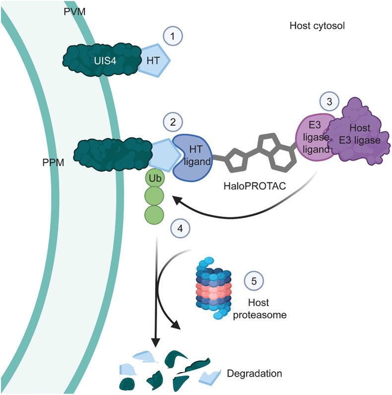
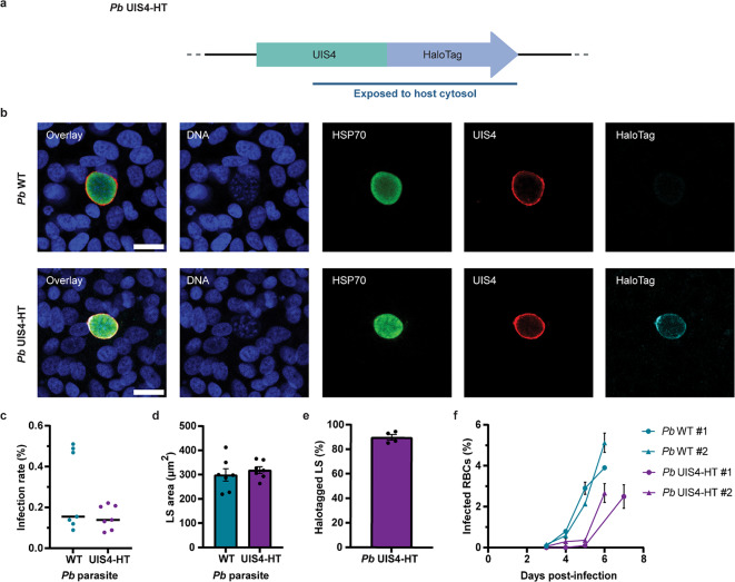
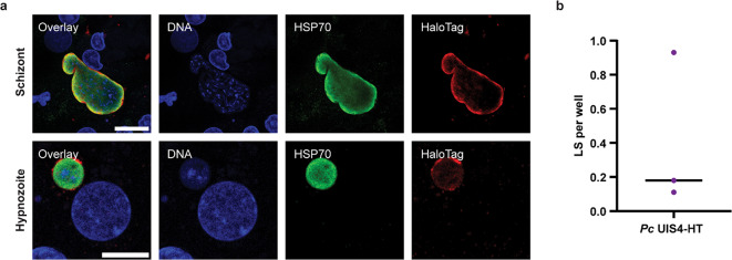
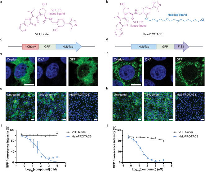
Targeted protein degradation (TPD) is a novel strategy for developing therapeutics against pathogens. Prior to causing malaria, Plasmodium parasites replicate within hepatocytes as liver stages, surrounded by a parasitophorous vacuole membrane (PVM). We hypothesized that TPD can be employed to trigger host-driven degradation of essential liver stage PVM proteins and lead to parasite death. To explore this, we took advantage of the proteolysis-targeting-chimera HaloPROTAC3, a molecule that recruits the host von Hippel-Lindau (VHL) E3 ligase to the HaloTag (HT). Parasites expressing HT fused to the host cytosol-exposed domain of the PVM protein UIS4 (UIS4-HT) were generated in Plasmodium berghei and Plasmodium cynomolgi, but only P. berghei UIS4-HT enabled productive liver stage infection experiments in vitro. Although HaloPROTAC3 triggered the degradation of HT proteins in host cells, it had no impact on the survival of P. berghei UIS4-HT liver stages. Furthermore, HaloPROTAC3 bound to P. berghei UIS4-HT but did not recruit VHL or trigger ubiquitination of the PVM. Overall, although this study did not establish whether host-driven TPD can degrade Plasmodium PVM proteins, it highlights the challenges of developing TPD approaches against novel targets and offers insights for advancing this therapeutic strategy against pathogens.

Keywords: Hypnozoite; Induced proximity; Infectious disease; Malaria; Schizont; Targeted protein degradation.


Conflict of interest statement

Declarations. Competing interests: M.L., A.P., L.T., A.A.L., M.E.F., J.R.K., M.S., A.T., S.H., C.M., E.L.F., D.M., B.N., Z.T., S.A.M., A.H. and G.M. were employed by and/or shareholders of Novartis Pharma AG during this study. All other authors have no competing interest in this study. Ethical approval: Female 6-8-week-old C57BL/6 (Jackson Laboratory) and Swiss Webster (Charles River Laboratories) mice were maintained in a facility accredited by the Association for Assessment and Accreditation of Laboratory Animal Care (AAALAC) at Novartis AG. Experiments were conducted in accordance with animal protocols approved by the Institutional Animal Care and Use Committee (IACUC, Novartis). Mice were anesthetized using isoflurane (3% isoflurane and 2 L/min oxygen) prior to retro-orbital injections and euthanized with CO2 followed by either cervical dislocation or bilateral thoracotomy for terminal blood collection. Euthanasia methods were in accordance with the recommendations set forth in the latest Panel Report of the American Veterinary Medical Association guidelines on euthanasia or pre-approved by the IACUC and the attending veterinarian. All procedures involving NHPs were reviewed and approved by the IACUC of OHSU / ONPRC or UGA. Both OHSU / ONPRC and UGA are certified by AAALAC International. For experiments performed at UGA, male Japanese macaques (Macaca fuscata) were obtained from ONPRC, transported to UGA for use in malaria-related studies, and underwent quarantine procedures upon arrival. For experiments performed at OSHU, Japanese macaques and rhesus macaques (Macaca mulatta) are purpose-bred at ONPRC. Prior to use, all NHPs underwent thorough physical exams and blood work to confirm their suitability for malaria-related studies. All NHPs were socially housed when compatible animals were available and when it did not interfere with the study goals and were provided environmental enrichment consisting of daily feeding enrichment, provision of manipulanda, and physical enrichment as described in the Guide for the Care and Use of Laboratory Animals. All procedures, including blood collections, inoculation procedures, parasitemia monitoring, sedation and analgesia protocols, were reviewed and approved by the IACUC prior to the beginning of an infection and were followed accordingly. The authors confirm that this study is reported in accordance with the ARRIVE guidelines.

Figures

Fig. 1
Fig. 1
Host-driven targeted degradation of a Plasmodium liver stage PVM protein using HaloTag technology. The illustration shows: (1) the host cytosol-exposed C-terminus of the PVM protein UIS4 fused to HaloTag (HT), (2) the binding of the reactive chloroalkane HaloTag ligand (HTL) moiety of a HaloPROTAC to the UIS4-HT fusion protein, (3) the binding of a host E3 ligase to the E3 ligase ligand moiety on the same HaloPROTAC, and (4) the ubiquitination and (5) the proteasomal degradation of UIS4-HT. PVM, parasitophorous vacuole membrane; PPM, parasite plasma membrane; Ub, ubiquitin; HaloPROTAC, HaloTag-targeted proteolysis targeting chimeras.
Fig. 2
Fig. 2
Generation and characterization of P. berghei (Pb) UIS4-HT. (a) Illustration of the P. berghei UIS4-HT locus. The blue line indicates the portion that is exposed to the host cytosol. (b) Confocal micrographs of Huh7 cells infected with P. berghei WT and P. berghei UIS4-HT for 2 days and stained for DNA (blue), Pb HSP70 (green), Pb UIS4 (red), and HaloTag (cyan). Scale bars are 20 μm. (c) Infection rates (% liver stages (LS) per host nuclei). Median infection rates were calculated from 2–4 technical replicates and pooled from 7 independent experiments (N = 7). No statistically significant difference was detected (P = 0.32, Mann-Whitney test). The solid black lines indicate medians. (d) Area sizes of liver stages at 2 days post-infection. Median area sizes were determined for each well and averaged from 2–4 technical replicates. No statistically significant difference was detected (P = 0.51, unpaired t test, N = 7). Results are expressed as means with SEMs. (e) Proportion of HaloTag-positive liver stages for Huh7 cells infected with P. berghei UIS4-HT. Median values were calculated from 2–3 technical replicates and expressed as means with SEMs (N = 4). (f) Blood stage parasitemia in mice infected with P. berghei WT and P. berghei UIS4-HT sporozoites. The graph shows data from two independent experiments, each including 2 (#1) or 3 (#2) mice per condition apart from one mouse for P. berghei WT #1 at 6 days post-infection (one mouse had to be euthanized a day earlier for this group). Results are expressed as means with SEMs. RBCs, red blood cells.
Fig. 3
Fig. 3
Characterization of P. cynomolgi (Pc) UIS4-HT liver stages. (a) Airyscan micrographs of primary simian hepatocytes infected with P. cynomolgi UIS4-HT for 8 days and stained for DNA (blue), Pc HSP70 (green), and HaloTag (red). A schizont (top row) and a hypnozoite (bottom row) are shown. Scale bars are 20 μm and 10 μm, respectively. (b) Infection rate of P. cynomolgi UIS4-HT presented as number of liver stages (LS) per well (≥ 80 wells per biological replicate). The solid black line indicates the median (N = 3).
Fig. 4
Fig. 4
VHL-recruiting HaloPROTAC3 triggers the degradation of soluble cytosolic and transmembrane HT fusion proteins in Huh7 cells. Chemical structure of (a) the VHL control binder and (b) HaloPROTAC3. Illustration of the (c) mCherry-GFP-HT and (d) HT-GFP-FIS1 constructs expressed in Huh7 cells. Airyscan micrographs showing DNA (blue) and GFP fluorescence (green) in (e) mCherry-GFP-HT and (f) HT-GFP-FIS1 Huh7 cells. Scale bars are 10 μm. Confocal micrographs of Huh7 (g) mCherry-GFP-HT and (h) HT-GFP-FIS1 cells treated with 1 µM of the VHL binder or HaloPROTAC3. Scale bars are 50 μm. Dose-response curves showing that HaloPROTAC3 triggers degradation of (i) soluble cytosolic mCherry-GFP-HT and (j) transmembrane HT-GFP-FIS1 proteins in Huh7 cells. Average fluorescence intensities from technical duplicates were normalized and presented as means with SEMs (N = 2–3). 100% corresponds to DMSO-treated samples. GFP, green fluorescent protein; FIS1, mitochondrial fission 1 protein.
Fig. 5
Fig. 5
HaloPROTAC3 does not influence the development of P. berghei UIS4-HT liver stages (LS). The impact of the VHL binder and HaloPROTAC3 on the (a) number of P. berghei liver stages and (b) number of host nuclei was determined in Huh7 cells 2 days post-infection. Results were normalized and median values were calculated from 2–3 technical replicates. Results are presented as means with SEMs (N = 4). The impact of compounds on (c) liver stage sizes and (d) the median UIS4 fluorescence intensity of P. berghei liver stages was also determined. Median area sizes and medians of median UIS4 fluorescence intensities of P. berghei liver stages were calculated for each well, normalized, and averaged from 2–3 technical replicates. 100% corresponds to infected samples treated with DMSO. Results are presented as means with SEMs (N = 4). Statistically significant differences between the VHL binder and HaloPROTAC3 were detected only for the number of P. berghei liver stages and UIS4 intensities at 10,000 nM of compounds (P < 0.05, two-way ANOVA with Bonferroni post-hoc test).
Fig. 6
Fig. 6
HaloPROTAC3 binds to P. berghei (Pb) UIS4-HT liver stages (LS). (a) Confocal micrographs of Huh7 cells infected with P. berghei WT and P. berghei UIS4-HT and stained for DNA (blue), parasite markers UIS4 (red) and HSP70 (cyan), and a fluorescent HaloTag ligand (HTL, green) 2 days post-infection. Scale bars are 20 μm. (b) Percentages of liver stages with bound fluorescent HTL were quantified for cells infected with P. berghei WT and P. berghei UIS4-HT. Median percentages were calculated from 4–8 technical replicates and expressed as means with SEMs (N = 2). (c) Dose-response curves showing the percentages of liver stages that bound to the fluorescent HTL as a function of HaloPROTAC3 concentration. During this assay, the binding of HaloPROTAC3 to UIS4-HT liver stages was evaluated by probing for remaining available binding sites with the fluorescent HTL. P. berghei WT and the VHL binder served as negative controls. Median percentages of liver stages with bound fluorescent HTL were calculated from technical duplicates and normalized. 100% corresponds to samples infected with P. berghei UIS4-HT and treated with DMSO. Results are means with SEMs (N = 2).
Fig. 7
Fig. 7
HaloPROTAC3 does not induce VHL recruitment and PVM ubiquitination during P. berghei liver stage development. (a) Illustration of the mCherry-VHL construct expressed in Huh7 cells. (b) Confocal micrographs of Huh7 mCherry-VHL cells infected with P. berghei UIS4-HT and stained for DNA (blue), mCherry (red), and parasite markers HSP70 (green) and UIS4 (cyan). Scale bar is 20 μm. (c) Illustration of the mCherry-ubiquitin (Ub) construct expressed in Huh7 cells. (d) Confocal micrographs of Huh7 mCherry-Ub cells infected with P. berghei UIS4-HT and stained for DNA (blue), mCherry (red), and parasite markers HSP70 (green) and UIS4 (cyan). Scale bar is 20 μm. Integrated mCherry-VHL (e) and mCherry-Ub (f) fluorescence intensities associated with P. berghei UIS4-HT liver stages in Huh7 cells treated with either the VHL binder or HaloPROTAC3. The median integrated fluorescence intensities of P. berghei liver stages were calculated for each well, normalized and averaged from 4 technical replicates. 100% corresponds to infected samples treated with DMSO. Results are presented as means with SEMs (N = 2). No statistically significant differences were detected at 100 or 500 nM (unpaired t tests).

References

    1. World Health Organization. Antimicrobial resistance: global report on surveillance (WHO, 2014).
    1. Brauner, A., Fridman, O., Gefen, O. & Balaban, N. Q. Distinguishing between resistance, tolerance and persistence to antibiotic treatment. Nat. Rev. Microbiol.14, 320–330. 10.1038/nrmicro.2016.34 (2016). - PubMed
    1. Wei, L. et al. Host-directed therapy, an untapped opportunity for antimalarial intervention. Cell. Rep. Med.2, 100423. 10.1016/j.xcrm.2021.100423 (2021). - PMC - PubMed
    1. Duffey, M. et al. Combating antimicrobial resistance in malaria, HIV and tuberculosis. Nat. Rev. Drug Discov. 23, 461–479. 10.1038/s41573-024-00933-4 (2024). - PubMed
    1. World Health Organization. World Malaria Report (WHO, 2023).

LinkOut - more resources