. 2025 Jun;27(6):1021-1034.
doi: 10.1038/s41556-025-01663-4. Epub 2025 May 26.

Dynamic regulation of integrin β1 phosphorylation supports invasion of breast cancer cells

Affiliations

Dynamic regulation of integrin β1 phosphorylation supports invasion of breast cancer cells

James R W Conway et al. Nat Cell Biol. 2025 Jun.

Abstract

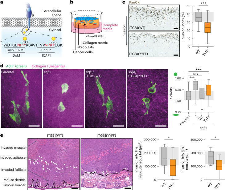
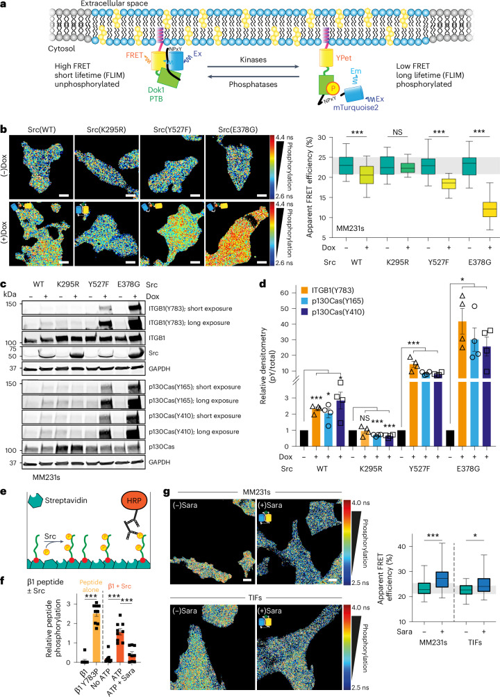
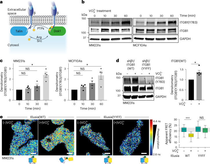
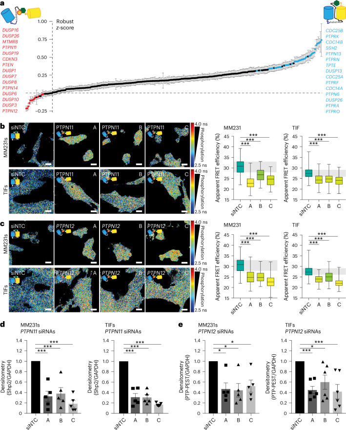
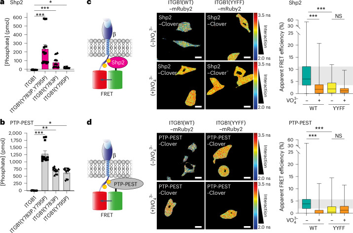
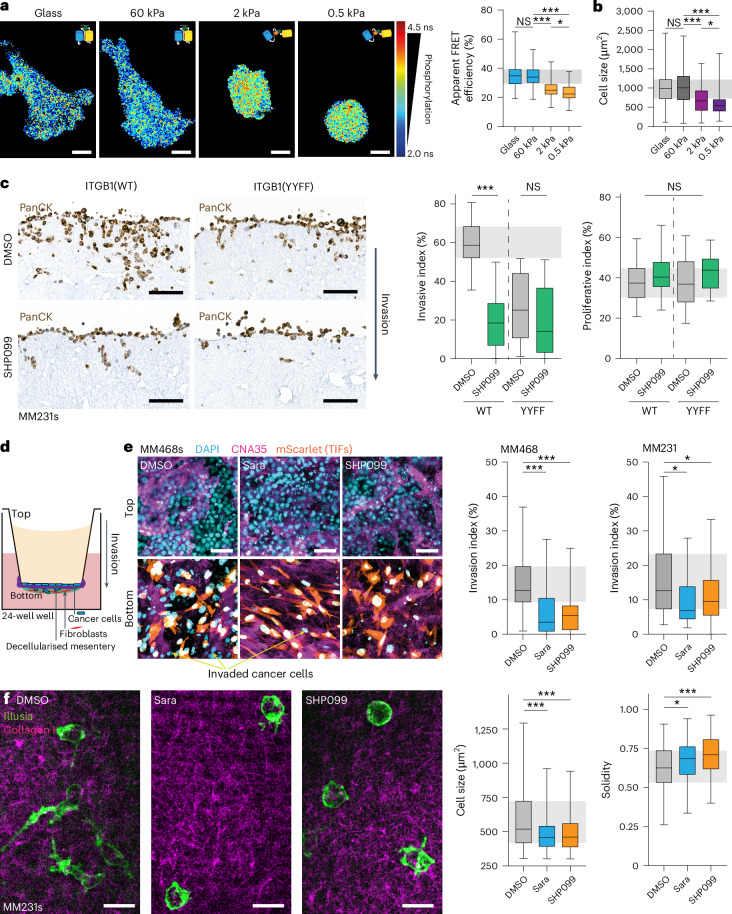
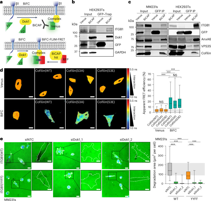
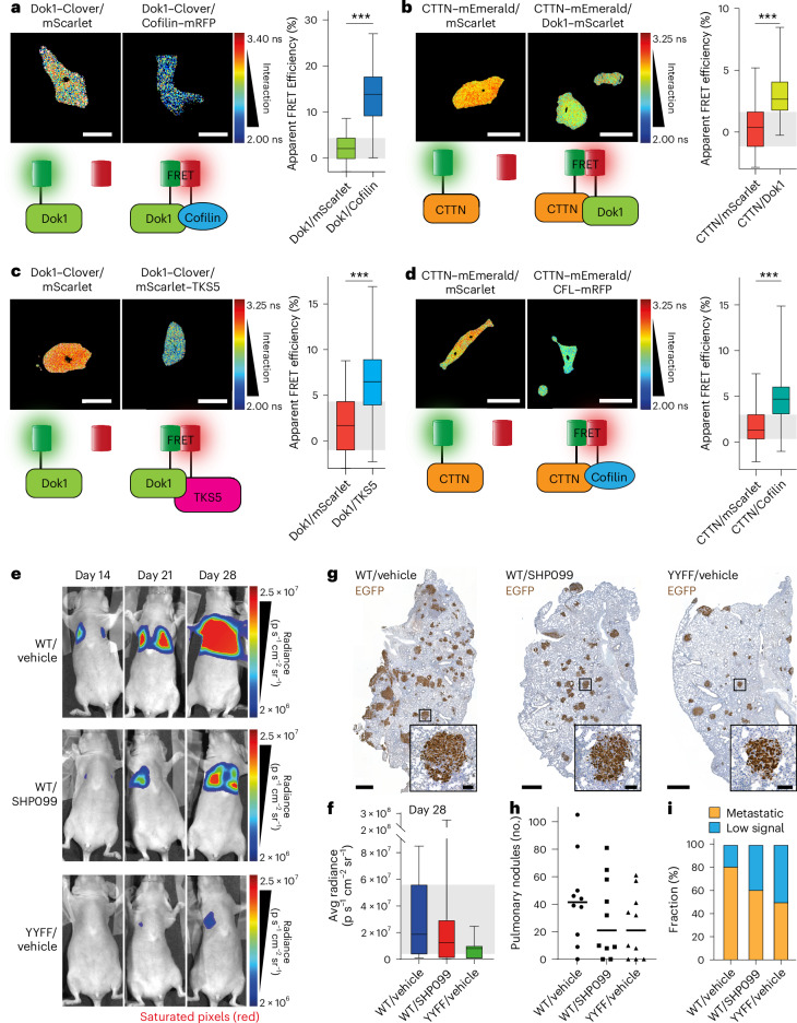
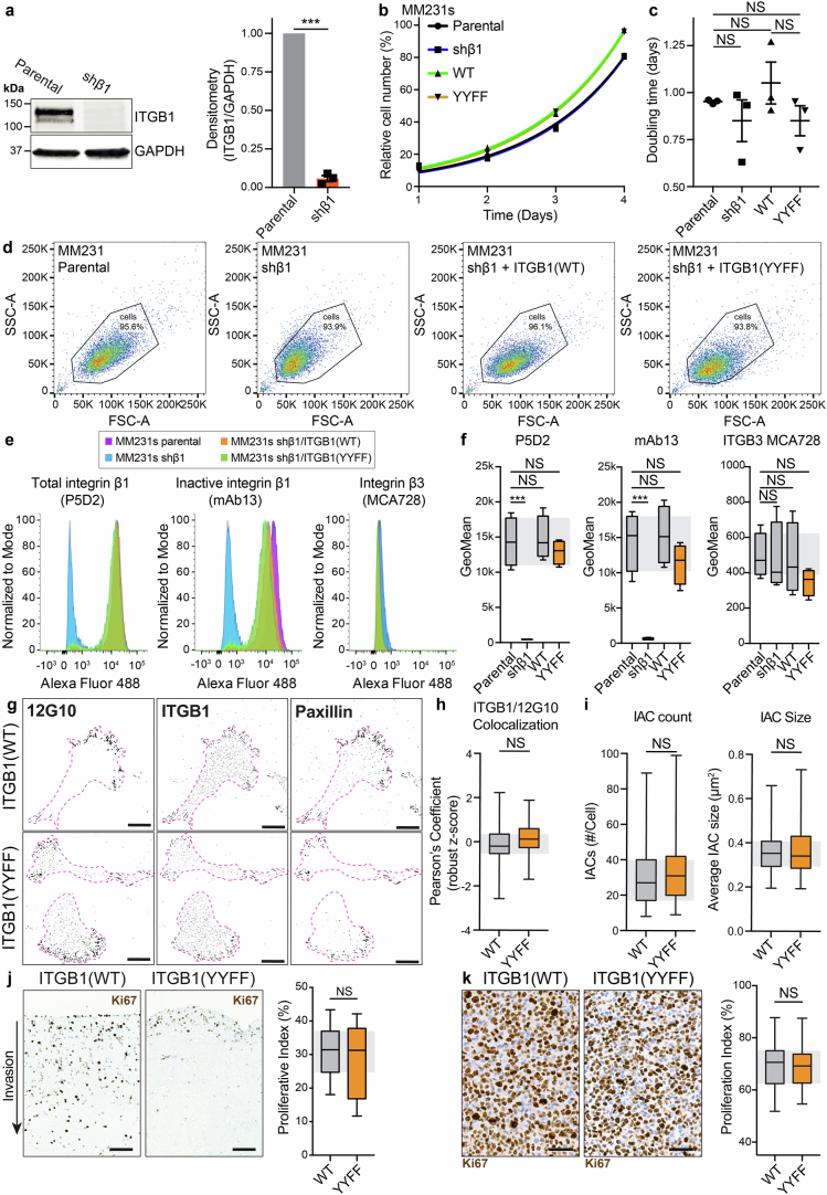
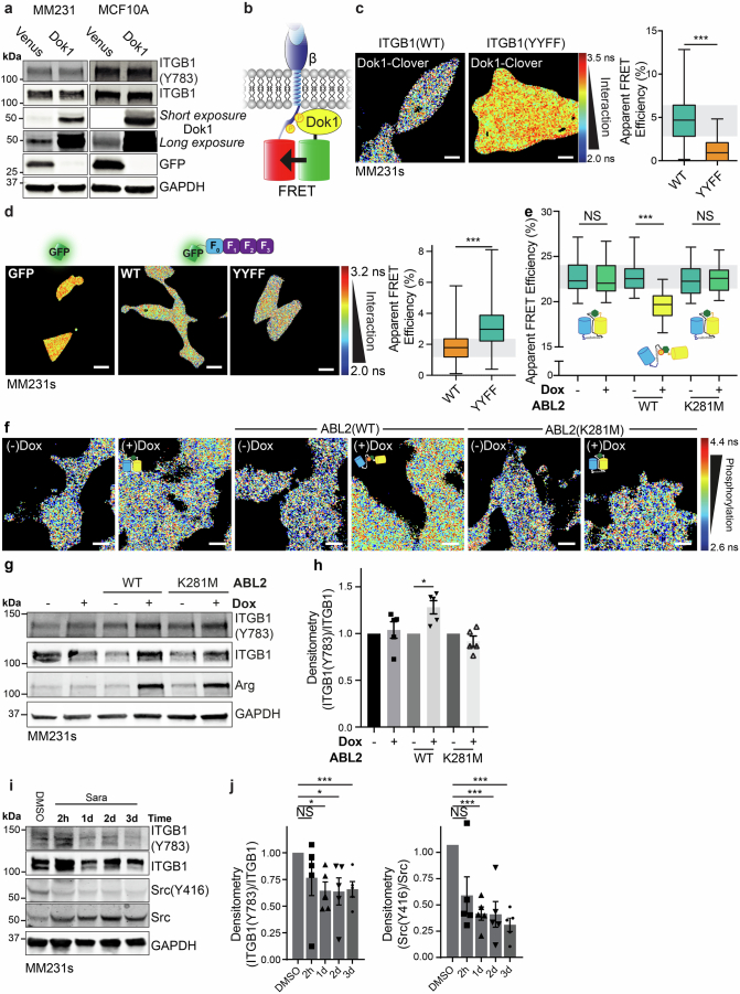
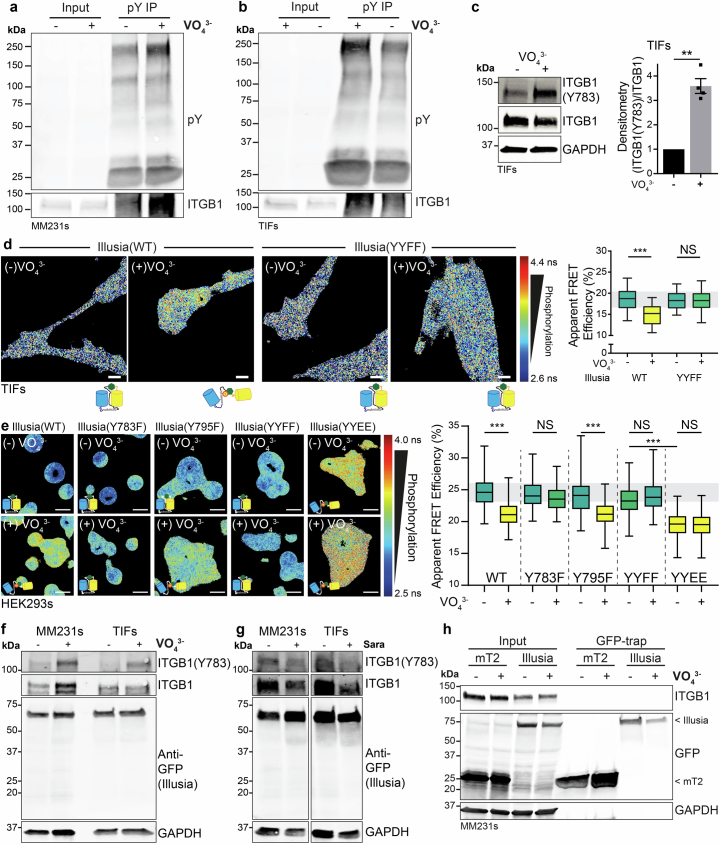
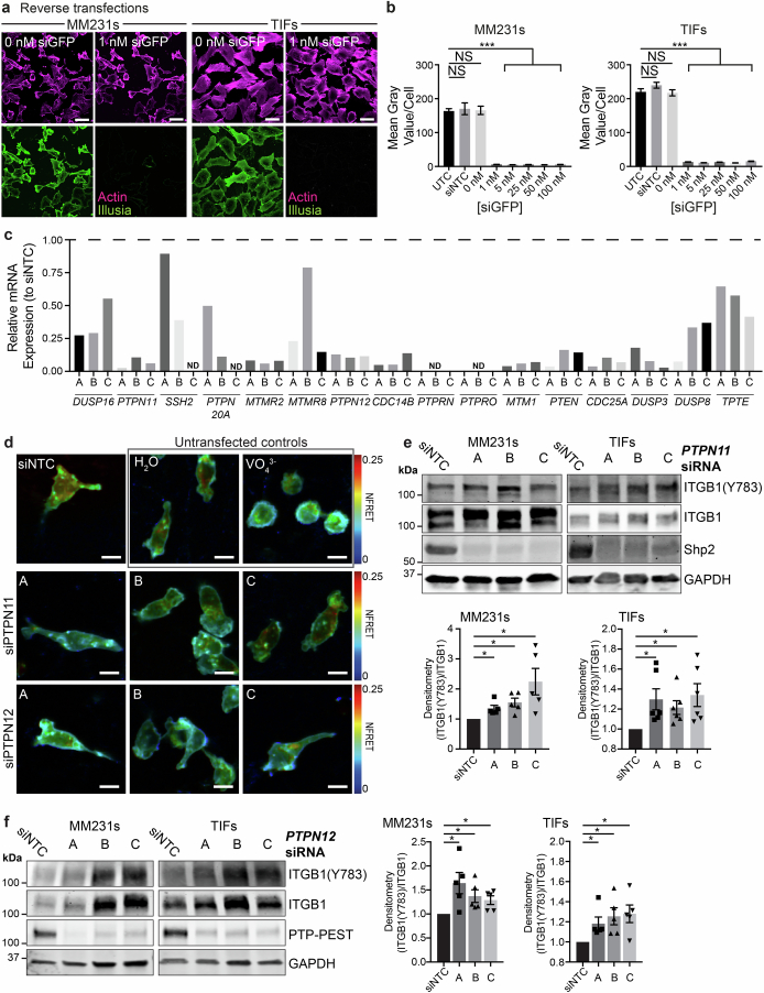
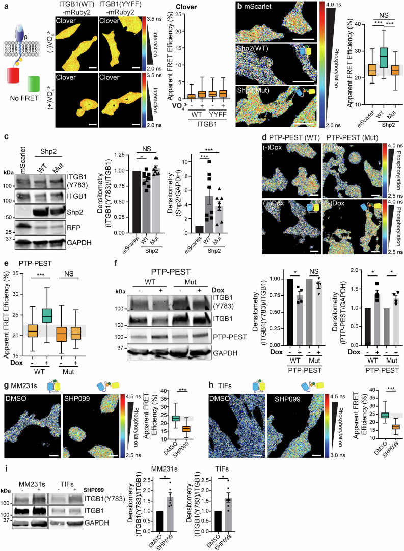
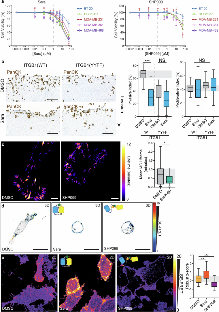
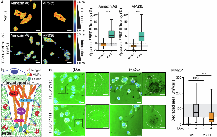
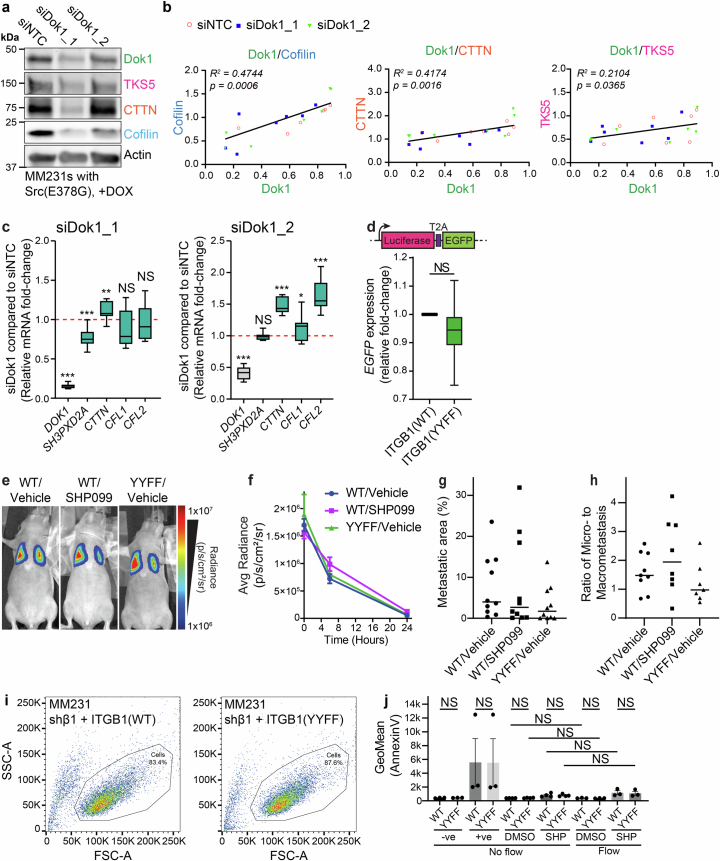
Integrins provide an essential bridge between cancer cells and the extracellular matrix, playing a central role in every stage of disease progression. Despite the recognized importance of integrin phosphorylation in several biological processes, the regulatory mechanisms and their relevance remained elusive. Here we engineer a fluorescence resonance energy transfer biosensor for integrin β1 phosphorylation, screening 96 protein tyrosine phosphatases and identifying Shp2 and PTP-PEST as negative regulators to address this gap. Mutation of the integrin NPxY(783/795) sites revealed the importance of integrin phosphorylation for efficient cancer cell invasion, further supported by inhibition of the identified integrin phosphorylation regulators Shp2 and Src kinase. Using proteomics approaches, we uncovered Cofilin as a component of the phosphorylated integrin-Dok1 complex and linked this axis to effective invadopodia formation, a process supporting breast cancer invasion. These data further implicate dynamic modulation of integrin β1 phosphorylation at NPxY sites at different stages of metastatic dissemination.


Conflict of interest statement

Competing interests: The authors declare no competing interests.

Figures

Fig. 1
Fig. 1. ITGB1 phosphorylation supports efficient cancer cell invasion.
a, A schematic highlighting the membrane-proximal and membrane-distal NPxY motifs of ITGB1. b, A schematic of the fibroblast-contracted 3D collagen matrix invasion assay platform. c, Left: representative images of MM231 triple-negative breast cancer cells expressing either ITGB1(WT or YYFF), invading into 3D fibroblast-contracted collagen I and stained with pan-cytokeratin (PanCK) to specifically detect the cancer cells and exclude fibroblasts from the analysis. Scale bars, 100 μm. Right: quantification of invasion beyond 100 μm, normalized to the total number of cells per region (n = 24 regions per cell line pooled from three biological replicates; unpaired two-tailed Student’s t-test with a Welch’s correction). d, Representative images (left) and quantification of cell shape (that is, solidity) (right) of MM231 cells (green is the actin staining) embedded in 3D collagen matrices overnight (magenta) (n = 104 (parental), 76 (shβ1), 84 (ITGB1(WT)) and 116 (ITGB1(YYFF)) cells pooled from three biological replicates; Kruskal–Wallis test with a Dunn’s correction for multiple comparisons). Scale bars, 20 μm. e, Representative H&E images (left) and quantification (right) of local invasion of MM231 ITGB1(WT or YYFF) cells into the mouse dermis from subcutaneous xenografts. Scale bars, 200 μm (n = 9 ITGB1(WT) or 11 ITGB1(YYFF) mice, respectively; unpaired two-tailed Student’s t-test with a Welch’s correction). The dotted line indicates the tumour boundary, where it meets the subcutaneous stromal cells. The boxplots represent the median and interquartile range (IQR). The whiskers extend to the minimum and maximum values. The grey areas on the boxplots highlight the IQR of the control conditions. NS, not significant. *P < 0.05, ***P < 0.001. Source data.
Fig. 2
Fig. 2. Src is a regulatory kinase for the ITGB1 NPxY motifs.
a, A schematic representation of the possible conformations for the ITGB1 intramolecular FRET biosensor (Illusia), where the mTurquoise2–YPet FRET pair is separated by the PTB from Dok1, a linker and the cytoplasmic domain (aa772–798) from ITGB1 (including the two NPxY motifs). Illusia is recruited to the membrane through an acylation substrate sequence derived from Lyn kinase. Ex, excitation; Em, emission. b, Representative FLIM images (left) and quantification of apparent FRET efficiency (right) of MM231 cells stably expressing Illusia after Dox-inducible overexpression of either Src(WT), kinase-dead Src(K295R) or constitutively active Src(Y527F)/Src(E378G) (n = 60 cells in all conditions with the exception of 70 for Src(Y527F) (−)Dox and 65 for Src(Y527F) (+)Dox pooled from three biological replicates; one-way analysis of variance (ANOVA) with a Šidák correction for multiple comparisons). Scale bars, 10 μm. c, Representative western blot of MM231 cells after Dox-inducible Src(WT), Src(K295R), Src(Y527F) or Src(E378G) overexpression. d, Densitometry analysis of western blots from c (n = 4 biological replicates; one-sample two-tailed t-test against the normalized control values for each cell line without Dox). The data are mean ± s.e.m. e, A scheme of an ELISA for pY. f, An ELISA for changes in ITGB1 phosphorylation using recombinant ITGB1 peptide and Src kinase in the absence or presence of ATP and the Src inhibitor Sara (1 μM; n = 3 biological replicates; triplicate wells/replicates; one-way ANOVA with a Šidák correction for multiple comparisons). Unphosphorylated ITGB1 and phosphorylated ITGB1 p(Y783) peptides were included as negative and positive controls, respectively. The data are the mean ± s.e.m. g, Representative FLIM–FRET images (left) and quantification (right) of MM231 and TIF cells with stable expression of Illusia and Sara treatment (1 μM) for 24 h (MM231s, n = 129 (DMSO) and 125 (Sara) cells pooled from five biological replicates; TIFs, n = 74 (DMSO) and 75 (Sara) cells pooled from three biological replicates; unpaired two-tailed Student’s t-test with a Welch’s correction). Scale bars, 10 μm. The boxplots represent the median and IQR. The whiskers extend to the minimum and maximum values. The grey areas on the boxplots highlight the IQR of the control conditions. NS, not significant. *P < 0.05, ***P < 0.001. Source data.
Fig. 3
Fig. 3. PTPs actively regulate phosphorylation of the ITGB1 NPxY motifs.
a, A schematic of phosphorylation-dependent Dok1 recruitment to ITGB1. b,c, Representative western blots (b) and densitometry (c) of MM231 (left) or MCF10A (right) cells treated with the broad-spectrum PTP inhibitor sodium orthovanadate (VO43; 100 μM, 2 h; n = 4 biological replicates; one-sample two-tailed t-test against the normalized control value without VO43). The data are the mean ± s.e.m. d, A representative western blot (left) of MM231 ITGB1(WT or YYFF) cells treated for 2 h with VO43 (100 μM) and densitometry analysis (right) of MM231 ITGB1(WT) cells (n = 5 biological replicates; one-sample two-tailed t-test against the normalized control value without VO43). The data are the mean ± s.e.m. e, Representative FLIM images (left) and quantification of apparent FRET efficiency (right) after VO43 treatment (100 μM, 2 h) of MM231 cells stably expressing Illusia(WT) or a non-phosphorylatable mutant reporter Illusia(YYFF) (n = 88 (Illusia(WT) (VO43)), 88 (Illusia(WT) (+VO43)), 93 (Illusia(YYFF) (VO43)) and 86 (Illusia(YYFF) (+VO43)) cells pooled from four biological replicates; one-way analysis of variance with a Šidák correction for multiple comparisons). n.s., not significant. Scale bars, 10 μm. The boxplot represents the median and IQR. The whiskers extend to the minimum and maximum values. The grey area on the boxplot highlights the IQR of the control condition. NS, not significant, *P < 0.05, ***P < 0.001. Source data
Fig. 4
Fig. 4. An RNAi FRET screen to identify regulatory PTPs for ITGB1 NPxY sites.
a, A waterfall plot from an RNAi sensitized emission (SE)-FRET screen of MM231 cells stably expressing Illusia to assess changes after KD of 96 out of the 108 PTPs in the human genome, using three siRNAs/target (A, B and C) (n = 3; one-way analysis of variance (ANOVA) with a Tukey correction for multiple comparisons after normalizing replicates into robust z-scores). PTPs that significantly (P < 0.01) increased phosphorylation upon KD are indicated with red text and circles (15 hits), while those that significantly (P < 0.01) decreased phosphorylation upon KD are indicated with blue text and circles (15 top hits). The data are the mean ± s.e.m. b,c, Representative FLIM–FRET images (left) and quantification (right) of MM231 and TIF cells with stable Illusia expression and transfected with siRNAs (A, B or C) against either PTPN11 (Shp2) (b) or PTPN12 (PTP-PEST) (c) (MM231 PTPN11 silencing, n = 100 (siNTC), 98 (siRNA A), 100 (siRNA B) and 100 (siRNA C); PTPN12 silencing, n = 99 (siNTC), 99 (siRNA A), 98 (siRNA B) and 97 (siRNA C) cells pooled from three biological replicates; TIF PTPN11 silencing, n = 75 (siNTC), 76 (siRNA A), 76 (siRNA B) and 78 (siRNA C); PTPN12 silencing, n = 74 (siNTC), 74 (siRNA A), 77 (siRNA B) and 74 (siRNA C) cells pooled from four biological replicates; one-way ANOVA with a Dunnett correction for multiple comparisons). n.s., not significant. Scale bars, 20 μm. The boxplots represent the median and IQR. The whiskers extend to the minimum and maximum values. The grey areas on the boxplots highlight the IQR of the control conditions. siNTC, non-targeting control siRNA. d,e, Densitometry from western blots of PTPN11 (representative western blot) (d) (Extended Data Fig. 4e) and PTPN12 (representative western blot) (e) (Extended Data Fig. 4f) siRNA KD in MM231 (n = 5 biological replicates) and TIF (n = 5 (Shp2) or 6 (PTP-PEST) biological replicates) cells. The data are the mean ± s.e.m. *P < 0.05, ***P < 0.001. Source data
Fig. 5
Fig. 5. ITGB1 is a substrate for PTP-PEST and Shp2.
a,b, A malachite green assay for free phosphate release after incubation of phosphorylated/non-phosphorylated ITGB1 peptides with recombinant Shp2 (n = 5 independent replicates, each performed in triplicate) (a) or PTP-PEST (n = 4 independent replicates, each performed in triplicate) (b). The significance was assessed using a Kruskal–Wallis test with a Dunn’s correction for multiple comparisons. The data are presented as the mean ± s.e.m. c,d, Schematics of FRET experiments (left) using mRuby2-tagged ITGB1 and Clover-tagged PTPs. Representative FLIM–FRET images (right) and quantification of apparent FRET efficiency of MM231 cells with stable expression of either ITGB1(WT)–mRuby2 or ITGB1(YYFF)–mRuby2 transfected with either Shp2–Clover (c) or PTP-PEST-Clover (d) and treated with VO43 (100 μM, 2 h) (for c, n = 73 (ITGB1(WT) ()VO43), 62 (ITGB1(WT) (+)VO43), 64 (ITGB1(YYFF) ()VO43) and 67 (ITBG1(YYFF) (+)VO43) cells pooled from three biological replicates; for d, n = 75 (ITGB1(WT) ()VO43), 58 (ITGB1(WT) (+)VO43), 58 (ITGB1(YYFF) ()VO43) and 65 (ITBG1(YYFF) (+)VO43) cells pooled from three biological replicates; one-way analysis of variance with a Tukey correction for multiple comparisons). Scale bars, 20 μm. The boxplots represent the median and IQR. The whiskers extend to the minimum and maximum values. The grey areas highlight the IQR of the control conditions. NS, not significant. *P < 0.05, **P < 0.01, ***P < 0.001. Source data.
Fig. 6
Fig. 6. Src or Shp2 inhibition of ITGB1 phosphorylation dynamics results in equivalent phenotypes.
a,b, Representative FLIM images (a, left) and apparent FRET efficiencies (a, right) and cell area (b) of MM231 cells stably expressing Illusia and seeded on either glass or hydrogels (60, 2 or 0.5 kPa) (for a, n = 110 (glass), 117 (60 kPa), 116 (2 kPa) and 113 (0.5 kPa) cells pooled from four biological replicates; for b, n = 121 (glass), 137 (60 kPa), 157 (2 kPa) and 165 (0.5 kPa) cells pooled from four biological replicates; one-way analysis of variance (ANOVA) with a Tukey correction for multiple comparisons). Scale bars, 10 μm. c, Representative images (left) of MM231 ITGB1(WT or YYFF) cells invading into 3D fibroblast-contracted collagen I treated with DMSO or SHP099 (100 nM). Pan-cytokeratin (PanCK) staining was used to mark cancer cells and exclude fibroblasts from the analysis. Quantification of invasion beyond 100 μm (right), normalized to the total number of cells/region, or proliferation, normalizing the number of Ki67-positive nuclei to the total number of cells/region (n = 24 regions per cell line pooled from four biological replicates; one-way ANOVA with a Tukey correction for multiple comparisons). Scale bars, 100 μm. d, A scheme for the basement membrane invasion assay. e, Left: representative images of MM231 and MM468 cells invading into the basement membrane matrix for 4 or 5 days, respectively, in the presence of SHP099 (100 nM) or Sara (1 μM) (collagen I is labelled with HaloTag-CNA35 (magenta), fibroblasts by mScarlet expression (red) and all nuclei stained with DAPI (cyan); the cancer cells are apparent by nuclei staining alone (cyan-positive, mScarlet-negative cells)). Quantification of basement membrane invasion (MM231 cells, n = 31 (from 11 basement membranes; DMSO), 24 (from 9 basement membranes; Sara) and 23 (from 8 basement membranes; SHP099) regions pooled from three biological replicates (right); MM468 cells, n = 31 (from 12 basement membranes; DMSO), 32 (from 12 basement membranes; Sara) and 27 (from 9 basement membranes; SHP099) regions pooled from four biological replicates; one-way ANOVA with a Dunnett’s correction for multiple comparisons). Scale bars, 50 μm. f, Illusia-expressing MM231s (green) embedded in collagen I (magenta) and treated with SHP099 (100 nM) or Sara (1 μM) for 24 h (cell size, n = 119 (DMSO), 93 (Sara) and 85 (SHP099); solidity, n = 122 (DMSO), 91 (Sara) and 94 (SHP099) cells from four biological replicates; one-way ANOVA with a Dunnett’s correction). The boxplots represent the median and IQR. The whiskers extend to the minimum and maximum values. The grey areas highlight the IQR of the control conditions. NS, not significant. *P < 0.05, ***P < 0.001. Source data.
Fig. 7
Fig. 7. Phosphorylation-sensitive recruitment of Dok1 supports invadopodia formation.
a, A schematic of the interaction between ITGB1-V1 and a V2-tagged adaptor or regulator, highlighting the resulting V1/V2 (Venus) protein complex as the restored epitope for BiCAP (top) or donor for BiFC–FLIM–FRET (bottom). The Venus tag alone was used as a control for the BiCAP and BiFC–FLIM–FRET experiments. b, A representative BiCAP immunoblot after HEK293T cell transfection with either Venus or ITGB1-V1/Dok1-V2 (n = 3 biological replicates). c, Representative BiCAP immunoblots from MM231 and HEK293T cells where annexin A6 (AnxA6), VPS35 and Cofilin coimmunoprecipitate with the Dok1/ITGB1 complex (n = 3 biological replicates). d, Representative BiFC–FLIM–FRET images (left) and quantification of apparent FRET efficiency (right) from MM231 cells transfected with RFP-tagged Cofilin mutants WT, S3A and S3E (Venus, n = 73 (WT), 72 (S3A) and 64 (S3E); BiFC, n = 92 (WT), 64 (S3A) and 64 (S3E) cells from three biological replicates; one-way analysis of variance (ANOVA) with a Tukey correction for multiple comparisons). Scale bars, 20 μm. e, Representative images (left) and quantification of gelatin degradation (right) by MM231 ITGB1(WT or YYFF) cells with Dox-inducible Src(E378G) expression. The MM231 cells were transfected with siRNAs against Dok1 (siDok1_1 and siDok1_2) or a NTC siRNA and treated with Dox for 24 h before being seeded on fluorescent gelatin (green) for 6 h (actin labelled with SiR-actin (white), nuclei with DAPI (blue)) (ITGB1(WT), n = 36 (NTC), 38 (siDok1_1) and 36 (siDok1_2); ITGB1(YYFF), n = 38 (NTC), 34 (siDok1_1) and 37 (siDok1_2) fields of view pooled from three biological replicates; one-way ANOVA with a Šidák correction for multiple comparisons). Scale bars, 20 μm; insets, 10 μm. The boxplots represent the median and IQR. The whiskers extend to the minimum and maximum values. The grey areas highlight the IQR of the control conditions. NS, not significant. ***P < 0.001. Source data.
Fig. 8
Fig. 8. The Dok1/ITGB1 complex recruits Cofilin and other invadopodia components to adhesion sites to mediate efficient cancer dissemination.
ad, Representative images (left) and quantification of apparent FRET efficiency (right) for intermolecular FLIM–FRET of the following tagged protein pairs, Dok1–Clover/Cofilin–mRFP (a), CTTN–mEmerald/Dok1–mScarlet (b), Dok1–Clover/mScarlet–TKS5 (c) and CTTN–mEmerald/Cofilin–mRFP (d). FRET between mScarlet and the donor-tagged protein was used as a negative control for all pairs (for a, n = 85 (Dok1/mScarlet) and 95 (Dok1/Cofilin) cells pooled from five biological replicates; for b, n = 65 (CTTN/mScarlet) and 68 (CTTN/Dok1) cells pooled from three biological replicates; for c, n = 62 cells for each condition pooled from three biological replicates; for d, n = 65 (CTTN/mScarlet) and 70 (CTTN/Cofilin) cells pooled from three biological replicates; unpaired two-tailed Student’s t-test with a Welch’s correction). Scale bars, 20 μm. e, Representative images of mice with MM231 ITGB1(WT or YYFF) cells stably expressing the luciferase/EGFP construct. Oral gavage of Vehicle or SHP099 (100 mg kg−1) proceeded for 5 days from the day of injection. f, A box and whisker plot highlighting the endpoint metastatic burden as an average (Avg) radiance value from the luciferase signal of the MM231 cells in e (n = 9 mice tracked per group). g, Representative lung sections stained for EGFP-positive MM231 cells. Scale bars, 2 mm; insets: 200 μm. h, Quantification of pulmonary nodule number (that is, clusters of greater than ten cells) in lungs from EGFP-positive MM231 cells (n = 10 mice per group). i, Quantitative real-time PCR of the RNA samples collected from the MM231 ITGB1(WT or YYFF) cells stably expressing the luciferase/EGFP construct. The mice were designated as either ‘metastatic’ or ‘low signal’ after setting a threshold for ‘metastatic’ as having an expression fold change >1 compared with the mean of the WT/vehicle control with human GAPDH normalized to mouse/human GAPDH (n = 10 mice/group). The boxplots represent the median and IQR. The whiskers extend to min and max values. The grey areas highlight the IQR of the control conditions. ***P < 0.001. Source data.
Extended Data Fig. 1
Extended Data Fig. 1. Mutation of the ITGB1 NPxY tyrosine (Y) residues to non-phosphorylatable phenylalanine (F) does not change surface integrin levels, or cancer cell proliferation.
a, Representative western blot (left) and densitometry (right) of ITGB1 levels after shRNA-mediated KD in MM231 cells (shβ1; n = 3 biological replicates; significance assessed using a one-sample two-tailed t-test against the normalised control value; ***p < 0.001). Data are mean ± s.e.m. b & c, Representative curves (b) and doubling times (c) from the relative cell density of parental and shβ1 MM231 cells, and shβ1 MM231s with stable ITGB1(WT or YYFF) reexpression (ITGB1(WT or YYFF)-mRuby2 transposon vectors, described in methods; n = 3 biological replicates; significance assessed using a one-way ANOVA with a Šidák correction for multiple comparisons; NS, not significant). d, Gating strategy for the flow cytometry data presented in e & f. e & f, Representative histograms (e) and quantification (f) of the surface expression of total ITGB1 (P5D2), inactive ITGB1 (mAb13) and total integrin β3 (MCA728) using flow cytometry in shβ1 MM231s with or without ITGB1 reexpression as indicated (n = 4 biological replicates; significance assessed using a one-way ANOVA with a Tukey correction for multiple comparisons; NS, not significant; ***p < 0.001). g & h, TIRF images (g) of MM231 shβ1 cells with ITGB1 WT or YYFF reexpression (cells outlined with pink dashed lines) and quantification of colocalization (h) between active integrin staining (12G10 antibody) and either mRuby2-tagged ITGB1 WT (average Pearson’s r of 0.4853) or YYFF (average Pearson’s r of 0.5012; n = 65 [ITGB1 (WT)] and 63 [ITGB1(YYFF)] cells pooled from three biological replicates; significance assessed using an unpaired two-tailed Student’s t-test with a Welch’s correction; NS, not significant). i, Analysis of paxillin staining in MM231 cells from (g) to compare IAC average number and size per cell (n = 87 [ITGB1 (WT)] and 85 [ITGB1 (YYFF)] cells pooled from four biological replicates; NS, not significant). j, Representative images of invading MM231 ITGB1(WT or YYFF) cells (left) and quantification of their proliferation (right) from the fibroblast-contracted 3D collagen I invasion assays in Fig. 1c. Cells are stained with the proliferation marker Ki67. Quantification was achieved by normalising the number of Ki67-positive nuclei to the total number of cells/region (n = 24 regions for each cell line pooled from three biological replicates; significance assessed using an unpaired two-tailed Student’s t-test with a Welch’s correction; NS, not significant). Scale bars, 100 μm. k, Representative images (left) and quantification (right) of Ki67-stained subcutaneous xenografts of MM231 cells with either ITGB1(WT or YYFF) from Fig. 1e. Quantification of positive (brown) to negative (blue) staining of Ki67 in 400 μm2 regions of interest from subcutaneous xenografts is shown (n = 9 mice (WT) or 11 mice (YYFF); 5 regions/mouse/condition); significance assessed using an unpaired two-tailed Student’s t-test with a Welch’s correction; NS, not significant). Scale bars, 50 μm. Source data and exact p-values are provided in the statistical source data file. Boxplots represent median and interquartile range. Whiskers extend to min and max values. Grey areas on boxplots highlight the IQR of the control conditions. Source data
Extended Data Fig. 2
Extended Data Fig. 2. Arg (ABL2) phosphorylates the ITGB1 NPxY sites.
a, Representative western blots of MM231 and MCF10A cells expressing either Venus or Dok1 (n = 4 biological replicates). b, A schematic of the intermolecular FRET approach between Dok1 and ITGB1 (WT or YYFF). c, Representative FLIM-FRET images (left) and quantification of apparent FRET efficiency (right) from MM231 shβ1 cells reexpressing mRuby2-tagged ITGB1(WT or YYFF) and transfected with Dok1-Clover (n = 60 cells in each condition pooled from three biological replicates; significance assessed using an unpaired two-tailed Student’s t-test with a Welch’s correction; ***p < 0.001). Scale bars, 10 μm. d, Representative FLIM-FRET images (left) and quantification of apparent FRET efficiency (right) from MM231 shβ1 cells reexpressing mRuby2-tagged ITGB1(WT or YYFF), and transfected with GFP-F0F3 talin head domain fragment (n = 62 cells in each condition pooled from three biological replicates; significance assessed using an unpaired two-tailed Student’s t-test with a Welch’s correction; ***p < 0.001). Scale bars, 20 μm. e & f, Apparent FRET efficiencies (e) and representative images (f) of MM231 cells stably expressing Illusia and Dox-inducible ABL2(WT) or kinase-dead ABL2(K281M) ± Dox treatment. Parental cells treated with Dox are used as an additional control (n = 60 cells in each condition pooled from three biological replicates; significance assessed using a one-way ANOVA with a Šidák correction for multiple comparisons; NS, not significant, ***p < 0.001). Scale bars, 10 μm. g & h, Representative western blot (g) and densitometry (h) analysis of ITGB1 phosphorylation levels in MM231 cells with Dox-induced ABL2(WT) or ABL2(K281M) overexpression (n = 5 biological replicates; significance assessed using a one-sample two-tailed t-test against the normalised control values for each cell line without Dox; NS, not significant, *p < 0.05). Data are mean ± SEM. i & j, Representative western blot (i) and densitometry (j) of MM231 cells treated with saracatenib (Sara) for 0, 2, 24, 48 and 72 h (1 μM; n = 5 biological replicates; significance assessed using a one-sample two-tailed t-test against the normalised DMSO control). Source data and exact p-values are provided in the statistical source data file. Boxplots represent median and interquartile range. Whiskers extend to min and max values. Grey areas on boxplots highlight the interquartile range of the control conditions. Source data
Extended Data Fig. 3
Extended Data Fig. 3. Validation of the phosphorylation-dependent changes of Illusia and the active dephosphorylation of ITGB1 in cancer and normal cells.
a & b, Representative immunoprecipitation (IP) of phosphorylated ITGB1 in MM231 (a) and TIF (b) cells using anti-pY beads after VO43 treatment (100 mM, 2 h; n = 3 biological replicates). c, Representative western blot and densitometry analysis of ITGB1 phosphorylation levels in TIF cells after VO43 treatment (100 mM, 2 h; n = 4 biological replicates; significance assessed using a one-sample two-tailed t-test against the normalised control value without VO43; **p < 0.01). Data are mean ± SEM. d, Representative images (left) and quantification of apparent FRET efficiency (right) of VO43- (100 mM, 2 h) treated TIF cells stably expressing Illusia (WT), or a non-phosphorylatable mutant (YYFF) (n = 60 cells in each condition pooled from three biological replicates; significance assessed using a one-way ANOVA with a Šidák correction for multiple comparisons; NS, not significant, ***p < 0.001). Scale bars, 10 μm. e, Representative FLIM-FRET images (left) and apparent FRET efficiencies (right) from HEK293 cells transfected with different Illusia variants (WT, Y783F, Y795F, YYFF and YYEE; n = 127 [WT ()VO43], 152 [WT (+)VO43], 113 [Y783F ()VO43], 120 [Y783F (+)VO43], 109 [Y795F ()VO43], 117 [Y795F (+)VO43], 115 [YYFF ()VO43], 121 [YYFF (+)VO43], 106 [YYEE ()VO43], and 109 [YYEE (+)VO43] cells pooled from four biological replicates; significance assessed using one-way ANOVA with a Šidák correction for multiple comparisons; NS, not significant, ***p < 0.001). Scale bars, 20 μm. f & g, Representative western blots of MM231 and TIF cells stably expressing Illusia and treated with VO43 (f; 100 mM, 2 h; n = 3 biological replicates) or Sara for 24 h (g; 1 μM; n = 4 biological replicates). h, Representative GFP-trap IP of mT2 or Illusia from MM231 cells stably expressing the reported constructs (n = 3 biological replicates). Source data and exact p-values are provided in the statistical source data file. Boxplots represent median and interquartile range. Whiskers extend to min and max values. Grey areas on boxplots highlight the interquartile range of the control conditions. Source data
Extended Data Fig. 4
Extended Data Fig. 4. Validation of siRNA knock-down of a subset of hits that showed significant changes in the Illusia FRET screen.
a & b, Representative images (a; Scale bars, 50 μm) and quantification of reverse transfections in MM231 and TIF cells using an siRNA against GFP; actin staining (SiR-Actin, magenta) and Illusia (Green) (UTC, untransfected control; siNTC, non-targeting control siRNA) (MM231s, n = 220 (UTC), 70 (siNTC), 122 (0 nM), 140 (1 nM), 79 (5 nM), 89 (25 nM), 117 (50 nM) and 98 (100 nM) | TIFs, n = 179 (UTC), 155 (siNTC), 174 (0 nM), 183 (1 nM), 224 (5 nM), 199 (25 nM), 118 (50 nM) and 198 (100 nM) cells pooled from three biological replicates; significance assessed using a one-way ANOVA with a Dunnett’s correction for multiple comparisons; NS, not significant, ***p < 0.001). Data are mean ± SEM. c, Parallel qRT-PCR from RNA samples collected from the RNAi FRET screen cells targeting the top 16 hits (n = 1 experiment, assessed in triplicate). ND, Not detected. d, Representative SE-FRET images of MM231s stably-expressing Illusia showing the normalised FRET (NFRET) from the untransfected controls with or without VO43-, and siRNAs targeting PTPN11 and PTPN12, against the NTC siRNA. Scale bars, 20 µm. e & f, Representative western blots validating siRNA KD efficiency and ITGB1 phosphorylation levels in MM231 (left; n = 5 biological replicates; densitometry for ITGB1(Y783)/ITGB1 is displayed here. Densitometry for Shp2/GAPDH is in Fig. 4e) and TIF (right; n = 6 biological replicates; densitometry for ITGB1(Y783)/ITGB1 is displayed here. Densitometry for Shp2/GAPDH is in Fig. 4f) cells transfected with three different siRNAs (A, B and C) against PTPN11 (e) or PTPN12 (f). Source data and exact p-values are provided in the statistical source data file. Source data
Extended Data Fig. 5
Extended Data Fig. 5. Inhibition or overexpression of Shp2 or PTP-PEST modulates ITGB1 phosphorylation.
a, Schematic of the FRET experiment (left), with representative FLIM-FRET images (middle) and quantification of apparent FRET efficiency (right) of MM231 cells with stable expression of either ITGB1(WT)-mRuby2 or ITGB1(YYFF)-mRuby2 transfected with Clover and treated with VO43- (n = 65 [ITGB1(WT) ()VO43], 70 [ITGB1(WT) (+)VO43], 72 [ITGB1(YYFF) ()VO43], and 66 [ITGB1(YYFF) (+)VO43]) cells pooled from three biological replicates. Scale bars, 20 μm. b, Representative FLIM-FRET images (left) and quantification of apparent FRET efficiency (right) in MM231 cells with stable Illusia expression and constitutive overexpression of PTPN11 (Shp2, WT) or a phosphatase-dead mutant (Shp2, Mut; PTPN11(D425A, C459S)) (n = 97 (mScarlet), 96 (WT) and 96 (Mut) cells pooled from four biological replicates; significance assessed using a one-way ANOVA with a Tukey correction for multiple comparisons; NS, not significant, ***p < 0.001). Scale bars, 20 μm. c, Representative western blot (left) and densitometry (right) of MM231 cells with stable Illusia expression and constitutive overexpression of PTPN11 (Shp2, WT) or a phosphatase-dead mutant (Shp2, Mut; PTPN11(D425A, C459S); n = 8 biological replicates; significance assessed using a one-sample two-tailed t-test against the normalised control value; *p < 0.05, ***p < 0.001; NS, not significant). d-e, Representative FLIM-FRET images (d) and quantification of apparent FRET efficiency (e) of MM231 cells with stable Illusia expression treated overnight with Dox to induce overexpression of PTP-PEST WT or a phosphatase-dead mutant (Mut, PTPN12(D199A, C231S) (n = 100 cells in each condition pooled from four biological replicates; significance assessed using a one-way ANOVA with a Šidák correction for multiple comparisons; NS, not significant, ***p < 0.001). Scale bars, 20 μm. f, Western blot (left) and densitometry (right) from parallel data in (d; n = 4 biological replicates; significance assessed using a one-sample two-tailed t-test against the normalised control value; *p < 0.05; NS, not significant). g & h, Representative FLIM-FRET images (left) and quantification of apparent FRET efficiency (right) of MM231 (g) and TIF (h) cells with stable Illusia expression and treated with SHP099 for 2 h (100 nM) (MM231, n = 96 (DMSO) and 95 (SHP099) | TIFs, n = 100 (DMSO) and 99 (SHP099) cells pooled from four biological replicates; significance assessed using an unpaired two-tailed Student’s t-test with a Welch’s correction (NS, not significant, ***p < 0.001). Scale bars, 20 μm. i, Representative western blots (left) and densitometry (right) of MM231 and TIF cells with stable Illusia expression and treated with SHP099 for 2 h (100 nM; n = 6 biological replicates; significance assessed using a one-sample two-tailed t-test against the normalised control value; *p < 0.05). Data are mean ± SEM. Source data and exact p-values are provided in the statistical source data file. Boxplots represent median and interquartile range. Whiskers extend to min and max values. Grey areas on boxplots highlight the interquartile range of the control conditions. Source data
Extended Data Fig. 6
Extended Data Fig. 6. Inhibition of Src or Shp2 reduces breast cancer invasion and IAC lifetime.
a, IC50 curves for human HCC1937, MM231, MDA-MB-361, MM468 and BT-20 cells treated with increasing concentrations of either Sara or SHP099 (n = 3 biological replicates; 6 wells analysed per treatment per cell line for each replicate). Dotted lines indicate concentrations used for later experiments in the article. Data are presented as mean values +/- SEM. b, Representative images (left) of MM231 ITGB1(WT or YYFF) cell invasion into 3D fibroblast-contracted collagen I in the presence of DMSO or Sara (1 μM), stained with pan-cytokeratin (PanCK) to exclude fibroblasts from the analysis. Quantification (right) of invasion beyond 100 μm, normalised to the total number of cells/region, or proliferation, normalising the number of Ki67-positive nuclei to the total number of cells/region (ITGB1(WT), n = 32 (DMSO) and 32 (Sara) | ITGB1(YYFF), n = 32 (DMSO) and 30 (Sara) regions pooled from four biological replicates; significance assessed using an unpaired two-tailed Student’s t-test with Welch’s correction; NS, not significant, ***p < 0.001). Scale bars, 100 μm. c, MM231 cells stably expressing paxillin-EGFP imaged on OMX TIRF for 30 min in the presence of SHP099 (100 nM) or DMSO control (n = 26 (DMSO) and 21 (SHP099) cells pooled from three biological replicates; significance assessed using a one-way ANOVA with a Tukey correction for multiple comparisons; *p < 0.05). d, Representative images of Illusia-expressing MM231 cells embedded in 3D collagen matrices and treated with DMSO or either SHP099 (100 nM) or Sara (1 μM; n = 3 biological replicates; 3-8 movies/replicate/condition). Scale bars, 20 μm. e, Illusia-expressing MM231 cells treated overnight with Sara (1 μM) or SHP099 (100 nM) and imaged for changes in FRET by sensitised emission (SE-FRET) on a confocal microscope (n = 30 (DMSO), 29 (Sara) and 27 (SHP099) fields of view pooled from three biological replicates; significance assessed using a one-way ANOVA with a Dunnett’s correction for multiple comparisons; **p < 0.01, not significant, ***p < 0.001). Scale bars, 10 μm. Source data and exact p-values are provided in the statistical source data file. Boxplots represent median and interquartile range. Whiskers extend to min and max values. Grey areas on boxplots highlight the interquartile range of the control conditions. Source data
Extended Data Fig. 7
Extended Data Fig. 7. Cofilin, VPS35 and Annexin A6 are recruited to the phosphorylated Dok1/ITGB1 complex.
a, Representative BiFC-FLIM-FRET images (left) and quantification of apparent FRET efficiency (right) from MM231 cells transfected with Venus or ITGB1-V1/ Dok1-V2 and either mScarlet-tagged Annexin A6 or VPS35 (n = 61 for each Annexin A6 condition, 58 (VPS35, Venus) and 60 (VPS35, BiFC) cells pooled from three biological replicates; significance assessed using an unpaired two-tailed Student’s t-test with a Welch’s correction; ***p < 0.001). Scale bars, 20 μm. b, Schematic of an invadopodium degrading the ECM. c, Representative images (left) and quantification (right) of MM231 ITGB1(WT or YYFF) cells with doxycycline(Dox)-inducible Src(E378G), treated (+/-)Dox overnight and then seeded on fluorescent gelatin (green) for 6 h (white, SiR-Actin stain; blue, DAPI nuclear stain; n = 30 [ITGB1 (WT) (-)Dox], 41 [ITGB1 (WT) ( + )Dox], 34 [ITGB1(YYFF) (-)Dox] and 41 [ITGB1(YYFF) ( + )Dox] fields of view pooled from three biological replicates; significance assessed using a one-way ANOVA with a Šidák correction for multiple comparisons; NS, not significant, ***p < 0.001). Scale bars, 20 μm. Source data and exact p-values are provided in the statistical source data file. Boxplots represent median and interquartile range. Whiskers extend to min and max values. Grey areas on boxplots highlight the interquartile range of the control conditions. Source data
Extended Data Fig. 8
Extended Data Fig. 8. Expression of invadopodia components is co-regulated at the protein level.
a, Representative immunoblot from MM231 ITGB1 (WT or YYFF) cells and doxycycline (Dox)-inducible Src(E378G) transfected with siRNAs against Dok1, treated with Dox for 24 h and blotted for Cofilin, TKS5, CTTN and Dok1. b, X-Y correlation plots from densitometric analysis of western blots in (a), normalising to the actin loading control for Cofilin, TKS5, CTTN and Dok1. Plotted are graphs showing correlations between Dok1-Cofilin (left), Dok1-TKS5 (middle) and Dok1-CTTN (right; n = 7 biological replicates; significance assessed using an X-Y linear regression). c, qPCR data from MM231 cells transfected with siRNAs against DOK1 (siDok1_1 and siDok1_2), or a NTC (siNTC), and treated with Dox for 24 h before processing for qRT-PCR. Plotted is the relative mRNA fold-change, normalised to GAPDH (n = 4 biological replicates, performed in duplicate or triplicate; significance assessed using a one sample t-test against the normalised siNTC control values; NS, not significant, ***p < 0.001, **p < 0.01, *p < 0.05). d, Schematic of luciferase/EGFP construct (top) and qRT-PCR (bottom) for EGFP fold-change between the MM231 ITGB1(WT or YYFF) cells stably-expressing the luciferase/EGFP construct, normalised to GAPDH (n = 4 biological replicates, performed in triplicate; significance assessed using a one-sample two-tailed t-test against the normalised control values; NS, not significant). e, Representative images of mice immediately after lateral tail vein injection with MM231 ITGB1(WT or YYFF) cells stably-expressing the luciferase/EGFP construct and pre-treated with either DMSO or SHP099 (100 nM). Oral gavage of Vehicle or SHP099 (100 mg/kg) proceeded for 5 days from the day of injection. f, Quantification of the average (avg) radiance from the luciferase signal during the colonisation stages of the MM231 cells (n = 9 mice tracked/group; mean ± SEM). g, Quantification of metastatic area (%) in lungs from EGFP-positive MM231 cells (n = 10 mice/group; scatter plot with a line at the median value). h, Assessment of the ratio of micro- to macrometastasis in lungs from clusters of EGFP-positive MM231 cells lesser or greater than 3,000 µm2 respectively; excluding mice where no micro- or macrometastasis were detected (n = 9, 8 & 7 mice for the WT/vehicle, WT/SHP099 and YYFF/vehicle groups respectively; scatter plot with a line at the median value). i & j, Gating strategy (i) and flow cytometric analysis (j) of annexinV-stained MM231s with ITGB1(WT or YYFF) and treated with either DMSO or SHP099 (SHP; 100 nM) while grown overnight in suspension on ultra-low attachment plates and either subjected to flow and/or treated with SHP099 (100 nM) or DMSO control (negative (-ve): unstained negative control samples; positive ( + ve): MM231s treated with a cell-death-inducing cocktail of doxorubicin (10 µg/ml), VO43 (50 µM), gemcitabine (10 µM); n = 4 biological replicates/cell line for DMSO and SHP099 “No flow” and n = 3 biological replicates for all other conditions; mean ± SEM; Statistics from a one-way ANOVA with a Tukey correction for multiple comparisons; NS, not significant). Source data and exact p-values are provided in the statistical source data file. Boxplots represent median and interquartile range. Whiskers extend to min and max values. Source data

Similar articles

References

    1. Chastney, M. R., Conway, J. R. W. & Ivaska, J. Integrin adhesion complexes. Curr. Biol.31, R536–R542 (2021). - PubMed
    1. Stephens, L. E. et al. Deletion of beta-1 integrins in mice results in inner cell mass failure and periimplantation lethality. Gene Dev.9, 1883–1895 (1995). - PubMed
    1. Fassler, R. et al. Lack of beta 1 integrin gene in embryonic stem cells affects morphology, adhesion, and migration but not integration into the inner cell mass of blastocysts. J. Cell Biol.128, 979–988 (1995). - PMC - PubMed
    1. Meves, A., Stremmel, C., Bottcher, R. T. & Fassler, R. Beta 1 integrins with individually disrupted cytoplasmic NPxY motifs are embryonic lethal but partially active in the epidermis. J. Investig. Dermatol.133, 2722–2731 (2013). - PMC - PubMed
    1. Czuchra, A., Meyer, H., Legate, K. R., Brakebusch, C. & Fassler, R. Genetic analysis of beta1 integrin “activation motifs” in mice. J. Cell Biol.174, 889–899 (2006). - PMC - PubMed

MeSH terms

Substances