

Detecting microcephaly and macrocephaly from ultrasound images using artificial intelligence
- PMID: 40419983
- PMCID: PMC12105205
- DOI: 10.1186/s12880-025-01709-x
Detecting microcephaly and macrocephaly from ultrasound images using artificial intelligence
Abstract
Background: Microcephaly and macrocephaly, which are abnormal congenital markers, are associated with developmental and neurologic deficits. Hence, there is a medically imperative need to conduct ultrasound imaging early on. However, resource-limited countries such as Ethiopia are confronted with inadequacies such that access to trained personnel and diagnostic machines inhibits the exact and continuous diagnosis from being met.
Objective: This study aims to develop a fetal head abnormality detection model from ultrasound images via deep learning.
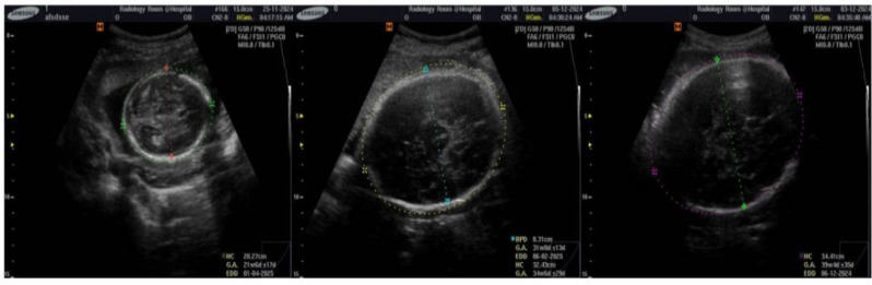
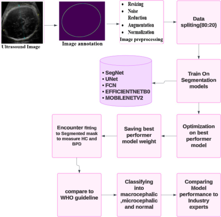
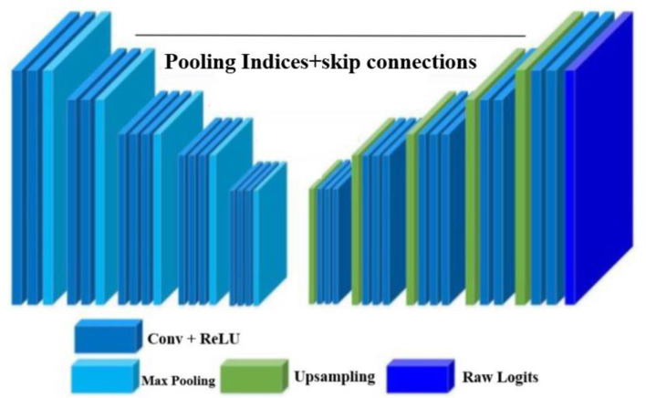
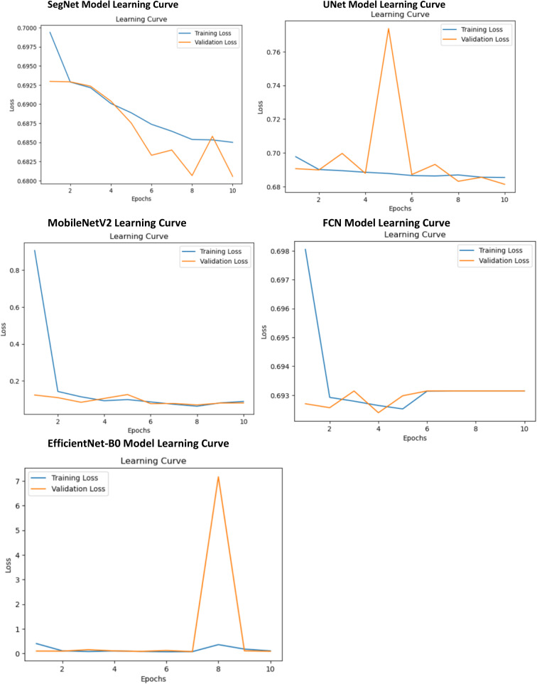
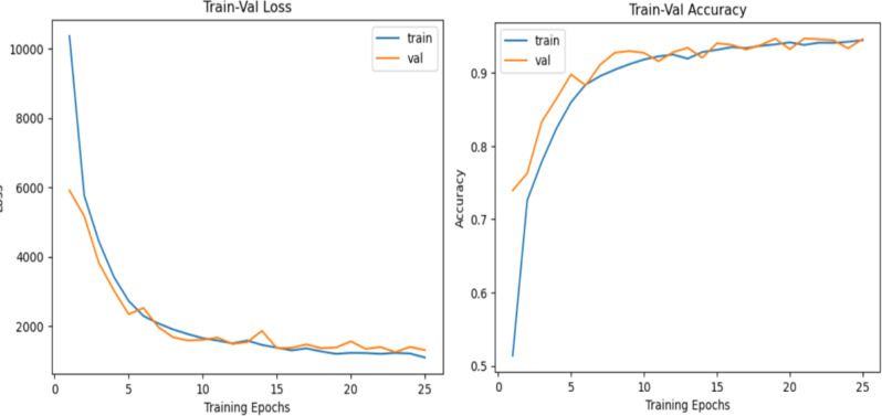
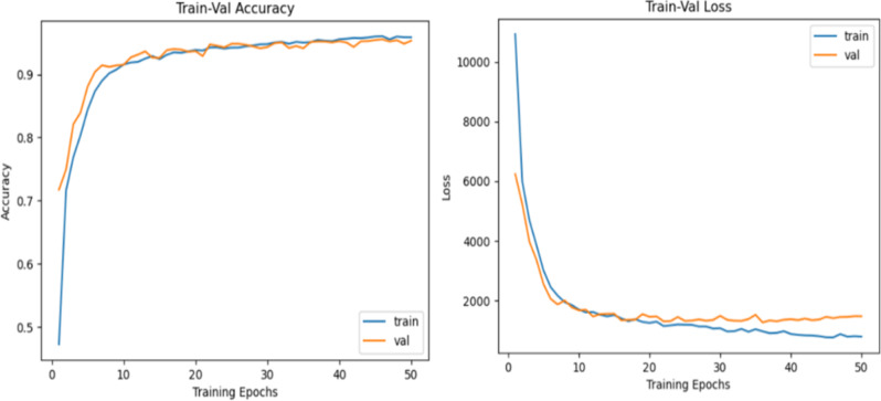
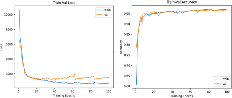
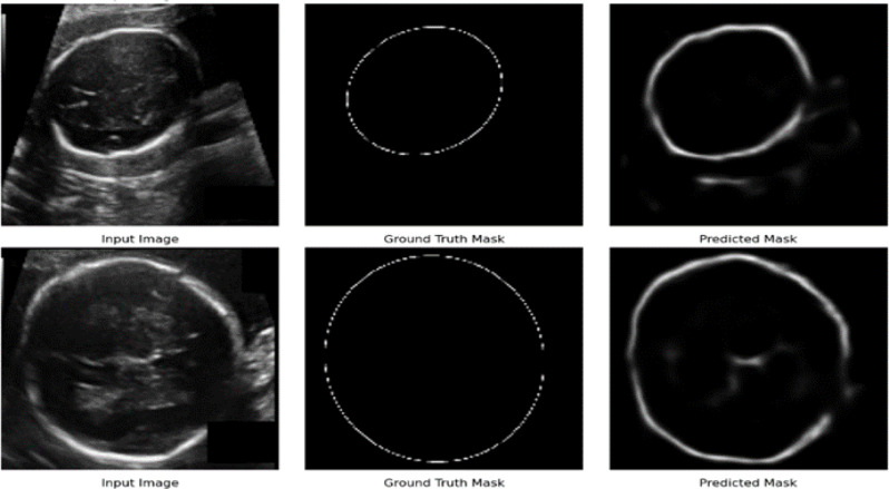
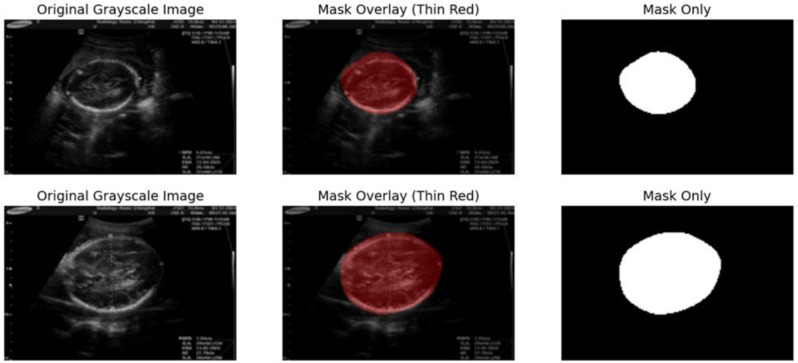
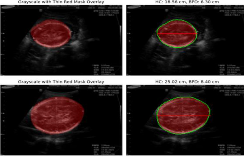
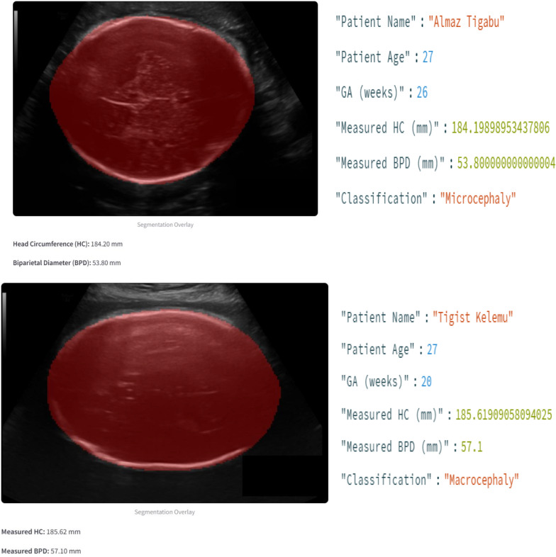
Methods: Data were collected from three Ethiopian healthcare facilities to increase model generalizability. The recruitment period for this study started on November 9, 2024, and ended on November 30, 2024. Several preprocessing techniques have been performed, such as augmentation, noise reduction, and normalization. SegNet, UNet, FCN, MobileNetV2, and EfficientNet-B0 were applied to segment and measure fetal head structures using ultrasound images. The measurements were classified as microcephaly, macrocephaly, or normal using WHO guidelines for gestational age, and then the model performance was compared with that of existing industry experts. The metrics used for evaluation included accuracy, precision, recall, the F1 score, and the Dice coefficient.
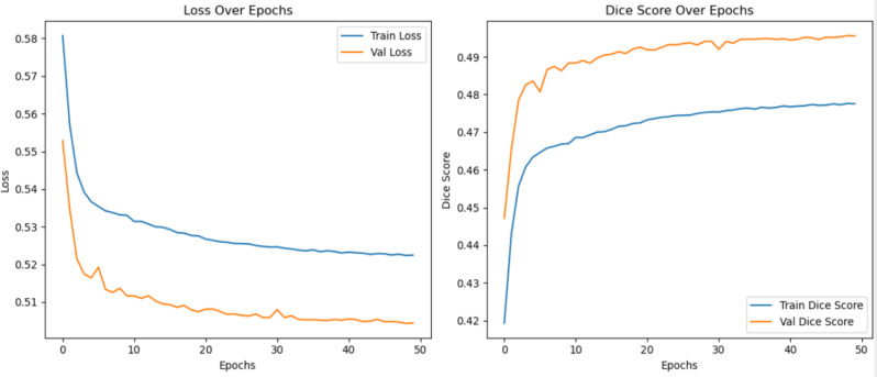
Results: This study was able to demonstrate the feasibility of using SegNet for automatic segmentation, measurement of abnormalities of the fetal head, and classification of macrocephaly and microcephaly, with an accuracy of 98% and a Dice coefficient of 0.97. Compared with industry experts, the model achieved accuracies of 92.5% and 91.2% for the BPD and HC measurements, respectively.
Conclusion: Deep learning models can enhance prenatal diagnosis workflows, especially in resource-constrained settings. Future work needs to be done on optimizing model performance, trying complex models, and expanding datasets to improve generalizability. If these technologies are adopted, they can be used in prenatal care delivery.
Clinical trial number: Not applicable.
Keywords: BPD; Congenital abnormality; HC; Macrocephaly; Microcephaly.
© 2025. The Author(s).
Conflict of interest statement
Declarations. Ethics approval and consent to participate: The study received ethical clearance from Debre Markos University CMHS (College of Medicine and Health Sciences) IRERC (Institutional Review and Ethics Review Committee) with Reference: RCSTTD/397/01/17, accredited under ICH-GCP and IORG standards, and our study adhered to the Declaration of Helsinki. Written informed consent was obtained from all adult participants before their inclusion. The consent process, conducted by the participating health institution, was documented and verified by the radiologist responsible for image collection, ensuring adherence to ethical protocols. No minors were included in the study. No personal identifiers were collected to safeguard privacy, and all ultrasound data were anonymized before analysis. Data integrity was maintained through secure protection systems that were compliant with international standards for medical imaging research. Collaborations with health institutions emphasized accountability and ethical rigor in handling imaging data, with oversight from the ethics committee to ensure full compliance. Consent for publication: Not applicable. No identifying details, images, or personal information of participants are included in this manuscript. All data were anonymized before analysis, and no individual consent for publication was required. Competing interests: The authors declare no competing interests.
Figures

Similar articles
-
[EXAMINING THE ASSOCIATION BETWEEN THE FETAL SUPRATENTORIAL BRAIN VOLUME AND THE SUBARACHNOID SPACE IN VARIOUS FETAL PATHOLOGIES USING MAGNETIC RESONANCE IMAGING].Harefuah. 2023 Dec;162(10):644-649. Harefuah. 2023. PMID: 38126147 Hebrew.
-
An adaptive convolution neural network model for tuberculosis detection and diagnosis using semantic segmentation.Pol J Radiol. 2025 Mar 14;90:e124-e137. doi: 10.5114/pjr/200628. eCollection 2025. Pol J Radiol. 2025. PMID: 40321710 Free PMC article.
-
Deep-learning model for prenatal congenital heart disease screening generalizes to community setting and outperforms clinical detection.Ultrasound Obstet Gynecol. 2024 Jan;63(1):44-52. doi: 10.1002/uog.27503. Ultrasound Obstet Gynecol. 2024. PMID: 37774040 Free PMC article.
-
Diagnostic approach to fetal microcephaly.Eur J Paediatr Neurol. 2018 Nov;22(6):935-943. doi: 10.1016/j.ejpn.2018.06.002. Epub 2018 Jun 30. Eur J Paediatr Neurol. 2018. PMID: 29970280 Review.
-
Diagnostic Accuracy of Ultrasound Scanning for Prenatal Microcephaly in the context of Zika Virus Infection: A Systematic Review and Meta-analysis.Sci Rep. 2017 May 23;7(1):2310. doi: 10.1038/s41598-017-01991-y. Sci Rep. 2017. PMID: 28536443 Free PMC article.
References
MeSH terms
LinkOut - more resources
Full Text Sources
Miscellaneous
