. 2025 May 26;20(1):71.
doi: 10.1186/s13020-025-01130-0.

Evodiamine induces ferroptosis in prostate cancer cells by inhibiting TRIM26-mediated stabilization of GPX4

Affiliations

Evodiamine induces ferroptosis in prostate cancer cells by inhibiting TRIM26-mediated stabilization of GPX4

Lanlan Li et al. Chin Med. .

Abstract

Background: Prostate cancer is a major global health challenge, characterized by high morbidity and mortality rates. Traditional treatment options, including androgen deprivation therapy and chemotherapy, often lead to drug resistance. In recent years, natural compounds have garnered attention for their potential therapeutic effects. Evodiamine, a bioactive alkaloid from Evodia rutaecarpa, has demonstrated promising anti-cancer properties in various malignancies, including oral squamous cell carcinoma, breast, colorectal, and ovarian cancers. This study explores the efficacy of evodiamine in prostate cancer cells and investigates the mechanisms underlying evodiamine-induced cell death.

Methods: To investigate the effects of evodiamine on prostate cancer cells, various cell lines, including both castration-sensitive and castration-resistant variants, were treated with different concentrations of evodiamine for various durations. Cell viability, proliferation, invasion ability, and colony formation were assessed using the CCK8 assay, EdU assay, 3D matrigel drop invasion assay, and colony formation assay, respectively. The effects of evodiamine on apoptosis were analyzed using FACS, Hoechst staining, and Western blot. To evaluate its effects on ferroptosis, malondialdehyde (MDA) and glutathione (GSH) assay kits, as well as DCFH-DA and the lipid peroxidation sensor BODIPY 581/501 C11 fluorescent probes, were employed. The molecular mechanisms through which evodiamine regulates GPX4 protein instability were investigated using Western blot and TRIM26 ectopic expression. Additionally, a mouse xenograft model derived from DU145 cells was established to evaluate the in vivo effects of evodiamine and its molecular mechanisms, utilizing hematoxylin and eosin (H&E) staining, immunohistochemistry (IHC), and Western blot analysis.

Results: Evodiamine significantly suppressed cell viability, proliferation, invasion, and colony formation in prostate cancer cells. Importantly, evodiamine-induced cell death in the PC3 and DU145 cell lines was independent of apoptosis pathway. Instead, evodiamine increased reactive oxygen species (ROS) production, lipid ROS levels and MDA levels, while decreasing GSH levels, indicating the induction of ferroptosis. The key role of ROS in evodiamine-induced ferroptosis was further confirmed by the partial reversal of cell death upon treatment with the ROS scavenger N-acetylcysteine (NAC). Mechanistically, evodiamine induced ferroptosis by destabilizing GPX4 protein in a TRIM26-dependent manner. Moreover, in vivo studies demonstrated that evodiamine significantly inhibited tumor growth and induced ferroptosis in tumor cells, highlighting its therapeutic potential.

Conclusion: This study demonstrates that evodiamine exerts potent antitumor effects against prostate cancer through inhibiting TRIM26-mediated stabilization of GPX4 protein and triggering ferroptosis. These findings suggest that evodiamine, a natural product derived from traditional Chinese medicine, could be a promising therapeutic agent for prostate cancer.

Keywords: Evodiamine; Ferroptosis; GPX4; Prostate cancer; TRIM26.


Conflict of interest statement

Declarations. Ethics approval and consent to participate: In this study, all animal experiment and procedures have been approved and performed in accordance with the Animal Care Welfare Committee of Lanzhou University Second Hospital Ethics approval and consent to participate (D2024-011). Consent for publication: Not applicable. Competing interests: The authors declare that they have no competing interests.

Figures

Fig. 1
Fig. 1
Evodiamine represses cell viability, invasion and colony formation in prostate cancer cells. A LNCaP, 22RV1, VCaP, PC3, and DU145 cells were treated with indicated concentration of evod (0, 0.5, 1.0, 2.0, 5.0 μM) for 24 h and 48 h, and cell viability was measured by CCK8 assay. B An EdU proliferation assay was performed on PC3 and DU145 cells treated with evod (0, 0.5, and 1.0 μM) at 24 h. C Representative images of cell colonies after 10 days treatment with evod (0, 0.5, 1.0 μM). D 3D matrigel drop invasion assay was conducted to evaluate the invasion ability in prostate cancer cells receiving evod treatment for 3 days. *P < 0.05; **P < 0.01; ***P < 0.001
Fig. 2
Fig. 2
Evodiamine induces prostate cancer cell death in an apoptotic independent manner. A PC3 cells and B DU145 cells were treated with indicated concentration (0, 0.5, and 1.0 μM) of evod for 48 h, and the apoptosis rates were detected by flow cytometry. Apoptosis related proteins in PC3 (C) and DU145 (D) cells were analyzed by western blot with GAPDH as the loading control. E PC3 and DU145 cells were treated with 1.0 μM evod with or without Z-VAD-FMK (20 μM) for 48 h, after which cell viability was assessed using the CCK8 assay. *P < 0.05; **P < 0.01; ***P < 0.001
Fig. 3
Fig. 3
Ferroptosis contributes to Evodiamine induced-cell death in prostate cancer cells. PC3 and DU145 cells were treated with evod at concentrations of 0, 0.5, and 1.0 μM for 48 h. Following treatment, intracellular ROS (A, B), MDA (C), GSH (D) level were detected, the protein level of SLC7A11 and GPX4 were assessed using western blot with GAPDH as the loading control (E), and lipid peroxidation was probed by C11-BODIPY.581/591 (F). *P < 0.05; **P < 0.01; ***P < 0.001
Fig. 4
Fig. 4
ROS scavenger inhibits the evodiamine-induced ferroptosis in prostate cancer cells. Cells were pretreated with 5.0 mM NAC for 1 h prior to treatment with 1.0 μM evod for 48 h, Following treatment, cell viability were assessed using the CCK8 assay (A), ROS levels were probed with DCFH-DA (B, C), MDA and GSH levels were quantitatively analyzed using commercial kits (D, E), The protein levels of SLC7A11 and GPX4 were evaluated by Western blot analysis with GAPDH as the loading control (F), and lipid peroxidation was probed by C11-BODIPY.581/591 (G). *P < 0.05; **P < 0.01; ***P < 0.001
Fig. 5
Fig. 5
Evodiamine triggers ferroptosis via targeting GPX4. A The protein levels of NCOA4, FTH, CD71, SLC7A11, SLC40A1, GPX4, P62, FTL, DMT1, LC3B, and FSP1 were examined by Western blot analysis with GAPDH as the loading control in PC3 cells and DU145 cells treated with evod at concentrations of 0, 0.5, and 1.0 μM for 48 h. B, C mRNA and protein levels of GPX4, SLC7A11 and FSP1 measured by qRT-PCR and western blot in indicated cells. D, E Following a 48 h treatment with 1.0 μM evod, indicated cells’ viability was assessed using the CCK8 assay. F 3D matrigel drop invasion assay was used to assess the invasion capability of cells receiving 1.0 μM evod treatment or not. *P < 0.05; **P < 0.01; ***P < 0.001
Fig. 6
Fig. 6
GPX4 overexpression reverses the inhibition of evodiamine in prostate cancer cells. A, B PC3 and DU145 cells were treated with 1.0 μM evod for 48 h. Subsequently, intracellular ROS levels were evaluated using the DCFH-DA probe. MDA levels was quantitatively analyzed using commercial kits (C, D), and lipid peroxidation was probed by C11-BODIPY.581/591 (E). *P < 0.05; **P < 0.01; ***P < 0.001
Fig. 7
Fig. 7
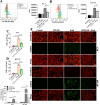
Evodiamine treatments caused GPX4 protein instability by reducing TRIM26 expression. A GPX4 protein levels were assessed by Western blot in PC3 and DU145 cells treated with 1.0 μM evod for 0, 12, 24 and 48 h. B PC3 and DU145 cells were treated with 1.0 μM evod, 10 μM MG132 or 200 nM Baf-A1 for 12 h, and GPX4 expression was measured by Western blot with GAPDH as the loading control. C The protein level of OTUB1, HSC70, USP14, USP7, USP25, USP10, and TRIM26 in PC3 and DU145 cells were evaluated following treatment with evod at concentrations of 0, 0.5, and 1.0 μM for 48 h. D The protein levels of GPX4 and TRIM26 were assessed in both control and trim26-overexpressing PC3 and DU145 cells, with or without a 48 h treatment with 1.0 μM evod. Following 48 h treatment with evod, cell viability was measured using the CCK8 assay (E) and the lipid peroxidation levels were investigated using C11-BODIPY.581/591 staining (F). *P < 0.05; **P < 0.01; ***P < 0.001
Fig. 8
Fig. 8
Evodiamine treatment suppresses cancer progression in DU145 xenograft model. Subcutaneous xenografts were established in nude mice by injecting DU145 cells into their flank. 10 days post-inoculation, the mice received intraperitoneal treatments with either evod at doses of 10 mg/kg and 20 mg/kg, or phosphate-buffered saline (PBS), administered every two days for a total of 12 days. A Photographic images of nude mice bearing xenograft tumors are presented. B The body weights of the mice were recorded throughout the experiment. C After euthanasia, tumor tissues were excised and photographed. D Tumor volumes and (E) tumor weights were subsequently calculated. F Representative images of H&E staining and IHC staining for Ki67 are shown. G Protein levels of GPX4, TRIM26, SLC7A11 and FSP1 were analyzed by western blot with GAPDH as the loading control. *P < 0.05; **P < 0.01; ***P < 0.001

References

    1. Bray F, Laversanne M, Sung H, Ferlay J, Siegel RL, Soerjomataram I, et al. Global cancer statistics 2022 GLOBOCAN estimates of incidence and mortality worldwide for 36 cancers in 185 countries. CA A Cancer J Clinic. 2024;74(3):229–63. - PubMed
    1. Siegel RL, Giaquinto AN, Jemal A. Cancer statistics, 2024. CA A Cancer Clinic. 2024;74(1):12–49. - PubMed
    1. Miyahira AK, Soule HR. The 30th annual prostate cancer foundation scientific retreat report. Prostate. 2024;84(14):1271–89. - PubMed
    1. Kaur P, Shankar E, Gupta S. EZH2-mediated development of therapeutic resistance in cancer. Cancer Lett. 2024;586: 216706. - PubMed
    1. Rawat C, Heemers HV. Alternative splicing in prostate cancer progression and therapeutic resistance. Oncogene. 2024;43(22):1655–68. - PMC - PubMed

LinkOut - more resources