. 2025 May 26;14(1):26.
doi: 10.1186/s40035-025-00484-7.

Development of human targeted extracellular vesicles loaded with shRNA minicircles to prevent parkinsonian pathology

Affiliations

Development of human targeted extracellular vesicles loaded with shRNA minicircles to prevent parkinsonian pathology

Maria Izco et al. Transl Neurodegener. .

Abstract

Background: Neurological disorders are the second leading cause of death and the leading cause of disability in the world. Thus, the development of novel disease-modifying strategies is clearly warranted. We have previously developed a therapeutic approach using mouse targeted rabies virus glycoprotein (RVG) extracellular vesicles (EVs) to deliver minicircles (MCs) expressing shRNA (shRNA-MCs) to induce long-term α-synuclein down-regulation. Although the previous therapy successfully reduced the pathology, the clinical translation was extremely unlikely since they were mouse extracellular vesicles.

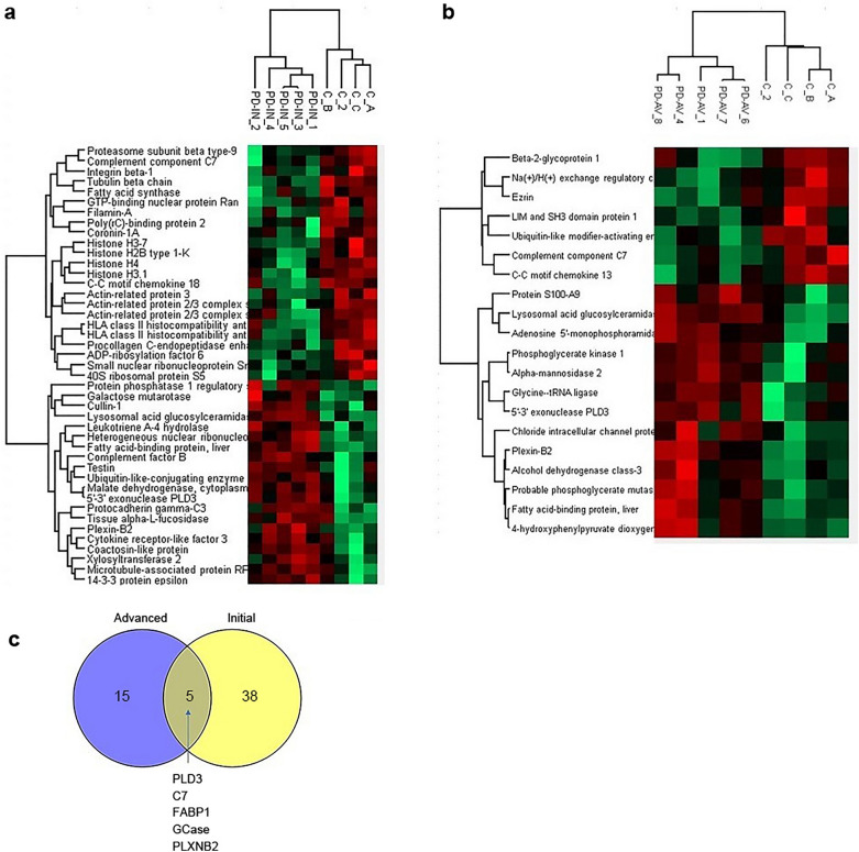
Methods: To overcome this limitation, we developed a source of human RVG-EVs compatible with a personalized therapy using immature dendritic cells. Human peripheral blood monocytes were differentiated in vitro into immature dendritic cells, which were transfected to express the RVG peptide. RVG-EVs containing shRNA-MCs, loaded by electroporation, were injected intravenously in the α-synuclein performed fibril (PFF) mouse model. Level of α-synuclein, phosphorylated α-synuclein aggregates, dopaminergic neurons and motor function were evaluated 90 days after the treatment. To confirm that EVs derived from patients were suitable as a vehicle, proteomic analysis of EVs derived from control, initial and advanced Parkinson's disease was performed.

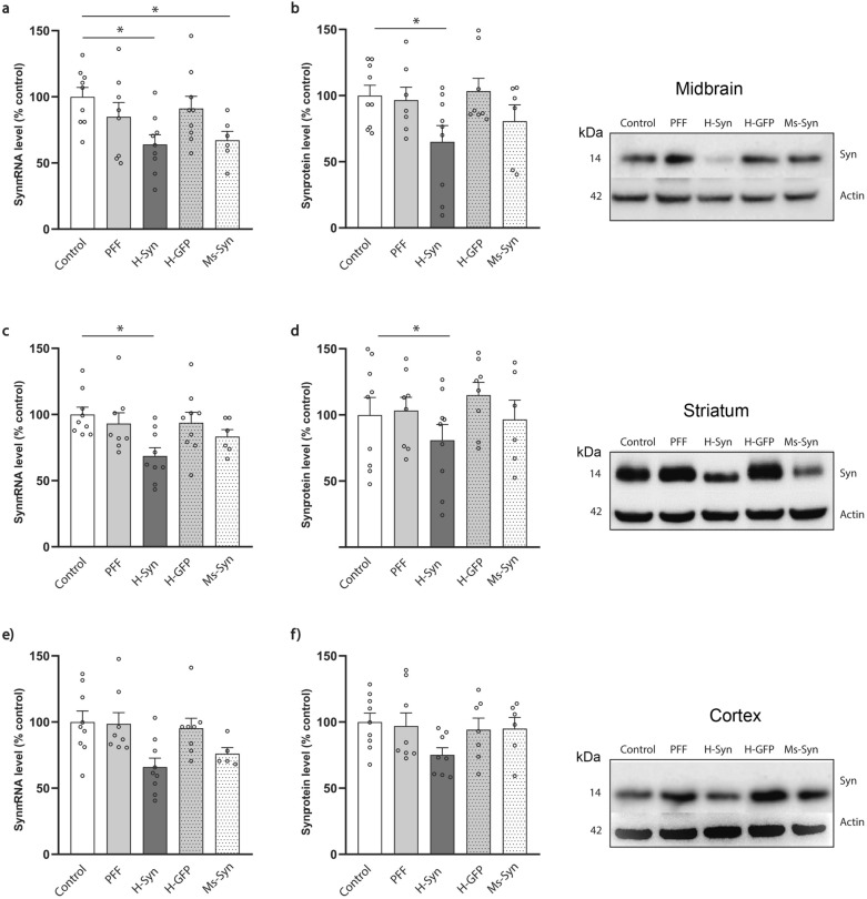
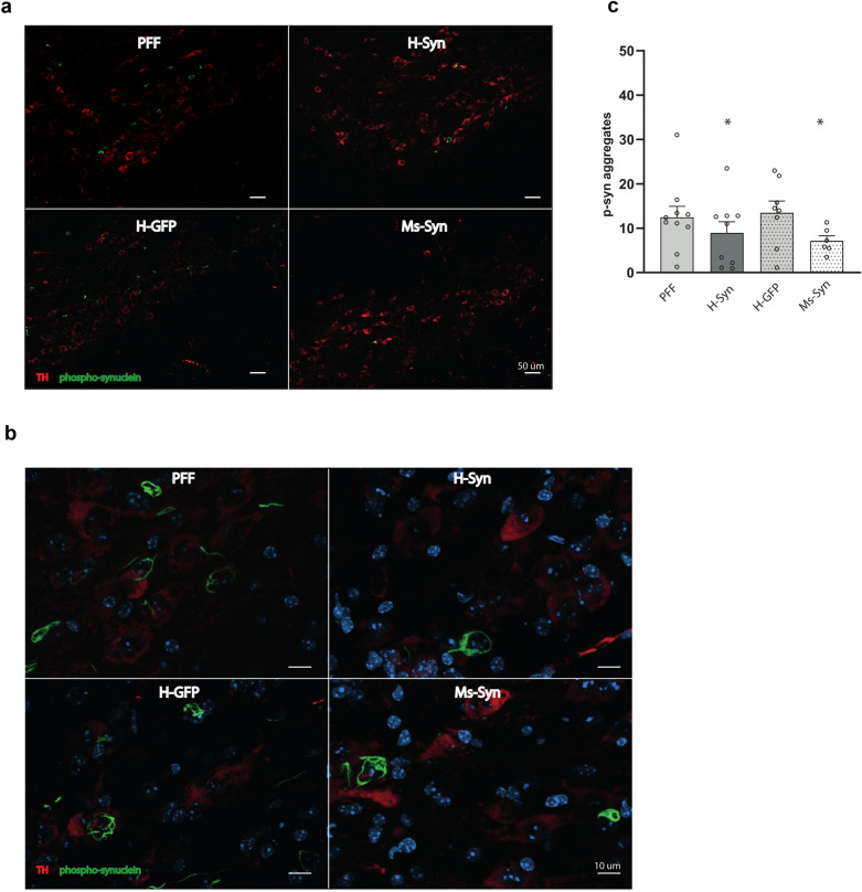
Results: The shRNA-MCs could be successfully loaded into human RVG-EVs and downregulate α-synuclein in SH-SY5Y cells. Intravenous injection of the shRNA-MC-loaded RVG-EVs induced long-term downregulation of α-synuclein mRNA expression and protein level, decreased α-synuclein aggregates, prevented dopaminergic cell death and ameliorated motor impairment in the α-synuclein PFF mouse model. Moreover, we confirmed that the EVs from PD patients are suitable as a personalized therapeutic vehicle.

Conclusion: Our study confirmed the therapeutic potential of shRNA-MCs delivered by human RVG-EVs for long-term treatment of neurodegenerative diseases. These results pave the way for clinical use of this approach.

Keywords: Gene therapy; Human targeted extracellular vesicles; Parkinson’s disease; ShRNA minicircles; α-Synuclein.


Conflict of interest statement

Declarations. Ethics approval and consent to participate: All studies involving human samples were approved by the Institutional Ethical Committee (CEImLar, protocol number: P.I.374). All animal studies involving mice followed the guidelines of the National Institutes of Health Guide for the Care and Use of Laboratory Animals and were approved by the ethical committee on animal welfare at our institution (Center for Biomedical Research of La Rioja, ref LAE-03). Consent for publication: Not applicable. Competing interests: LAE, EC and MI have filled a patent application related to the work in this paper. The remaining authors declare no competing interests.

Figures

Fig. 1
Fig. 1
Optimization of loading and validation of h-RVG-EVs loaded with anti-α-synuclein shRNA-MCs. a, b Nanoparticle tracking analysis characterization of EVs isolated from control hDCs (a) and hDCs electroporated with hLamp2b-RVG mRNA (b). c Western blot analysis to assess expression of markers in control EV and RVG-EV. d Representative scanning electron microscopy images of EV isolated from control hDCs and hDCs electroporated with hLamp2b-RVG mRNA. Scale bar, 200 nm. e, f SH-SY5Y cells overexpressing α-synuclein were treated with anti α-synuclein shRNA-MC using a transfection reaction (TR), or loaded into control h-EVs (EV) and h-RVG-EV (RVG-EV). α-synuclein mRNA (e) and protein (f) levels were quantified and normalized to control cells. Typical western blots are shown. Data are expressed as mean ± SEM (n = 4). *P < 0.05, non-parametric Kruskal–Wallis test, statistical analyses compared to untreated control cells
Fig. 2
Fig. 2
α-synuclein downregulation in several brain regions of mice treated with syn-MCs delivered by h-RVG-EVs. Analyses of α-synuclein mRNA expression (a, c, e) and protein (b, d, f) levels normalized to actin in the ipsilateral midbrain (a, b), striatum (c, d) and cortex (e, f) of α-synuclein PFF-treated mice (PFF) after treatment with h-RVG-EVs loaded with anti-α-synuclein (H-Syn) or GFP (H-GFP) shRNA-MCs. A group of mice was treated with anti-α-synuclein shRNA-MCs delivered by Ms-RVG-EV (Ms-Syn). Typical western blots are shown. Data are expressed as mean ± SEM (n = 6–8). *P < 0.05 compared to control mice, one-way ANOVA
Fig. 3
Fig. 3
Effect of h-RVG-EVs loaded with syn-MCs on α-synuclein pathology in SNc. a Immunofluorescent images of midbrain sections stained with antibodies to S129 phospho-α-synuclein (green) and TH (red). Scale bar, 50 μm. b Magnified images of stained midbrain section showing co-localization of α-synuclein aggregates (green) in dopaminergic neurons (red). Scale bar, 10 μm. c Quantitation of α-synuclein-positive aggregates per section in the ipsilateral SNc of α-synuclein PFF-treated mice following treatment with h-RVG-EVs loaded with anti-α-synuclein (H-Syn) or GFP (H-GFP) shRNA-MCs or after treatment with Ms-RVG-EVs loaded with anti-α-synuclein shRNA-MCs (Ms-Syn). Data are expressed as mean ± SEM (n = 6–10). *P < 0.05 compared to PFF mice, one-way ANOVA
Fig. 4
Fig. 4
α-Synuclein pathology in the striatum, cortex and amygdala after treatment with h-RVG-EVs loaded with syn-MCs. Quantification of aggregates in the ipsilateral striatum (a), cortex (b) and amygdala (c) of α-synuclein PFF-injected mice (PFF) following treatment with h-RVG-EVs loaded with anti-α-synuclein (H-Syn) or GFP (H-GFP) shRNA-MCs or after treatment with anti-α-synuclein shRNA-MCs loaded in Ms-RVG-EVs (Ms-Syn). Representative immunohistochemical images of intraneuronal phospho-α-synuclein-positive aggregates in the ipsilateral striatum, cortex and amygdala are shown. Scale bar, 100 µm. Data are expressed as mean ± SEM (n = 6–10), *P < 0.05, one-way ANOVA compared to PFF mice
Fig. 5
Fig. 5
Syn-MC h-RVG-EV therapy prevented dopaminergic dysfunction and motor impairments. a TH staining of dopaminergic neurons in coronal midbrain sections of α-synuclein PFF-injected mice (PFF) following treatment with h-RVG-EVs loaded with anti-α-synuclein (H-Syn) or GFP (H-GFP) shRNA-MCs or after treatment with anti-α-synuclein shRNA-MCs loaded in Ms-RVG-EVs (Ms-Syn). Arrows point to area of decreased dopaminergic neurons. Numbers of nigral dopaminergic neurons were quantified by unbiased stereology on each brain hemisphere (I, ipsilateral; C, contralateral). b TH quantitation by optical density in ipsilateral sections of anterior, medial and posterior striatum normalized to contralateral striatum. Representative striatal section showing TH staining are shown. c Quantitative analysis of the hind limb clasping scores. d Wire hanging performance. e Time to turn around and move up toward the top of the platform in the negative geotaxis test. If the mouse was unable to turn, the default value of 30 s was taken as maximal severity of impairment. Data are expressed as mean ± SEM (n = 6–10). *P < 0.05, ***P < 0.001 compared to control mice, one-way ANOVA
Fig. 6
Fig. 6
Effect on α-synuclein mRNA and protein levels in the spinal cord, intestine and olfactory bulb. Quantitation of α-synuclein mRNA (a, c, e) and protein (b, d, f) levels in the olfactory bulb (a and b), spinal cord (c and d) and intestine (e and f) from α-synuclein PFF-treated mice (PFF) at 90 days following treatment with h-RVG-EVs loaded with anti-α-synuclein (H-Syn) or GFP (H-GFP) shRNA-MCs or after treatment with anti-α-synuclein shRNA-MCs loaded in Ms-RVG-EVs (Ms-Syn). Typical western blots are shown. Data are expressed as mean ± SEM (n = 6–8). *P < 0.05 compared to control mice, one-way ANOVA
Fig. 7
Fig. 7
Proteomic analysis of EVs produced by DCs isolated from control, initial PD and advance PD. a, b Heat map analysis showing differentially expressed proteins between initial PD (a) or advanced PD (b) and control patients. c Venn diagram of differentially expressed proteins detected in EVs derived from initial and advanced PD

References

    1. Hirsch L, Jette N, Frolkis A, Steeves T, Pringsheim T. The Incidence of Parkinson’s disease: A systematic review and meta-analysis. Neuroepidemiology. 2016;46(4):292–300. - PubMed
    1. Dorsey ER, Sherer T, Okun MS, Bloem BR. The emerging evidence of the Parkinson pandemic. J Parkinsons Dis. 2018;8(s1):S3–8. - PMC - PubMed
    1. Simón-Sánchez J, Schulte C, Bras JM, Sharma M, Gibbs JR, Berg D, et al. Genome-wide association study reveals genetic risk underlying Parkinson’s disease. Nat Genet. 2009;41(12):1308–12. - PMC - PubMed
    1. Lee VM, Trojanowski JQ. Mechanisms of Parkinson’s disease linked to pathological α-synuclein: new targets for drug discovery. Neuron. 2006;52(1):33–8. - PubMed
    1. Li JY, Englund E, Holton JL, Soulet D, Hagell P, Lees AJ, et al. Lewy bodies in grafted neurons in subjects with Parkinson’s disease suggest host-to-graft disease propagation. Nat Med. 2008;14(5):501–3. - PubMed

LinkOut - more resources