. 2025 May 26;23(1):381.
doi: 10.1186/s12951-025-03421-y.

Sequential nanoparticle therapy targeting neutrophil hyperactivation to prevent neutrophil-induced pulmonary fibrosis

Affiliations

Sequential nanoparticle therapy targeting neutrophil hyperactivation to prevent neutrophil-induced pulmonary fibrosis

Hye-Jin Lee et al. J Nanobiotechnology. .

Abstract

Background: Pulmonary fibrosis, a major complication of severe COVID-19 and post-acute sequelae of SARS-CoV-2 infection (PASC), is driven by excessive neutrophil activation and the formation of neutrophil extracellular trap (NET).

Results: This study presents a sequential nanoparticle-based therapy combining DNase-I-loaded polydopamine nanoparticles (DNase-I@PDA NPs) with Sivelestat-encapsulated PLGA nanoparticles (Siv@PLGA NPs) to target both NETs and neutrophil elastase (NE) activity. DNase-I@PDA NPs were aerosolized to the lungs, facilitating NET clearance and reducing the fibrotic microenvironment, followed by intravenous administration of Siv@PLGA NPs to inhibit NE activity and prevent neutrophil hyperactivation. In a murine model of lipopolysaccharide (LPS)-induced pulmonary fibrosis, this dual approach significantly decreased fibrotic lesions, collagen deposition, and myofibroblast activation. Notably, treatment with the nanoparticles led to substantial improvements in pulmonary function. In neutrophils isolated from COVID-19 patients, the combined nanoparticle therapy reduced circulating cell-free DNA, NET, NE, and myeloperoxidase (MPO) levels, while enhancing neutrophil viability and reducing inflammatory responses.

Conclusions: These findings highlight the efficacy of DNase-I@PDA NPs and Siv@PLGA NPs in addressing both acute inflammation and chronic fibrosis by simultaneously targeting NET formation and neutrophil hyperactivation. This dual nanoparticle therapy represents a promising clinical strategy for treating COVID-19-associated pulmonary complications, including PASC, by preventing long-term fibrotic progression and promoting lung recovery.

Keywords: Aerosolized drug delivery; Chronic inflammation; Neutrophil extracellular traps; Post-acute sequelae of SARS-CoV-2 (PASC); Pulmonary fibrosis.


Conflict of interest statement

Declarations. Ethics approval and consent to participate: This study was approved by the Ethics Committee of Yeungnam University Medical Center, and all participants provided consent to participate (YUH 2020-03-057 and 2020-05-031-001). This research received approval from the Ethics Committee of Sungkyunkwan University, and all procedures with animal models followed established ethical standards (IACUC No.: 202111291). Consent for publication: Consent for publication is not applicable. Competing interests: The authors declare no competing interests.

Figures
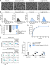
Fig. 1
Fig. 1
Characterization of NPs. A SEM image (n = 3, Scale bar = 500 nm) B Size and C Zeta potential (n = 3) of PDA, DNase-I@PDA, PLGA, and Siv@PLGA NPs. D Drug release profile of Siv@PLGA NPs (n = 4). E In vitro targeting efficiency of Neutrophil targeted PLGA NPs (n = 5). Statistical analysis was performed using a two-tailed unpaired t-test. Data are presented as mean ± SEM. * P < 0.05, ** P < 0.01, *** P < 0.001, **** P < 0.0001 (v.s. LPS)
Fig. 2
Fig. 2
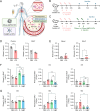
Early administration of DNase-I ameliorates acute immune response in the case of COVID-19 infection. A Schematic of DNase-I@PDA NPs and Siv@PLGA NPs administration. B Schematic of LPS injection and sacrifice schedule. Mice were intratracheally (i.t.) administered three times with LPS (3 mg/kg) and sacrificed at every 24 h interval. C Schematic of LPS injection and treatment schedule. Mice were intratracheally (i.t.) administered three times with LPS (3 mg/kg) and were intratracheally (i.t.) administered twice with free DNase-I (D group) or DNase-I @NP (D@P group) at 24 h after the third injection of LPS. Mice were sacrificed at 12 h after the final administration of free DNase-I or DNase-I@NP at every 24 h interval. D Concentration of DNase-I in blood and BALF. E Neutrophil count in blood and BALF. F Immune cell populations in BALF at 72 h post-administration. G Immune cell populations in BALF at 96 h post-administration. Statistical analysis was performed using a two-tailed unpaired t-test. Data are presented as mean ± SEM. * P < 0.05, **P < 0.01, ***P < 0.001, ****P < 0.0001 (v.s. LPS), ##P < 0.01, ###P < 0.001 (v.s. D + S)
Fig. 3
Fig. 3
Coadministration of DNase-I and Sivelestat nanoparticle and drug release. A Ex vivo imaging of dissected organs after the i.v. administration of Cy7- Siv@PLGA NP and the i.t. administration of Cy7- DNase-I PDA@NP. B Schematic of LPS injection and treatment schedule. Mice were intratracheally (i.t.) administered three times with LPS (3 mg/kg). DNase-I PDA@NP was administrated intratracheally and Siv@PLGA NP was administrated intravenously, alternating 12 h apart. Mice were sacrificed at 48 h after the final administration of Siv@PLGA NP. C Heatmap illustrating the top 50 cytokines identified from a cytokine array analysis across the treatment conditions: LPS, DNase-I combined with Sivelestat (D + S), and each nanoparticle treatment (NP). The color scale denotes relative expression levels, with red indicating upregulation. Pathway enrichment analysis revealed significant involvement of inflammatory and immune-related pathways, including cytokine-cytokine receptor interaction, chemokine signaling, and the MAPK signaling pathway. D Neutrophil counts, E NET ratio, F DNase-I activity, G Concentration of NE, H MPO activity, and I cfDNA in murine BALF. Statistical analysis was performed using a two-tailed unpaired t-test. Data are presented as mean ± SEM. *P < 0.05, **P < 0.01, ***P < 0.001, ****P < 0.0001 (v.s. LPS), # P < 0.05, ## P < 0.01, ### P < 0.001 (v.s. D + S)
Fig. 4
Fig. 4
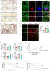
Improved respiratory function by administrating of DNase-I@PDA NPs and Siv@PLGA NPs. A H&E analysis of lung tissue from LPS-induced pulmonary fibrosis mouse model. (Scale bar: 200 μm). B Orange G staining images. (Scale bar: 200 μm). C Immunohistochemistry (IHC) analysis (Cit-H3, NE, and Sytox-Green™) of lung sections of LPS-induced pulmonary fibrosis mouse model. (Scale bar: 40 μm). D Trichrome stained images of murine lung tissue. (Scale bar: 200 μm). E IHC analysis of fibrotic markers (collagen I and α-SMA). (Scale bar: 100 μm). F Measurement of hydroxyproline content. G Inspiratory capacity, lung resistance, compliance, and elastance ascertained by FlexiVent forced oscillation technique at endpoint. H PV curve. I Body weight of LPS-induced pulmonary fibrosis mouse model. J Survival rate of LPS-induced pulmonary fibrosis mouse model. Statistical analysis was performed using a two-tailed unpaired t-test. Data are presented as mean ± SEM. *P < 0.05, **P < 0.01, ****P < 0.0001 (v.s. LPS), ##P < 0.01, ####P < 0.0001 (v.s. D + S)
Fig. 5
Fig. 5
Evaluation of NETosis following coadministration of free drugs and NPs in the blood of SARS-CoV-2 patients. A Circulating cfDNA. B DNase-I enzymatic activity. C NET formation index in PBMCs. D NE activity. E MPO activity. F Viability of the neutrophils. Statistical analysis was performed using a two-tailed unpaired t-test. Data are presented as mean ± SEM. *P < 0.05, ****P < 0.0001 (v.s. PBS), #P < 0.05, ####P < 0.0001 (v.s. D + S)

References

    1. Raslan AA, Pham TX, Lee JS, Kontodimas K, Tilston-Lunel A, Schmottlach J, Hong JM, Dinc T, Bujor AM, Caporarello N, et al. Lung injury-induced activated endothelial cell states persist in aging-associated progressive fibrosis. Nat Commun. 2024. 10.1038/s41467-024-49545-x. - PMC - PubMed
    1. Bonella F, Spagnolo P, Ryerson C. Current and future treatment landscape for idiopathic pulmonary fibrosis. Drugs. 2023;83:1581–93. - PMC - PubMed
    1. Liu SA, Su XL, Pan PH, Zhang LM, Hu YB, Tan HY, Wu DD, Liu B, Li HT, Li HS, et al. Neutrophil extracellular traps are indirectly triggered by lipopolysaccharide and contribute to acute lung injury. Sci Rep. 2016. 10.1038/srep37252. - PMC - PubMed
    1. Ambardar SR, Hightower SL, Huprikar NA, Chung KK, Singhal A, Collen JF. Post-COVID-19 pulmonary fibrosis: novel sequelae of the current pandemic. J Clin Med. 2021;10:2452. - PMC - PubMed
    1. Brinkmann V, Reichard U, Goosmann C, Fauler B, Uhlemann Y, Weiss DS, Weinrauch Y, Zychlinsky A. Neutrophil extracellular traps kill bacteria. Science. 2004;303:1532–5. - PubMed

MeSH terms