. 2025 Aug;12(4):3179-3182.
doi: 10.1002/ehf2.15319. Epub 2025 May 26.

Alterations of myocardial ketone metabolism in heart failure with preserved ejection fraction (HFpEF)

Affiliations

Alterations of myocardial ketone metabolism in heart failure with preserved ejection fraction (HFpEF)

Qiuyu Sun et al. ESC Heart Fail. 2025 Aug.

Abstract

Introduction: Cardiac energy metabolism is disrupted in heart failure with preserved ejection fraction (HFpEF), as characterized by a switch from glucose oxidation towards fatty acid oxidation. However, although oxidation of ketones is an important source of ATP it remains unclear how the heart oxidizes ketones in HFpEF. It is also unclear whether elevating ketone supply to the heart can improve cardiac energetics and/or provide functional benefit for the hearts in HFpEF.

Aims: The present study investigated the effects of increasing ketone supply to the heart via ketone supplementation or SGLT2 inhibitor treatment in a mouse model of HFpEF.

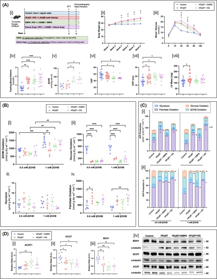
Methods: HFpEF was induced in 13-month-old C57BL/6N female mice with 60% high-fat diet and L-NAME (0.5 g/L/day in the drinking water) for 6 weeks. In parallel, two other groups of mice were maintained on the HFpEF protocol while also receiving either a ketone ester supplement (1-3 butanediol 1 g/kg/day) or SGLT2 inhibitor (empagliflozin 10 mg/kg/day) for 6 weeks. Control mice were fed with regular low-fat diet and regular drinking water. Hearts of the mice were excised and perfused in the isolated working mode aerobically with 5-mM glucose, 0.8-mM palmitate, 100-μU/mL insulin, with either low (0.6 mM) or high (1 mM) levels of β-hydroxybutyrate. Metabolic rates of the hearts were measured with radiolabelled [U-14C] glucose, [9,10-3H] palmitate and [3-14C] β-hydroxybutyrate.

Results: In HFpEF mouse hearts, glucose oxidation was significantly decreased with a parallel increase in fatty acid oxidation. Increasing β-hydroxybutyrate levels from 0.6 to 1 mM in the perfusate resulted in a rise in ketone oxidation rates in control hearts (from 861 ± 63 to 1377 ± 94 nmol g dry wt-1 min-1), which was muted in HFpEF hearts (from 737 ± 68 to 897 ± 134 nmol g dry wt-1 min-1). Following ketone ester supplement or SGLT2 inhibitor treatment, HFpEF mice presented with restored ketone oxidation rates (from 674 ± 36 to 1181 ± 115 nmol g dry wt-1 min-1 with ketone ester supplement and from 797 ± 121 to 1240 ± 120 nmol g dry wt-1 min-1 with SGLT2i). Yet, this was not associated with improvement in cardiac function.

Conclusions: In HFpEF mice, the heart switches from glucose oxidation to fatty acid oxidation, with ketone oxidation being impaired. Increasing ketone supply to the heart via ketone ester supplementation or SGLT2 inhibitor treatment increases myocardial ketone oxidation rates but was not associated with functional improvements. Unlike HFrEF, ketone supplementation strategies may be less effective in HFpEF due to an impairment of myocardial ketone oxidation in HFpEF.


Figures

Figure 1
Figure 1
Myocardial ketone oxidation is impaired in heart failure with preserved ejection fraction (HFpEF). (A) HFpEF protocol and empagliflozin (EMPA) and ketone supplement (KS) protocol (i), body weight changes over the HFpEF protocol (ii), blood glucose changes over the 2‐h course of intraperitoneal glucose‐tolerance test (GTT), which were performed by injection of glucose (2 g kg−1 in saline) after 16‐h fasting (iii), fasting blood ketone levels assessed by sampling blood from mice tail and read with FreeStyle precision ß‐Ketone test strips paired with its analyser (n = 14–17) (iv), cardiac tissue levels of βOHB assessed with a colorimetric assay kit from Abcam (ab83390) (n = 6) (v), transthoracic echocardiographic analysis of LV ejection fraction (%EF) (n = 11–17) (vi), isovolumic relaxation time (IVRT) (n = 11–17) (vii) and LV mass (LV mass) (n = 11–17) (viii). (B) βOHB/ketone oxidation rates (n = 5–8) (i), glucose oxidation rates (n = 4–6) (ii), glycolysis (n = 4–8) (iii) and palmitate/fatty acid oxidation rates (n = 4–5) (iv) in isolated working hearts perfused with appropriately radiolabelled 5‐mM glucose, 0.8‐mM palmitate, 3% bovine serum albumin (BSA), 100 μU/mL insulin and either 0.6‐ or 1‐mM βOHB. (C) ATP production rates were calculated based on the oxidation rates from glucose (31 moles of ATP from glucose oxidation), fatty acid (104 moles of ATP from palmitate oxidation) and βOHB (21.25 moles of ATP from βOHB oxidation) (i), %ATP production is calculated by matching the ATP production rates attributed to each of the four metabolic pathways normalized to the sum of ATP production rates (ii). (D) Quantification of western blotting showing expression of enzymes involved in myocardial ketone oxidation (ACAT1 [i], BDH1 [ii] and SCOT [iii]), and representative blots (n = 6–8) (iv). Data are mean ± SEM. Statistics were performed in GraphPad Prism 10. Significant differences were determined by using a one‐way or two‐way analysis of variance (ANOVA) followed by a Tukey's multiple comparisons post hoc test. For Panel C, *P < 0.05 compared with control group under the same condition, #P < 0.05 compared with HFpEF under the same condition. For all other panels, *P < 0.05 compared with corresponding group with bracket. Mouse studies were approved by the University of Alberta Animal Care and Use Committee and comply with Animal Research: Reporting of In Vivo Experiments (ARRIVE) and National Institutes of Health guidelines. Abbreviations: ACAT1: acetyl‐CoA Acetyltransferase 1; ATP, adenosine triphosphate; βOHB, β‐hydroxybutyrate; BDH1, β‐hydroxybutyrate dehydrogenase 1; EMPA, empagliflozin; EF, ejection fraction; HFD, high‐fat diet; HFpEF, heart failure with preserved ejection fraction; IVRT, isovolumetric relaxation time; KS, ketone supplement; L‐NAME, L‐NG‐nitroarginine methyl ester, N(G)‐nitro‐L‐arginine methyl ester; LV, left ventricle; SCOT, succinyl‐CoA:3‐ketoacid CoA transferase; Supp, supplement.

References

    1. Dunlay SM, Roger VL, Redfield MM. Epidemiology of heart failure with preserved ejection fraction. Nat Rev Cardiol 2017;14:591–602. doi: 10.1038/nrcardio.2017.65 - DOI - PubMed
    1. Sun Q, Güven B, Wagg CS, Almeida de Oliveira A, Silver H, Zhang L, et al. Mitochondrial fatty acid oxidation is the major source of cardiac adenosine triphosphate production in heart failure with preserved ejection fraction. Cardiovasc Res 2024;120:360–371. doi: 10.1093/cvr/cvae006 - DOI - PubMed
    1. Aubert G, Martin OJ, Horton JL, Lai L, Vega RB, Leone TC, et al. The failing heart relies on ketone bodies as a fuel. Circulation 2016;133:698–705. doi: 10.1161/CIRCULATIONAHA.115.017355 - DOI - PMC - PubMed
    1. Nielsen R, Møller N, Gormsen LC, Tolbod LP, Hansson NH, Sorensen J, et al. Cardiovascular effects of treatment with the ketone body 3‐hydroxybutyrate in chronic heart failure patients. Circulation 2019;139:2129–2141. doi: 10.1161/CIRCULATIONAHA.118.036459 - DOI - PMC - PubMed
    1. Schiattarella GG, Altamirano F, Tong D, French KM, Villalobos E, Kim SY, et al. Nitrosative stress drives heart failure with preserved ejection fraction. Nature 2019;568:351–356. doi: 10.1038/s41586-019-1100-z - DOI - PMC - PubMed

Substances

LinkOut - more resources