. 2025 May 26;9(5):e70148.
doi: 10.1002/hem3.70148. eCollection 2025 May.

Genetic evolution and relapse-associated mutations in adult T-cell acute lymphoblastic leukemia patients treated in PETHEMA trials

Affiliations

Genetic evolution and relapse-associated mutations in adult T-cell acute lymphoblastic leukemia patients treated in PETHEMA trials

Celia González-Gil et al. Hemasphere. .

Abstract

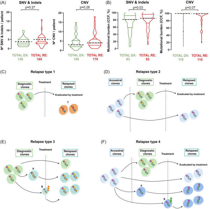
Relapse is the main cause of treatment failure in T-cell acute lymphoblastic leukemia (T-ALL). Despite this, data from adult T-ALL patients treated with specific chemotherapeutic regimens that examine predictive markers and describe relapse mechanisms are scarce. In this study, we studied 74 paired diagnosis-relapse samples from 37 patients homogeneously treated with three consecutive measurable residual disease-oriented trials to identify genetic determinants involved in relapse in adult T-ALL. Analysis of single-nucleotide variants and copy number alterations consistently found N/KRAS mutations (20% relapsed cases) at diagnosis and at relapse (resistance profile). N/KRAS mut patients frequently relapse early during consolidation treatment. Relapse-specific mutations in NT5C2, NR3C1, SMARCA4, and TP53 (40% relapse cases) were not detected at diagnosis by conventional molecular techniques (relapse profile). However, single-cell-based analysis revealed a very minor clone containing the NT5C2(p.R367Q) variant at diagnosis. Patients with the NT5C2(p.R367Q) variant mostly relapse later during maintenance treatment. Tracking the NT5C2 variant by digital PCR confirm the expansion of the NT5C2 clone at maintenance treatment. Overall, our exploratory analysis suggests a role for these genetic events, most of which have already been described in pediatric cases, driving resistance associated to specific chemotherapeutic agents, contributing to the relapse of a high proportion of adult T-ALL patients (60%).


Conflict of interest statement

The authors declare no conflicts of interest.

Figures

Figure 1
Figure 1
Variant distribution and clonal evolution models in bulk leukemia. (A) Differences in the number of variants per patient between diagnosis and relapse (Mann–Whitney U test). Differences in single‐nucleotide variants (SNVs) and indels, and in copy number variants (CNVs) are shown on the left and right, respectively. (B) Differences in the mutational burden quantified by the cancer cell fraction (CCF), considering only common variants whose CCF differs between DX and RE (Wilcoxon test). Differences in SNVs and indels and in CNVs are shown on the left and right, respectively. (A, B) The median CCF is indicated by the dashed line. The number of variants assessed are indicated below each graph. Clonal evolution models in bulk leukemia: (C) Relapse leukemia has an origin independent of diagnosis. The diagnosis clone is represented in green with its specific variants shown in yellow and pink; the relapse clone is represented in orange with its specific variants shown in red and blue. (D) Relapse leukemia evolves from an ancestral clone. The diagnosis clone is represented in green with specific variants shown in yellow and pink; the relapse clone evolves from an ancestral clone, with fewer variants than the main diagnosis clone. (E) The relapse clone evolves from the main diagnosis clone by acquiring a new variant at diagnosis (A) or during treatment (B). The most common diagnosis clone is represented in green with common diagnosis and relapse variants shown in yellow and pink; the relapse clone is represented in turquoise with the specific relapse variants shown in orange. (F) The relapse clone evolves from an ancestral clone through the acquisition of new variants at diagnosis (A) or during treatment (B). The diagnosis clone is represented in green with a diagnosis‐specific variant shown in yellow; the ancestral clone is represented in light blue with an ancestral variant shown in pink; the relapse clone is represented in dark blue with the specific relapse variants shown in green.
Figure 2
Figure 2
Genetic profiles associated with relapse in adult T‐ALL. (A) Genetic landscape of the paired samples. Only genes mutated in at least four patients are shown. Diagnosis‐specific variants are shown in green, relapse‐specific variants are shown in red, and common variants at diagnosis and relapse are shown in orange. Relapse characteristics for each patient are indicated above each figure. The frequency of each type of variant per gene (diagnosis‐specific, relapse‐specific, or common diagnosis‐relapse) is shown on the right. Gene/genetic alteration with more than 75% of its variants detected at DX and RE are assigned to resistance profile group, and those with more than 75% of its variants only detected at RE are classified as having a relapse profile. The frequency of each type of variant per patient is shown at the bottom. (B) Pairs of associations of the frequent mutated genes and the defined relapse profiles. Positive associations (log10 odds ratio >0) are represented in blue; negative associations (log10 odds ratios <0) are shown in red. (C) Cohort classification according to the defined relapse profiles. Patients with the resistance profile are shown in yellow; those with the relapse‐specific profile are identified in red; and unclassified patients are shown in gray.
Figure 3
Figure 3
dPCR‐based identification of the NT5C2(p.R367Q) mutation versus NGFC‐MRD levels in T‐ALL samples obtained at early treatment times. (A–C) The sensitivity of the dPCR (Y‐axis) considers the number of DNA copies with the mutation relative to the total number of DNA copies (purple line). Measurable residual disease (MRD), quantified by the next‐generation flow cytometry (NGFC), is represented by a green line. The limit of detection (LOD) is indicated with a horizontal dashed line. The days elapsed between diagnosis and relapse are shown on the X‐axis. The different treatment blocks are represented by different colors.
Figure 4
Figure 4
Identification of the NT5C2(p.R367Q) variant at diagnosis by single‐cell analysis. (A–C, Left) The fish plot shows the clonal evolution from diagnosis to relapse of three T‐ALL patients carrying relapse‐specific variants: (A) NR3C1(p.A578T) and SMARCA4(p.R425Q); (B) NR3C1(p.G290E); and (C) NT5C2(p.R176Q) and TP53 del . Each color represents a clone. The minimum clone size was five cells. The percentages show the proportion of cells that made up each clone at diagnosis (middle of fish plot) and relapse (right side of fish plot). Only percentages ≥3 are shown. (A–C, Right) Phylogenetic trees illustrating the maximum likelihood of the order in which mutations were acquired during T‐ALL development in the indicated patients. The colors used to represent each clone are the same as those used in the fish plots (left). Mutational history was reconstructed from the genotype data from the samples obtained at diagnosis and relapse. The size of the circle denotes the relative clone size in the samples (integrating the information from diagnosis and relapse). The new variant acquired at each step is noted. The CNVs and SNVs are indicated in blue and black, respectively. The dashed line separates the last DX clone and the first RE clone.

References

    1. Huguet F, Chevret S, Leguay T, et al. Intensified therapy of acute lymphoblastic leukemia in adults: report of the randomized GRAALL‐2005 clinical trial. J Clin Oncol. 2018;36(24):2514‐2523. - PubMed
    1. Ribera J‐M, Morgades M, Ciudad J, et al. Chemotherapy or allogeneic transplantation in high‐risk Philadelphia chromosome‐negative adult lymphoblastic leukemia. Blood. 2021;137(14):1879‐1894. - PubMed
    1. Rowntree CJ, Kirkwood AA, Clifton‐Hadley L, et al. First analysis of the UKALL14 randomized trial to determine whether the addition of nelarabine to standard chemotherapy improves event free survival in adults with T‐cell acute lymphoblastic leukaemia (CRUK/09/006). Blood. 2021;138(suppl 1):366.
    1. Beldjord K, Chevret S, Asnafi V, et al. Oncogenetics and minimal residual disease are independent outcome predictors in adult patients with acute lymphoblastic leukemia. Blood. 2014;123(24):3739‐3749. - PubMed
    1. Petit A, Trinquand A, Chevret S, et al. Oncogenetic mutations combined with MRD improve outcome prediction in pediatric T‐cell acute lymphoblastic leukemia. Blood. 2018;131(3):289‐300. - PubMed