

Dual-functional titanium implants via polydopamine-mediated lithium and copper co-incorporation: synergistic enhancement of osseointegration and antibacterial efficacy
- PMID: 40421118
- PMCID: PMC12104301
- DOI: 10.3389/fbioe.2025.1593545
Dual-functional titanium implants via polydopamine-mediated lithium and copper co-incorporation: synergistic enhancement of osseointegration and antibacterial efficacy
Abstract
Introduction: Orthopedic implant failure due to inadequate osseointegration and infection remains a critical challenge. To address this, we engineered a polydopamine (PDA)-mediated dual-functional platform for lithium (Li+) and copper (Cu2+) co-incorporation on titanium alloy (Ti6Al4V) implants, aiming to synergize osteogenic and antibacterial properties through a scalable surface modification strategy.
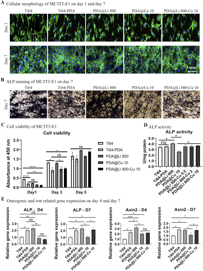
Methods: PDA coatings were polymerized onto polished Ti64 substrates, followed by sequential immersion in LiCl (800 μM) and CuCl2 (10 μM) solutions to construct Li+/Cu2+ co-doped surfaces (PDA@Li 800-Cu 10). In vitro assays assessed MC3T3-E1 pre-osteoblast proliferation (CCK-8), osteogenic differentiation (ALP activity, RT-PCR for ALP/Axin2), and antibacterial activity against S. aureus and E. coli (live/dead staining, CFU assays). In vivo efficacy was evaluated in a rat femoral defect model via micro-CT and histology.
Results and discussion: Li+-functionalized surfaces (PDA@Li 800) enhanced osteoblast proliferation and osteogenesis via Wnt/β-catenin activation. Cu2+-loaded coatings (PDA@Cu 10) eradicated >99% bacteria but moderately suppressed osteogenic markers. The dual-doped PDA@Li 800-Cu 10 surface resolved this bioactivity conflict, maintaining antibacterial efficacy comparable to PDA@Cu 10 while elevating the osteogenic capacity of Cu2+-only modified surfaces. In vivo, dual-modified implants eliminated bacterial colonization within 72 h and significantly increased peri-implant bone volume (BV/TV) in comparison to Ti64 controls, outperforming PDA-only counterparts. By harmonizing Li-driven osteoinduction and Cu-mediated bactericidal action through a scalable PDA platform, this work advances a transformative strategy for next-generation orthopedic and dental implants, simultaneously addressing infection risks and bone regeneration demands.
Keywords: Ti6Al4V; antibacterial; metal ions; osseointegration; surface modification.
Copyright © 2025 Li, Jiang, Yang, Zhang, Wu, Yang, Yang, Wang, Chen, Zhang, Huang, Zhang and Zhang.
Conflict of interest statement
The authors declare that the research was conducted in the absence of any commercial or financial relationships that could be construed as a potential conflict of interest.
Figures


Similar articles
-
Elimination of methicillin-resistant Staphylococcus aureus biofilms on titanium implants via photothermally-triggered nitric oxide and immunotherapy for enhanced osseointegration.Mil Med Res. 2023 May 4;10(1):21. doi: 10.1186/s40779-023-00454-y. Mil Med Res. 2023. PMID: 37143145 Free PMC article.
-
Multidynamic Osteogenic Differentiation by Effective Polydopamine Micro-Arc Oxide Manipulations.Int J Nanomedicine. 2022 Oct 11;17:4773-4790. doi: 10.2147/IJN.S378387. eCollection 2022. Int J Nanomedicine. 2022. PMID: 36246934 Free PMC article.
-
Molecular mechanisms of osteogenesis and antibacterial activity of Cu-bearing Ti alloy in a bone defect model with infection in vivo.J Orthop Translat. 2020 Dec 28;27:77-89. doi: 10.1016/j.jot.2020.10.004. eCollection 2021 Mar. J Orthop Translat. 2020. PMID: 33437640 Free PMC article.
-
Advanced surface modification techniques for titanium implants: a review of osteogenic and antibacterial strategies.Front Bioeng Biotechnol. 2025 Mar 19;13:1549439. doi: 10.3389/fbioe.2025.1549439. eCollection 2025. Front Bioeng Biotechnol. 2025. PMID: 40177619 Free PMC article. Review.
-
[Progress in antibacterial/osteogenesis dual-functional surface modification strategy of titanium-based implants].Zhongguo Xiu Fu Chong Jian Wai Ke Za Zhi. 2023 Oct 15;37(10):1300-1313. doi: 10.7507/1002-1892.202306025. Zhongguo Xiu Fu Chong Jian Wai Ke Za Zhi. 2023. PMID: 37848328 Free PMC article. Review. Chinese.
References
-
- Australian Orthopaedic Association National Joint Replacement Registry (2018). Hip, knee and shoulder arthroplasty: 2018 annual report. Adelaide: AOA.
LinkOut - more resources
Full Text Sources