. 2025 May 22;53(10):gkaf447.
doi: 10.1093/nar/gkaf447.

SARS-CoV-2 point mutations are over-represented in terminal loops of RNA stem-loop structures that can be resolved by Nsp13 helicase in a unique manner with respect to nucleotide dependence

Affiliations

SARS-CoV-2 point mutations are over-represented in terminal loops of RNA stem-loop structures that can be resolved by Nsp13 helicase in a unique manner with respect to nucleotide dependence

Adaira J Dumm et al. Nucleic Acids Res. .

Abstract

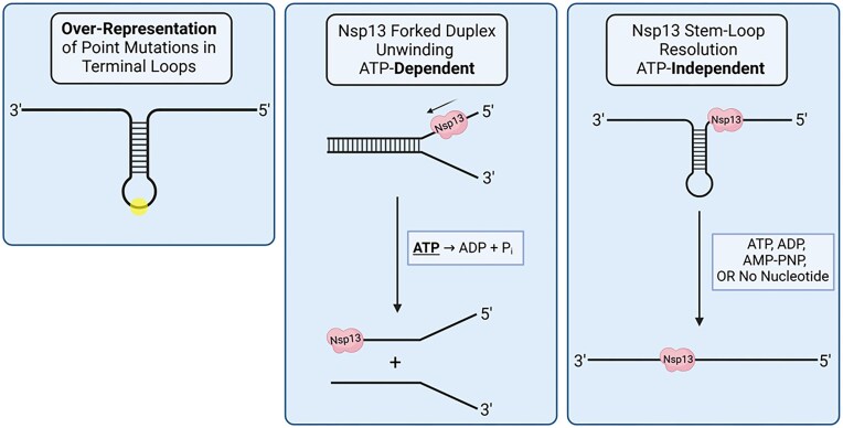
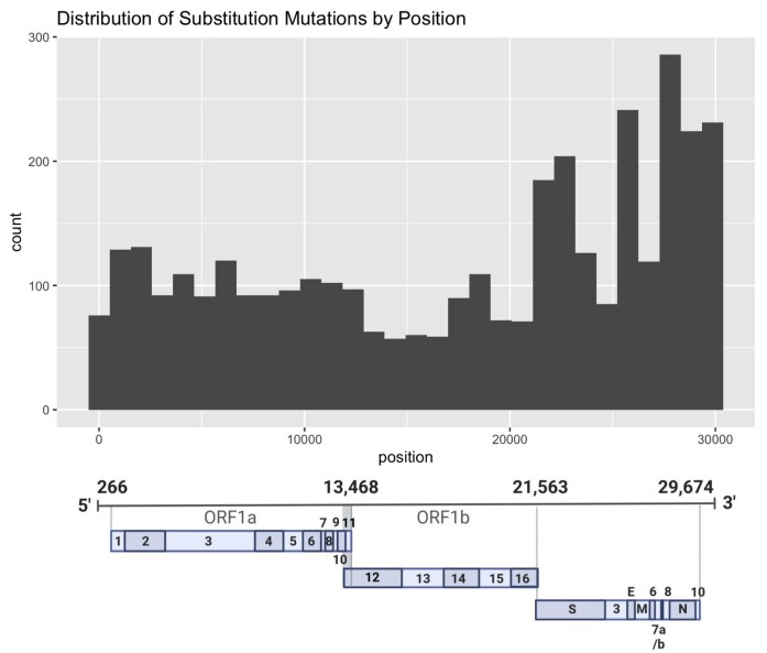
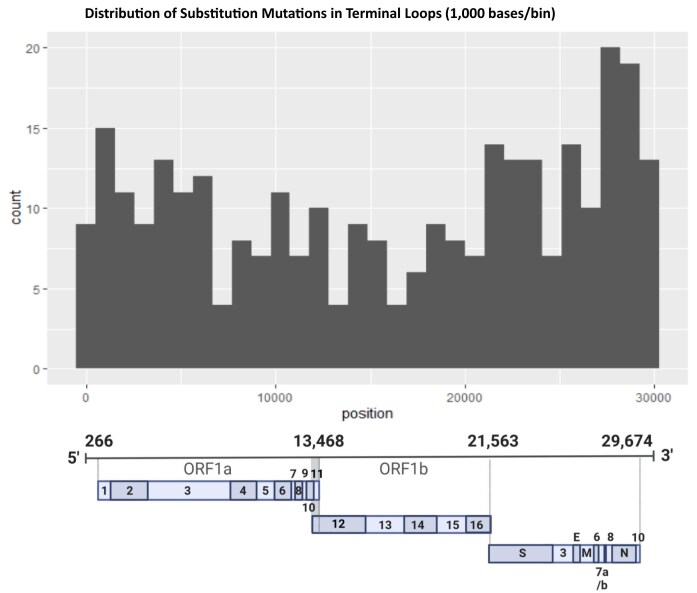
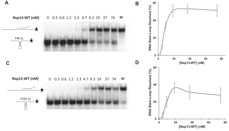
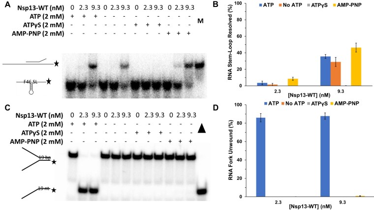
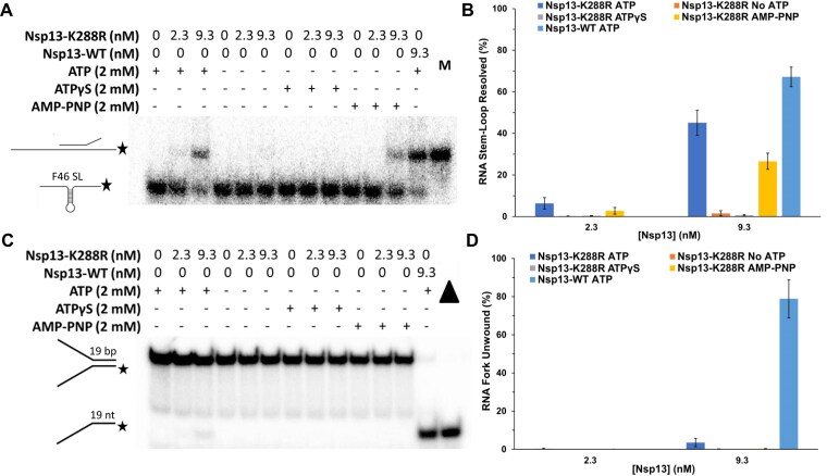
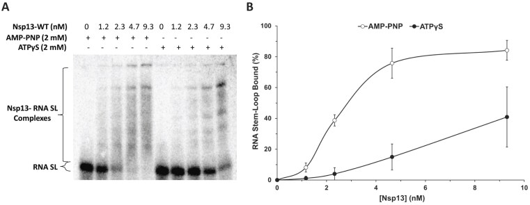
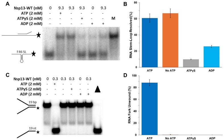
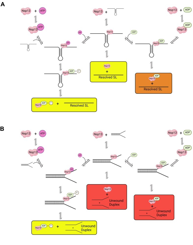
To improve health outcomes for COVID-19 (coronavirus disease 2019) patients, the factors that influence coronavirus genome variation need to be ascertained. The SARS-CoV-2 (severe acute respiratory syndrome coronavirus 2) genome is rich in predicted RNA secondary structures, particularly stem-loops (SLs) formed by intramolecular base pairing within palindromic sequences. We analyzed the NCBI Virus collection of SARS-CoV-2 genome sequences from COVID-19 individuals to map variants relative to SL structural elements. Point mutations in the SARS-CoV-2 genome, with a C-to-U transition bias, were over-represented in unpaired nucleotides and, more specifically, within the terminal loops of RNA SL structures. As the sole helicase encoded by SARS-CoV-2, Nsp13 may operate in the timely resolution of secondary RNA structures to facilitate SARS-CoV-2 RNA copying or processing. We characterized Nsp13 to resolve SARS-CoV-2 sequence-derived unimolecular RNA SL substrates and determined that it does so in a functionally cooperative manner. In addition to ATP, Nsp13 resolves the unimolecular RNA SL structure in the absence of nucleotide, in contrast to the strict ATP requirement for a bimolecular RNA forked duplex. We suggest a model in which a series of binary and ternary complex interactions of Nsp13 with nucleotide and/or RNA SL pose mechanistic implications for RNA SL resolution.


Conflict of interest statement

None declared.

Figures

Graphical Abstract
Graphical Abstract
Figure 1.
Figure 1.
Purified recombinant SARS-CoV-2 Nsp13-WT and Nsp13-K288R proteins used for biochemical assays. (A) Cartoon schematic of recombinant Nsp13 protein overexpressed using a baculovirus system. During purification, the N-terminal histidine (His) tag is cleaved off, as notated by arrowhead. The C-terminal possesses a Flag epitope. The position of the engineered Walker A box (motif I) ATPase domain mutation of the invariant lysine (K) residue replaced with an arginine (R) is shown in the conserved helicase subdomain 1A. 6xHis, six-histidine tag; ZBD, zinc binding domain; 1B, connector region between stalk domain and helicase subdomain 1A; 2A, helicase subdomain 2A; TEV, TEV protease site. (B) Nsp13 protein (1,000 ng) was resolved by SDS–PAGE and detected by Coomassie staining. Molecular weight markers (MW) are shown with size in kilodaltons (kDa) indicated.
Figure 2.
Figure 2.
Distribution of substitution mutations across the SARS-CoV-2 genome. The substitution mutation counts are plotted according to nucleotide position in bins of 300 bases, with 0.1% thresholding. For reference, the SARS-CoV-2 genome coding and noncoding sequences are depicted below. BioRender was used for SARS-CoV-2 genome depiction.
Figure 3.
Figure 3.
Distribution of substitution mutations in the TLs of the SARS-CoV-2 genome. The substitution mutation counts are plotted according to nucleotide position in bins of 1,000 bases, with 0.1% thresholding. For reference, the SARS-CoV-2 genome coding and noncoding sequences are depicted below. BioRender was used for SARS-CoV-2 genome depiction.
Figure 4.
Figure 4.
Biochemical analysis of RNA SL resolution by Nsp13-WT as a function of enzyme concentration. Representative native polyacrylamide gel images showing products from helicase reactions as a function of the indicated Nsp13-WT enzyme concentration in the presence of 2 mM ATP on the F46 RNA SL substrate (0.25 nM) (A) or F593 RNA SL substrate (0.25 nM) (C), as described in the “Materials and methods” section. M represents marker substrate with oligonucleotide used to make an SL substrate that was annealed to a complementary oligonucleotide to create a forked duplex. (B,D) Quantitative assessment of Nsp13-WT resolution of the F46 RNA SL substrate and F593 RNA SL substrate, respectively. Data represent the average of at least three independent experiments with standard deviations (SDs) indicated by error bars.
Figure 5.
Figure 5.
Biochemical analysis of RNA SL resolution and forked duplex unwinding by Nsp13-WT as a function of nucleotide. Representative native polyacrylamide gel images showing products from helicase reactions for the indicated Nsp13-WT concentration with the indicated nucleotide (2 mM) on F46 RNA SL substrate (0.25 nM) (A) or RNA forked duplex substrate (0.25 nM) (C), as described in the “Materials and methods” section. In panel (A),M represents the marker substrate with oligonucleotide used to make an SL substrate that was annealed to a complementary oligonucleotide to create a forked duplex. In panel (C), the filled triangle represents heat-denatured RNA substrate control. Quantitative assessment of Nsp13-WT resolution of the RNA F46 SL (B) and forked duplex (D) substrates, respectively. Data represent the average of at least three independent experiments with SDs indicated by error bars.
Figure 6.
Figure 6.
Biochemical analysis of RNA SL resolution and forked duplex unwinding by Nsp13-K288R as a function of nucleotide. Representative native polyacrylamide gel images showing products from helicase reactions for the indicated Nsp13-K288R or Nsp13-WT concentration as a function of nucleotide status on F46 RNA SL substrate (0.25 nM) (A) or RNA forked duplex substrate (0.25 nM) (C), as described in the “Materials and methods” section. Substrate was incubated with Nsp13-K288R or Nsp13-WT and the indicated nucleotide (2 mM), as described under the “Materials and methods” section. In panel (A),M represents the marker substrate with oligonucleotide used to make the SL substrate that was annealed to a complementary oligonucleotide to create a forked duplex. In panel (C), the filled triangle represents heat-denatured RNA substrate control. Quantitative assessment of Nsp13-K288R resolution of the RNA F46 SL (B) and forked duplex (D) substrates, respectively. Data represent the average of at least three independent experiments with SDs indicated by error bars.
Figure 7.
Figure 7.
Biochemical analysis of the effect of ATP analogs on Nsp13 binding to RNA SL. (A) Representative native polyacrylamide gel image showing by EMSA Nsp13 binding to F46 RNA SL substrate as a function of indicated enzyme concentration in the presence of the indicated nucleotide (2 mM). The indicated concentrations of Nsp13 were incubated with the F46 RNA SL (0.25 nM) for 15 min at 30°C with AMP-PNP or ATPγS and resolved on 5% polyacrylamide (0.5× TBE) gels at 200 V for 2 h 15 min. (B) Quantitative assessment of Nsp13 binding to F46 RNA SL. Data represent the average of at least three independent experiments with SDs indicated by error bars.
Figure 8.
Figure 8.
Biochemical analysis of the effect of ADP on RNA SL resolution and forked duplex unwinding by Nsp13-WT. Representative native polyacrylamide gel images showing products from helicase reactions for the indicated Nsp13-WT concentration with the specified nucleotide (2 mM) on F46 RNA SL substrate (0.25 nM) (A) or forked RNA duplex substrate (0.25 nM) (C) as described in the “Materials and methods” section. In panel (A), M represents the marker substrate with oligonucleotide used to make the SL substrate that was annealed to a complementary oligonucleotide to create a forked duplex. In panel (C), the filled triangle represents heat-denatured RNA substrate control. Quantitative assessment of Nsp13-WT resolution of the RNA F46 SL (B) and forked duplex (D) substrates, respectively. Data represent the average of at least three independent experiments with SDs indicated by error bars.
Figure 9.
Figure 9.
Model for binary and ternary complex interactions between Nsp13, ATP/ADP, and unimolecular RNA SL or bimolecular RNA forked duplex substrates. (A) Resolved RNA SL products reached through the efficient pathways (in the presence of ATP or absence of nucleotide) are indicated by a yellow box for “Resolved RNA-SL,” whereas less efficiently generated product (in the presence of ADP) is denoted by an orange box. (B) Resolved RNA fork products reached through the ATP-dependent pathway are indicated by a yellow box for “Resolved RNA-Fork.” Undetectable product (in the presence of ADP or absence of nucleotide) is denoted by a pink box. Figure created using BioRender. (A), Created in BioRender. Sommers, J. (2025) https://biorender.com/xh3hg7t (B), Created in BioRender. Sommers, J. (2025) https://biorender.com/ulh5buj.

Similar articles

References

    1. Carvajal JJ, García-Castillo V, Cuellar SV et al. New insights into the pathogenesis of SARS-CoV-2 during and after the COVID-19 pandemic. Front Immunol. 2024; 15:1363572. 10.3389/fimmu.2024.1363572. - DOI - PMC - PubMed
    1. Donald J, Bilasy SE, Yang C et al. Exploring the complexities of long COVID. Viruses. 2024; 16:1060. 10.3390/v16071060. - DOI - PMC - PubMed
    1. Huston NC, Wan H, Strine MS et al. Comprehensivein vivosecondary structure of the SARS-CoV-2 genome reveals novel regulatory motifs and mechanisms. Mol Cell. 2021; 81:584–98. 10.1016/j.molcel.2020.12.041. - DOI - PMC - PubMed
    1. Lan TCT, Allan MF, Malsick LE et al. Secondary structural ensembles of the SARS-CoV-2 RNA genome in infected cells. Nat Commun. 2022; 13:1128. 10.1038/s41467-022-28603-2. - DOI - PMC - PubMed
    1. Yang SL, DeFalco L, Anderson DE et al. Comprehensive mapping of SARS-CoV-2 interactionsin vivo reveals functional virus–host interactions. Nat Commun. 2021; 12:5113. 10.1038/s41467-021-25357-1. - DOI - PMC - PubMed