Review
. 2025 Apr 29;15(5):277.
doi: 10.3390/bios15050277.

Aptamer and Oligonucleotide-Based Biosensors for Health Applications

Affiliations
Review

Aptamer and Oligonucleotide-Based Biosensors for Health Applications

Beatriz Mayol et al. Biosensors (Basel). .

Abstract

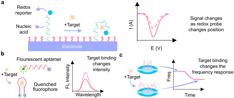
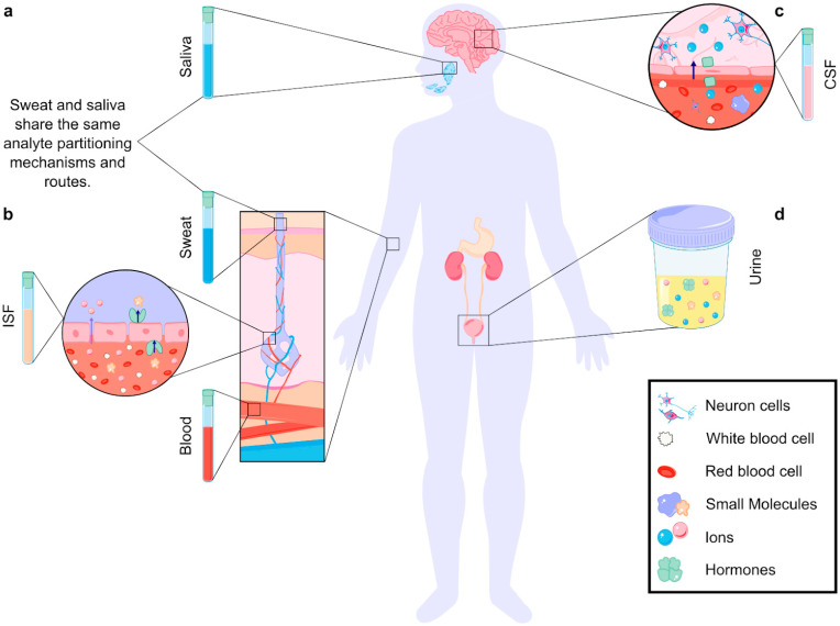
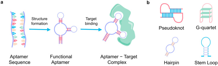
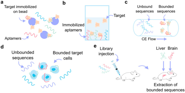
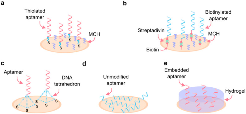
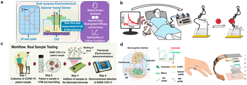
Aptamers have emerged as powerful molecular recognition elements for biosensing applications, offering high specificity, stability, and adaptability. This review explores key considerations in designing aptamer-based sensors (aptasensors), with a focus on biomarker selection, aptamer design, and detection and immobilization strategies. However, challenges such as biofluid stability and reversibility must be addressed to improve biosensor performance. In this study, the potential of aptamer-based platforms in diagnostics is explored, emphasizing their advantages and future applications. Looking ahead, advances in multifunctional aptamers, integration with nanomaterials, and computational optimization are highlighted as promising directions for enhancing their effectiveness in biosensing.

Keywords: aptamer biosensor; electrochemical aptasensor; health monitoring.


Conflict of interest statement

The authors declare no conflicts of interest.

Figures

Figure 5
Figure 5
Signal transduction techniques using aptamers. (a) Electrochemical aptasensor. In the presence of the target molecule, the aptamer undergoes a conformational change, which is transduced by the redox reporter and measured by electrochemical techniques. (b) Fluorescent aptasensor. Binding with the target molecule produces a change in the optical tag (fluorophore). (c) Quartz crystal microbalance aptasensor, which correlates with the mass accumulated on the quartz crystal resonator with the change in its frequency [99].
Figure 1
Figure 1
Analyte partitioning in different biofluids. (a) In biofluids produced by glands (sweat, saliva), analytes travel from the blood, through ISF, and finally to the glands. Analytes are filtered in each step, making the analyte concentrations in blood differ from those in the biofluids [23]. (b) Analytes enter biofluids through paracellular (purple arrow) and transcellular (dark blue arrow, transporter-mediated) transport. The size of the analyte, cellular structure, and partitioning mechanism define the final concentration in the fluid [23]. (c) Analytes can travel from the blood to the cerebrospinal fluid (CSF) through the blood–brain barrier (BBB). Partitioning mechanisms include paracellular and transcellular transport (ion transport depicted) [24,25]. (d) Analytes can also be found in other noninvasive biofluids, such as urine.
Figure 2
Figure 2
Aptamer basics. (a) The primary aptamer sequence, folding into its three-dimensional structure and binding with its specific target. (b) The four main secondary structures of aptamers.
Figure 3
Figure 3
Aptamer design. The Systematic Evolution of Ligands by Exponential Enrichment (SELEX) process allows for aptamer design and selection based on the specific target molecule. It starts with the creation of an aptamer library where the sequences have a central random region flanked by constant primer regions. The process involves 1 to 16 cycles of binding, washing, elution, and amplification. In the last round, the high-affinity aptamers are identified for cloning and sequencing.
Figure 4
Figure 4
SELEX variants. (a) Bead-based SELEX, where the target molecule is immobilized on the beads and the aptamer remains free in solution. (b) Target in solution SELEX, where the aptamers are immobilized to the substrate. (c) Capillary electrophoresis (CE) SELEX, where the unbounded and bounded sequences are separated based on their different flow rates over the electrophoresis gel. (d) Cell SELEX allows for the aptamer-based recognition of specific cells. (e) In vivo SELEX, where the aptamer library is injected into the animal model and the bound sequences are collected from organ tissue.
Figure 6
Figure 6
Immobilization of aptamers. (a) High-density covalent binding to the surface by thiol-metal interaction. (b) High-density affinity-based binding by streptavidin/avidin-biotin interactions. (c) Low-density DNA tetrahedron aptamer surface binding. (d) Adsorption binding of unmodified aptamers. (e) Immobilization by embedding aptamers in a hydrogel matrix. MCH: 6-Mercapto-1-hexanol.
Figure 7
Figure 7
Diagnostic applications of aptamers. (a) Electrochemical aptamer-based sensor for pH and metabolite monitoring in cell culture media for up to 72 h in multiple cell lines. Adapted from [155]. (b) Therapeutic drug monitoring with electrochemical aptamer-based sensor for vancomycin detection. Adapted from [157]. (c) Point-of-care electrochemical aptamer-based sensor for COVID-19 detection. Adapted from [158]. (d) Skin-interfaced electrochemical aptamer-based sensor for picomolar detection of oestradiol in sweat. Adapted from [126].

Similar articles

Cited by

References

    1. Bhalla N., Jolly P., Formisano N., Estrela P. Introduction to Biosensors. Essays Biochem. 2016;60:1–8. doi: 10.1042/EBC20150001. - DOI - PMC - PubMed
    1. Arroyo-Currás N., Dauphin-Ducharme P., Scida K., Chávez J.L. From the Beaker to the Body: Translational Challenges for Electrochemical, Aptamer-Based Sensors. Anal. Methods. 2020;12:1288–1310. doi: 10.1039/D0AY00026D. - DOI
    1. Nimjee S.M., White R.R., Becker R.C., Sullenger B.A. Aptamers as Therapeutics. Annu. Rev. Pharmacol. Toxicol. 2017;57:61–79. doi: 10.1146/annurev-pharmtox-010716-104558. - DOI - PMC - PubMed
    1. Yang L.F., Ling M., Kacherovsky N., Pun S.H. Aptamers 101: Aptamer Discovery and in Vitro Applications in Biosensors and Separations. Chem. Sci. 2023;14:4961–4978. doi: 10.1039/D3SC00439B. - DOI - PMC - PubMed
    1. Gelinas A.D., Davies D.R., Janjic N. Embracing Proteins: Structural Themes in Aptamer–Protein Complexes. Curr. Opin. Struct. Biol. 2016;36:122–132. doi: 10.1016/j.sbi.2016.01.009. - DOI - PubMed

LinkOut - more resources