Review
. 2025 Apr 30;15(5):283.
doi: 10.3390/bios15050283.

Advancements in Single-Molecule Fluorescence Detection Techniques and Their Expansive Applications in Drug Discovery and Neuroscience

Affiliations
Review

Advancements in Single-Molecule Fluorescence Detection Techniques and Their Expansive Applications in Drug Discovery and Neuroscience

Jing Yan et al. Biosensors (Basel). .

Abstract

Single-molecule fluorescence technology stands at the forefront of scientific research as a sophisticated tool, pushing the boundaries of our understanding. This review comprehensively summarizes the technological advancements in single-molecule fluorescence detection, highlighting the latest achievements in the development of single-molecule fluorescent probes, imaging systems, and biosensors. It delves into the applications of these cutting-edge tools in drug discovery and neuroscience research, encompassing the design and monitoring of complex drug delivery systems, the elucidation of pharmacological mechanisms and pharmacokinetics, the intricacies of neuronal signaling and synaptic function, and the molecular underpinnings of neurodegenerative diseases. The exceptional sensitivity demonstrated in these applications underscores the vast potential of single-molecule fluorescence technology in modern biomedical research, heralding its expansion into other scientific domains.

Keywords: biosensor; drug discovery; neuroscience; single-molecule fluorescent probe.


Conflict of interest statement

Authors Lin Cheng, Yitong Li and Ru Wang were emplayed by Holosensor Medical Technology Ltd., Suzhou 215000, China. The remaining authors declare that the research was conducted in the absence of ant commercial or financial relationships that could be construed as a potential conflict of interest.

Figures
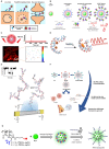
Figure 1
Figure 1
Nanoparticles utilized as single-molecule fluorescent probes. (A) Schematic representation of various molecular labeling approaches evaluated using quantum dots of diverse sizes as fluorescent probes. Reprinted with permission from ref. [29]. Copyright © 2020 American Chemical Society. (B) A biotin–streptavidin–biotin approach is depicted for the assembly of upconverting fluorescent nanoparticle (UCNP)-based single-particle tracking (SPT) labels. Reprinted with permission from ref. [24]. Copyright © 2024 American Chemical Society. (C) Near-infrared blinking carbon dots (CDs) specifically engineered for applications in single-molecule localization microscopy (SMLM). Reprinted with permission from ref. [30]. Copyright © 2022 American Chemical Society. (D) Schematic of the IR-Emitting C12-Agn formation, which involves the complexation of C12DNA with silver cations, followed by reduction in the mixture with sodium borohydride (NaBH4). Reprinted from ref. [31]. Copyright © 2007 by The National Academy of Sciences of the USA. (E) Scaled schematic of a SWCNT-based single-molecule field-effect transistor (smFET) device structure. Reprinted from ref. [32]. (F) The functional nucleic acid delivery (FND) system designed for the delivery of microRNA-34a (miR-34a). Reprinted from ref. [33]. (G) Nanoprobes based on plasmonic nanoparticles (PNPs). Reprinted with permission from ref. [34]. Copyright © 2021 American Chemical Society.
Figure 2
Figure 2
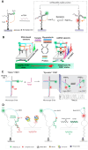
Total internal reflection fluorescence. (A) Principle of TIRF. Reprinted from ref. [54]. (B) Schematic diagram of mRNP-SiMPull procedure for isolating mRNPs from yeast cells and single-molecule imaging of RBP components. Reprinted from ref. [60].
Figure 3
Figure 3
Stimulated emission depletion. (A) Schematic diagram of STED nanoscopy. Reprinted from ref. [65]. (B) Schematic diagram of event-triggered STED. Reprinted from ref. [69].
Figure 4
Figure 4
Principles of three single-molecule localization microscopy. (A) Principle of PALM. The PA-FP (photoactivatable fluorescent protein) molecules attach to proteins of interest. (B) Principle of STORM. Improved clarity by separating the roles of the green and red lasers and refining the description of the imaging process. (C) Principles of DNA-PAINT. Reprinted with permission from ref. [76].
Figure 5
Figure 5
Single-molecule fluorescent biosensors based on the FRET principle. (A) A DNA-based smFRET sensor for high-confidence detection of miRNAs. Reprinted with permission from ref. [90]. Copyright © 2022 American Chemical Society. (B) Principles of nucleic acid detection using the interconvertible hairpin-based sensor of smFRET. Reprinted with permission from ref. [91]. Copyright © 2019 American Chemical Society. (C) Working principle of a smFRET-based dynamic DNA sensor. Reprinted with permission from ref. [92]. Copyright © 2021 American Chemical Society. (D) SmFRET-based aptamer sensors detect lysozyme selectively and sensitively. Reprinted from ref. [95].
Figure 6
Figure 6
SMLM bioimages based on burst-blinking nanographenes. (A) SMLM of amyloid fiber in air. (B) SMLM of neuron in PBS. (b) Reconstructed SMLM image of global nascent proteins in neurons. Imaging was performed in PBS solution. (c) Magnification of SMLM image and conventional wide-field fluorescence image for the yellow box region in (b), respectively. (C) Conventional wide-field fluorescence image of networks in neurons (left) and corresponding Voronoi diagram image (right) of the same position. Reprinted from ref. [133].
Figure 7
Figure 7
STORM images of senile plaques and neurofibrillary tangles in AD patient brains. (A) Shown from left to right are immunohistochemical staining, conventional fluorescence microscopy imaging, and super-resolution STORM images of a senile plaque located in the neocortex of an Alzheimer’s disease (AD) patient. (A1) Representative image of a senile plaque in the neocortex of an AD patient (immunohistochemical detection of Ab). (A2) Conventional fluorescence microscopy image of a whole senile plaque in a neocortex section of the same patient immunostained for Ab. (A3) STORM image of the same area. The insets (1 and 2) show close-up details of the distribution and size of aggregated Ab branches. (A4) Comparative TEM image of Ab fibrils (black arrowheads) in a senile plaque. (B) Displayed from left to right are representative images of neurofibrillary tangles in the neocortex of AD patients, captured using immunohistochemical staining, conventional fluorescence microscopy, and a combined approach that integrates conventional fluorescence microscopy for β-amyloid (Aβ) detection with STORM imaging for phosphorylated tau (p.Tau) visualization. Reprinted from ref. (B1) Representative image of neurofibrillary tangles in the neocortex of an AD patient (immunohistochemical detection of p.Tau). (B2) Conventional fluorescence microscopy image of neurofibrillary tangles within the soma of a whole degenerating neuron surrounded by Ab deposition in a neocortex section of the same patient. (B3) Same neuron imaged by combining conventional fluorescence microscopy (Ab) and STORM (p.Tau). The insets (3 and 4) show close-up details of the honeycombed structure of p.Tau aggregates in the soma and the filamentous organization in the axon. (B4) Comparative TEM image of Tau filaments (white arrowhead) in neurofibrillary tangles [145].

Similar articles

References

    1. Ha T., Kaiser C., Myong S., Wu B., Xiao J. Next Generation Single-Molecule Techniques: Imaging, Labeling, and Manipulation in Vitro and in Cellulo. Mol. Cell. 2022;82:304–314. doi: 10.1016/j.molcel.2021.12.019. - DOI - PMC - PubMed
    1. Blank K., De Cremer G., Hofkens J. Fluorescence-Based Analysis of Enzymes at the Single-Molecule Level. Biotechnol. J. 2009;4:465–479. doi: 10.1002/biot.200800262. - DOI - PubMed
    1. Ray S., Widom J.R., Walter N.G. Life under the Microscope: Single-Molecule Fluorescence Highlights the RNA World. Chem. Rev. 2018;118:4120–4155. doi: 10.1021/acs.chemrev.7b00519. - DOI - PMC - PubMed
    1. Ma F., Li Y., Tang B., Zhang C.-Y. Fluorescent Biosensors Based on Single-Molecule Counting. Acc. Chem. Res. 2016;49:1722–1730. doi: 10.1021/acs.accounts.6b00237. - DOI - PubMed
    1. Betzig E., Chichester R.J. Single Molecules Observed by Near-Field Scanning Optical Microscopy. Science. 1993;262:1422–1425. doi: 10.1126/science.262.5138.1422. - DOI - PubMed

Substances

LinkOut - more resources