. 2025 May 15;15(5):317.
doi: 10.3390/bios15050317.

One-Pot Detection of miRNA by Dual Rolling Circle Amplification at Ambient Temperature with High Specificity and Sensitivity

Affiliations

One-Pot Detection of miRNA by Dual Rolling Circle Amplification at Ambient Temperature with High Specificity and Sensitivity

Wenhua Sun et al. Biosensors (Basel). .

Abstract

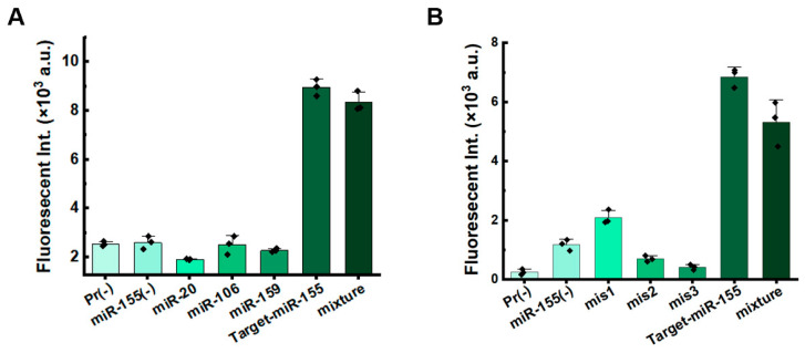
Rolling circle amplification (RCA) at ambient temperature is prone to false positive signals during nucleic acid detection, which makes it challenging to establish an efficient RCA detection method. The false positive signals are primarily caused by binding of non-target nucleic acids to the circular single-stranded template, leading to non-specific amplification. Here, we present an RCA method for miRNA detection at 37 °C using two circular ssDNAs, each of which is formed by ligating the intramolecularly formed nick (without any splint) in a secondary structure. The specific target recognition is realized by utilizing low concentrations (0.1 nM) of circular ssDNA1 (C1). A phosphorothioate modification is present at G*AATTC on C1 to generate a nick for primer extension during the primer self-generated rolling circle amplification (PG-RCA). The fragmented amplification products are used as primers for the following RCA that serves as signal amplification using circular ssDNA2 (C2). Notably, the absence of splints and the low concentration of C1 significantly inhibits non-target binding, thus minimizing false positive signals. A high concentration (10 nM) of C2 is used to carry out linear rolling circle amplification (LRCA), which is highly specific. This strategy demonstrates a good linear response to 0.01-100 pM of miRNA with a detection limit of 7.76 fM (miR-155). Moreover, it can distinguish single-nucleotide mismatch in the target miRNA, enabling the rapid one-pot detection of miRNA at 37 °C. Accordingly, this method performs with high specificity and sensitivity. This approach is suitable for clinical serum sample analysis and offers a strategy for developing specific biosensors and diagnostic tools.

Keywords: circular ssDNA; dual RCA; microRNA detection; splint; ssDNA circularization.


Conflict of interest statement

The authors declare no conflicts of interest.

Figures

Scheme 1
Scheme 1
Schematic diagram for the dual RCA strategy (A), and the example for detecting the miRNA of miR-155 (B). The phosphate bond between G and A in the recognition site (GAATTC) of EcoRI-HF (a restriction enzyme) is replaced by phosphorothioate to avoid its cleavage. The prepared circular ssDNA samples do not involve a splint which can serve as a primer for further RCA. The green line represents the complementary region of the target, and the yellow line represents the extension of the target to the EcoRI-HF enzyme cleavage site region.
Figure 1
Figure 1
Comparison of PG-RCA between two methods for circularization of circular ssDNA templates. (A) Real-time record of PG-RCA at various concentrations of miR-155 using splint-aided circularization. (B) Schematic diagrams of two (splint-aided or splint-free) approaches for template circularization. (C) The mechanism of PG-RCA by using restriction enzymes (circular ssDNA is prepared by splint-free approach). “*” represents phosphorothioate modification. The green line represents the complementary region of the target, and the yellow line represents the extension of the target to the EcoRI-HF enzyme cleavage site region. (D,E) Real-time record of PG-RCA at various concentrations of miR-155 using splint-free circularization. PG-RCA conditions: 1 nM C1 for (B,D) or 0.1 nM for (E), 25 U/mL phi29, 50 U/mL EcoRI-HF, 1× SYBR Green II, 0.5× phi29 buffer, 0.5× rCutsmart buffer, 400 μM each dNTP, and incubation at 37 °C. (F) Calibration curve showing the linear relationship between the threshold time (Tt) and the logarithmic concentration (lgM) of miR-155, within the range of 10−13 to 10−10 M. Tt is the threshold time for amplification, defined as the time when fluorescent intensity exceeds the calculated threshold, like Ct values for RT-PCR.
Figure 2
Figure 2
Optimization of conditions for dual RCA. (A) Efficiency of dual RCA compared with various controls. The label of (-) indicates the corresponding component is absent. (B) Effect of EcoRI-HF concentration on the threshold time (Tt) values. The dark green bars represent the Tt values for the 1 pM target group, while the light green bars correspond to the Tt values for the negative control group. The line graph illustrates the difference in Tt values between the target group and the negative control group. (C) Effect of phi29 concentration on Tt values. (D) Impact of various buffers on Tt values. Buffer A corresponds to the phi29 buffer, while buffer B corresponds to the rCutsmart buffer. (E) Effect of C1 concentration on Tt values. (F) Effect of C2 concentration on Tt values.
Figure 3
Figure 3
Sensitivity of the dual ssDNA ring system for miRNA detection. (A) Fluorescence intensity at various concentrations of miR-155. (B) Calibration curve showing the linear relationship between the threshold time (Tt) and the logarithmic concentration (lgM) of miR-155, within the range of 10−14 to 10−10 M. (C) Fluorescence intensity at various concentrations of miR-106. (D) Calibration curve depicting the linear correlation between Tt and lgM for miR-106, within the range of 10−13 to 10−10 M. Other reaction conditions: 0.1 nM C1, 10 nM C2, 25 U/mL phi29, 50 U/mL EcoRI-HF, 1× SYBR Green II, 0.5× phi29 buffer, 0.5× rCutsmart buffer, 400 μM each dNTP, and 37 °C.
Figure 4
Figure 4
Sensitivity evaluation of specificity for dual RCA. (A) Fluorescence intensity for targeting miR-155 in the presence of various other miRNAs. Pr (-) denotes the absence of miRNA; miR-155 (-) indicates the presence of 1 pM miR-20, miR-106, and miR-159, but not target-miR-155; and “mixture” refers to a reaction containing all miRNAs. (B) Fluorescence intensity of miR-155 with various numbers of mismatches. Pr (-) denotes the absence of miRNA; miR-155 (-) indicates the presence of 1 pM of mis1, mis2, and mis3, but not target-miR-155; and “mixture” refers to a reaction containing all the miRNAs. Other conditions: 0.1 nM C1, 10 nM C2, 1.0 pM miRNA, 25 U/mL phi29, 50 U/mL EcoRI-HF, 1× SYBR Green II, 0.5× phi 29 buffer, 0.5× rCutsmart buffer, 400 μM each dNTP, and incubation at 37 °C for 120 min.

Similar articles

References

    1. Michael G.M., Eric T.K. The discovery of rolling circle amplification and rolling circle transcription. Acc. Chem. Res. 2016;49:2540–2550. - PMC - PubMed
    1. Cao A.P., Zhang C.Y. Sensitive and label-free DNA methylation detection by ligation-mediated hyperbranched rolling circle amplification. Anal. Chem. 2012;84:6199–6205. doi: 10.1021/ac301186j. - DOI - PubMed
    1. Wang T., Tian S., Tikhonova E.B., Karamyshev A.L., Wang J.J., Zhang F., Wang D. The enrichment of miRNA-targeted mRNAs in translationally less active over more active polysomes. Biology. 2023;12:1536. doi: 10.3390/biology12121536. - DOI - PMC - PubMed
    1. Lu T.X., Rothenberg M.E. MicroRNA. J. Allergy Clin. Immunol. 2018;141:1202–1207. doi: 10.1016/j.jaci.2017.08.034. - DOI - PMC - PubMed
    1. Xu W.D., Feng S.Y., Huang A.F. Role of miR-155 in inflammatory autoimmune diseases: A comprehensive review. Inflamm. Res. 2022;71:1501–1517. doi: 10.1007/s00011-022-01643-6. - DOI - PubMed

LinkOut - more resources