. 2025 Jul 9;16(7):e0046725.
doi: 10.1128/mbio.00467-25. Epub 2025 May 27.

The sulfur assimilation pathway mitigates redox stress from acidic pH in Salmonella Typhi H58

Affiliations

The sulfur assimilation pathway mitigates redox stress from acidic pH in Salmonella Typhi H58

Marion Fernandez et al. mBio. .

Abstract

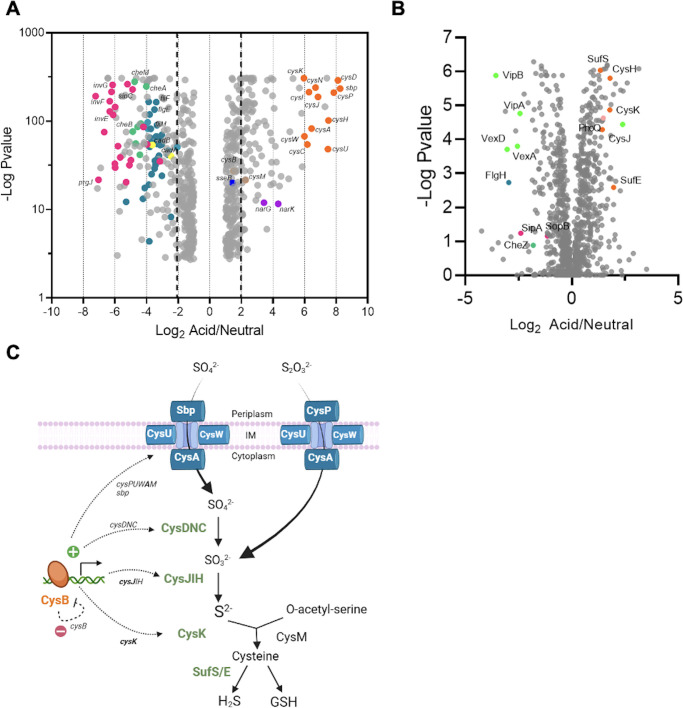
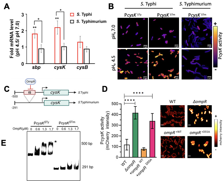
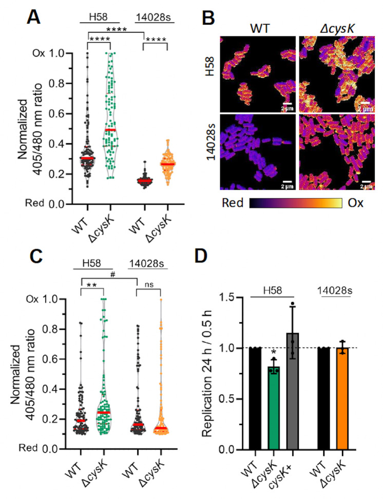
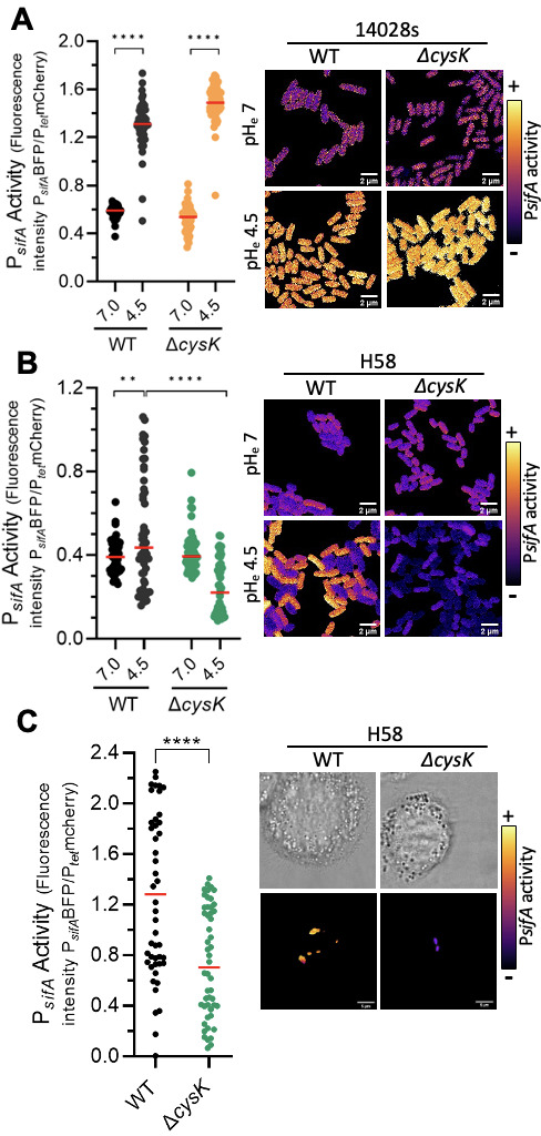
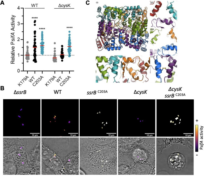
Salmonella enterica serovar Typhi is the causative agent of typhoid fever, a human-restricted systemic infection. The rapidly disseminating multi-drug resistant H58 subclade is endemic in Africa, Asia, and Oceania, yet little is known regarding its intracellular behavior and virulence properties. It was of interest to understand the H58 response to host cell infection in terms of its response to acid stress and subsequent changes in gene regulation. We compared the H58 response in vitro and during infection of THP-1 human macrophages with the well-characterized response of Salmonella Typhimurium, which causes gastroenteritis. In S. Typhimurium infections, bacteria reside in an acidic intracellular vacuole and become acidified, driving the expression of pathogenicity island 2 genes (SPI-2). SPI-2 virulence factors modify the vacuole and enable bacterial replication. In response to acid stress, the sulfur assimilation pathway is highly upregulated and unique to H58. Replacing the Typhi cysK promoter with the Typhimurium promoter resulted in a cysK gene that was upregulated by acid stress in the H58 background, highlighting the differential regulation between the two serovars. In acidic conditions, H58 experienced much greater redox stress compared with S. Typhimurium, and the sulfur assimilation pathway was required to mitigate the redox stress. Higher redox stress modified the transcriptional regulator SsrB, resulting in diminished secretion of the SPI-2 virulence factor SifA. Our results highlight significant differences between S. Typhi H58 and S. Typhimurium and emphasize the importance of studying S. Typhi strains directly to understand their unique behavior during pathogenesis.

Importance: In this study, we examined the clinically relevant, multi-drug resistant Salmonella Typhi strain H58, which is rapidly disseminating across Southeast Asia, Africa, and Oceania. It has heretofore been uncharacterized in terms of its gene regulation. Using human THP-1 macrophages, we discovered that S. Typhi strongly activates the sulfur utilization pathway in response to acid stress encountered in the vacuole once Typhi is inside host cells. Our novel findings were that S. Typhi experiences substantially higher redox stress compared with Typhimurium, and it requires the sulfur utilization pathway to mitigate this stress. This pathway is not upregulated in Typhimurium and represents a divergence in the response of these two serovars. We emphasize that S. Typhimurium is not a reasonable model for understanding H58, a serovar that is seriously impacting human health.

Keywords: H58; Salmonella Typhi; Salmonella pathogenicity island 2; SifA; SsrB; acid stress response; redox; sulfur assimilation.


Conflict of interest statement

The authors declare no conflict of interest.

Figures

Fig 1
Fig 1
Analysis of S. Typhi identifies an upregulation of the sulfur assimilation pathway in response to acid pH. Salmonella Typhi was grown in LB at neutral (pHe = 7) or acidic pH (pHe = 4.5), and cells were harvested for RNAseq (A) or mass spectrometry analysis (B). The data were analyzed and plotted as fold change of the acidic condition versus the neutral condition. The left side of both panels indicates downregulated in acid pH, whereas the right side is upregulated in acid pH. The genes are color-coded by categories (purple: nitrogen pathway; dark blue: SPI-2; magenta: SPI-1; light blue: flagella; light green: capsule; dark green: chemotaxis; orange: cysteine pathway. (C). The sulfur assimilation pathway in Salmonella.
Fig 2
Fig 2
Regulation of sulfur metabolism differs in STy and STm. (A) Salmonella strains were grown in LB at neutral (pHe = 7) or acid pH (pHe = 4.5) to late exponential phase, and cells were harvested for RNA extraction and subsequent qRT-PCR (see Materials and Methods). The mRNA levels of sbp, cysK, and cysB at pHe 4.5 were compared with pHe 7.0 in the WT S. Typhi (red bars) and S. Typhimurium strains (black bars). The mRNA expression levels of the target genes were normalized relative to 16S rRNA. The error bars represent the mean ± standard deviation (n = 3). **P < 0.005, *P < 0.05, Student’s t-test. (B) Salmonella strains expressing a PcysK-mCherry of S. Typhimurium (PcysKSTm) or S. Typhi (PcysKSTy) compared with constitutively expressed ceruleans. Bacteria were grown at neutral (pHe = 7) or acid pH (pHe = 4.5). PcysK activity was obtained as the ratio of mCherry/ceruleans fluorescence. Representative images from three independent experiments are shown. (C) PcysK of S. Typhi includes an insertion sequence (IS, Top panel). Note the scales differ due to the 209 bp IS. (D) In the absence of ompR, PcysK activity is increased (green column) compared with the WT (white column). Complementation with ompR in trans decreases PcysK activity (orange). Repression is abolished when the ompR null strain is complemented with a substitution (D55A) that eliminates phosphorylation (magenta). Bacteria were grown in LB at pHe 7.0 and harvested for confocal analysis. PcysK activity was obtained by direct measurement of mCherry fluorescence ****P < 0.0001, Student’s t-test. (E) OmpR represses PcysK by direct binding. Electrophoretic mobility shift assays were performed with purified OmpR protein incubated with PcysK from S. Typhi (left) vs S. Typhimurium (right). OmpR does not bind to the PcysK from S. Typhimurium.
Fig 3
Fig 3
S. Typhi experiences higher redox stress compared with S. Typhimurium. (A, B) Salmonella strains expressing RoGFP2 were grown under acidic (pHe = 4.5, SPI-2-inducing) conditions or (C) were used to infect THP-1 cells and then analyzed after 24 hpi. Cells were incubated with NEM and fixed as described in Materials and Methods and then analyzed by confocal microscopy. The emission at 550 nm was collected (excitation = 405 and 480 nm) for each individual bacterium, and the 405/480 ratio was plotted. (A and C) A total of 50 bacteria from two separate experiments were analyzed for each sample. **P < 0.001, ****P < 0.0001, Student’s t-test, the red bar is the median. The results were normalized according to the maximum oxidized and reduced values (see Materials and Methods). (B) Representative images in vitro are color coded for the redox state. Scale bar is 2 µm. (D) Deletion of cysK results in a bacterial replication defect in THP-1 cells for S. Typhi (green column) but not for S. Typhimurium (orange). After infection, THP-1 cells were lysed, and the intracellular bacterial load was determined by agar plating. Bacterial replication between 0.5 and 24 hpi was plotted relative to the corresponding WT strain. *P < 0.01, Student’s t-test.
Fig 4
Fig 4
SPI-2 is downregulated in an S. Typhi cysK null strain. SPI-2 activity was analyzed in S. Typhi (A) or S. Typhimurium (B) using the transcriptional fusion PsifA-mTagBFP, expressed from a plasmid that also contained a constitutively expressed PTet-mCherry fusion. In S. Typhi H58, sifA expression was stimulated by acid stress (black circles, left). In the cysK null mutant, the activity of PsifA actually decreased in acid pH compared with neutral pH (green circles). In S. Typhimurium 14028, there was essentially no difference in PsifA activity between the WT and the cysK null strain. Bacteria were analyzed by confocal microscopy for fluorescence emission from PsifA-mTagBFP and PTet-mCherry. The fluorescence from mTagBFP was divided by the mCherry fluorescence for each individual bacterium to determine the PsifA activity. Representative images are shown. One dot represents one bacterium; 50 bacteria were analyzed with the median shown as a red bar. **P < 0.05, ****P < 0.0001, Student’s t-test (right panel). Representative images were color-coded for PsifA activity (left panel). Scale bar, 2 µm. (C) Expression of SPI-2 was monitored in infected THP-1 cells at 24 hpi. Infected THP-1 cells were imaged by confocal microscopy, and mCherry and mTagBFP2 were recorded for each individual bacterium (n = 50). ****P < 0.0001, Student’s t-test. Representative images are shown; the scale bar is 5 µm.
Fig 5
Fig 5
Cysteine mutants of SsrB restore sifA activation in THP-1 macrophages. (A) SsrB variants were expressed after arabinose induction in H58 strains expressing or lacking cysK. The K179A variant of SsrB was used as a negative control. SPI-2 activity was analyzed in S. Typhi H58 after growth in LB pH 4.5 using the transcriptional fusion PsifA-mTagBFP2 that also contained a constitutively-expressed PTet-mCherry fusion. The fluorescence from mTagBFP2 was divided by the mCherry fluorescence for each individual bacterium to determine the PsifA activity. The results were normalized to the fluorescence average of the H58 WT strain expressing SsrB K179A. One dot represents one bacterium; 50 bacteria were analyzed with the median shown as a red bar. ****P < 0.0001 by ANOVA test. (B) A C203A mutant of SsrB restores PsifA activity in a cysK null background. THP-1 macrophages were infected with S. Typhi H58. At 24 hpi, cells were imaged by confocal microscopy, and mCherry and mTagBFP2 fluorescence intensities were recorded for each individual bacterial cell expressing the transcriptional fusion PsifA-mTagBFP from a plasmid, also containing a constitutively expressed PTet-mCherry fusion. The fluorescence from mTagBFP was divided by the mCherry fluorescence for each individual cell to determine the PsifA activity. Representative images (out of 50) are shown, color-coded for PsifA activity. Scale bar, 10 µm. (C) The SsrB hexamer model after 10 ns of Molecular Dynamics. (i) The SsrB hexamer, the cysteine Sg atoms are highlighted as large yellow spheres. The disulfide bond, between dimers (Cys203_A and Cys203_B, etc.), was visible along the vertical centerline of the hexamer. This inter-chain disulfide is internal to the three SsrB dimers that form the threefold symmetric SsrB hexamer. (ii) The threefold hexameric axis (left side) showing the large pore formed by cysteine 46 from chains “B,” “D,” and “F.” The partially hidden Cys46 residues are buried in a hydrophobic pocket that stabilizes the N-terminal domain. (iii) The threefold hexameric axis (right side) showing the large pore formed by Cys46 from chains “A,” “C,” and “E.” (iv, v, vi) View of the disulfide between Cys203 at the dimer interfaces from top to bottom of panel i: (iv) chains “A” and “B,” (v) chains “E” and “F,” (vi) chains “C” and “D.” Colors by chain: A: green; B: turquoise; C: blue; D: purple; E: orange; F: red.

Similar articles

References

    1. Stanaway JD, Reiner RC, Blacker BF, Goldberg EM, Khalil IA, Troeger CE, Andrews JR, Bhutta ZA, Crump JA, Im J, et al. 2019. The global burden of typhoid and paratyphoid fevers: a systematic analysis for the global burden of disease study 2017. Lancet Infect Dis 19:369–381. doi: 10.1016/S1473-3099(18)30685-6 - DOI - PMC - PubMed
    1. Wong VK, Baker S, Connor TR, Pickard D, Page AJ, Dave J, Murphy N, Holliman R, Sefton A, Millar M, Dyson ZA, Dougan G, Holt KE, International Typhoid C. 2016. An extended genotyping framework for Salmonella enterica serovar Typhi, the cause of human typhoid. Nat Commun 7:12827. doi: 10.1038/ncomms12827 - DOI - PMC - PubMed
    1. Basnyat B, Baker S. 2015. Typhoid carriage in the gallbladder. Lancet 386:1074. doi: 10.1016/S0140-6736(15)60029-0 - DOI - PubMed
    1. Desai SK, Zhou Y, Dilawari R, Routh AL, Popov V, Kenney LJ. 2023. RpoS activates Salmonella Typhi biofilms and drives persistence in a Zebrafish model. bioRxiv. doi: 10.1101/2023.10.26.564249 - DOI
    1. Rowe B, Ward LR, Threlfall EJ. 1997. Multidrug-resistant Salmonella typhi: a worldwide epidemic. Clin Infect Dis 24 Suppl 1:S106–9. doi: 10.1093/clinids/24.supplement_1.s106 - DOI - PubMed

MeSH terms

LinkOut - more resources