

Spontaneous Dramatic Regression of Clear Cell Renal Cell Carcinoma After Pazopanib-Induced Severe Systemic Inflammatory Syndrome: A Case Report and Literature Review
- PMID: 40422519
- PMCID: PMC12110578
- DOI: 10.3390/curroncol32050260
Spontaneous Dramatic Regression of Clear Cell Renal Cell Carcinoma After Pazopanib-Induced Severe Systemic Inflammatory Syndrome: A Case Report and Literature Review
Abstract
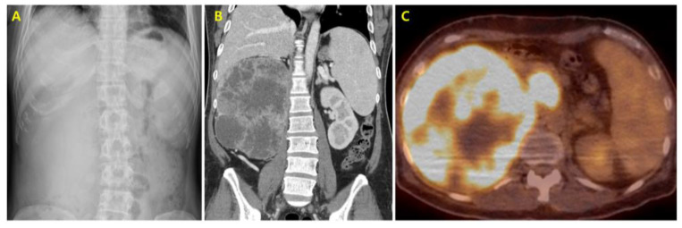
Renal cell carcinoma (RCC) is the most common type of kidney cancer, accounting for a significant proportion of all cancer cases in Korea. This case report presents a unique instance of spontaneous dramatic tumor regression in a 42-year-old Korean male diagnosed with clear cell RCC. The patient initially presented with right lower back pain, weight loss, and a loss of appetite. Following systemic immunotherapy with nivolumab and ipilimumab, and right radical nephrectomy, the patient was diagnosed with metastatic clear cell RCC, with new metastatic lesions detected in the liver, and on the chest wall on follow-up imaging. Second-line systemic treatment with pazopanib was initiated. Shortly thereafter, the patient developed severe systemic inflammatory syndrome, resulting in a mental stupor and acute kidney failure. Intensive care, including continuous renal replacement therapy and high-dose immunosuppressants, was administered. The patient's condition improved significantly with the intensive care regimen, leading to unintended tumor regression. These potentially fatal side effects occurred without infection, as confirmed by negative blood and urine cultures, and were attributed to the recent introduction of pazopanib. Follow-up imaging showed a significant reduction in hepatic metastatic lesions and the disappearance of chest wall nodules. This is the first reported case of RCC tumor regression following the side effects of pazopanib, underscoring the need for further studies into the immune mechanisms involved in RCC treatment and highlighting potential therapeutic strategies that leverage innate immune responses.
Keywords: inflammatory syndrome; pazopanib; renal cell carcinoma; spontaneous tumor regression.
Conflict of interest statement
The authors declare no conflicts of interest.
Figures


Similar articles
-
[RENAL CELL CARCINOMA METASTASIS TO BLADDER DURING MOLECULAR TARGETED THERAPY WITH PAZOPANIB: REPORT OF TWO CASES].Nihon Hinyokika Gakkai Zasshi. 2020;111(2):58-61. doi: 10.5980/jpnjurol.111.58. Nihon Hinyokika Gakkai Zasshi. 2020. PMID: 33883361 Japanese.
-
Disseminated intravascular coagulation induced by pazopanib following combination therapy of nivolumab plus ipilimumab in a patient with metastatic renal cell carcinoma.Anticancer Drugs. 2022 Jan 1;33(1):e818-e821. doi: 10.1097/CAD.0000000000001230. Anticancer Drugs. 2022. PMID: 34486537
-
An Italian, multicenter, real-world, retrospective study of first-line pazopanib in unselected metastatic renal-cell carcinoma patients: the 'Pamerit' study.Jpn J Clin Oncol. 2021 Mar 3;51(3):484-491. doi: 10.1093/jjco/hyaa193. Jpn J Clin Oncol. 2021. PMID: 33212499
-
Pazopanib for the treatment of metastatic renal cell carcinoma.Clin Ther. 2012 Mar;34(3):511-20. doi: 10.1016/j.clinthera.2012.01.014. Epub 2012 Feb 16. Clin Ther. 2012. PMID: 22341567 Review.
-
Pazopanib: the newest tyrosine kinase inhibitor for the treatment of advanced or metastatic renal cell carcinoma.Drugs. 2011 Mar 5;71(4):443-54. doi: 10.2165/11588960-000000000-00000. Drugs. 2011. PMID: 21395357 Review.
References
-
- Kang M.J., Jung K.-W., Bang S.H., Choi S.H., Park E.H., Yun E.H., Kim H.-J., Kong H.-J., Im J.-S., Seo H.G., et al. Cancer statistics in Korea: Incidence, mortality, survival, and prevalence in 2020. Cancer Res. Treat. Off. J. Korean Cancer Assoc. 2023;55:385–399. doi: 10.4143/crt.2023.447. - DOI - PMC - PubMed
-
- Raman G., Tarafdar S. Nephrology: A Comprehensive Guide to Renal Medicine. John Wiley & Sons; Hoboken, NJ, USA: 2020. Kidney Cancer; p. 67.
Publication types
MeSH terms
Substances
LinkOut - more resources
Full Text Sources
Medical
