

Type 2C Protein Phosphatase MoPtc6 Plays Critical Roles in the Development and Virulence of Magnaporthe oryzae
- PMID: 40422669
- PMCID: PMC12113234
- DOI: 10.3390/jof11050335
Type 2C Protein Phosphatase MoPtc6 Plays Critical Roles in the Development and Virulence of Magnaporthe oryzae
Abstract
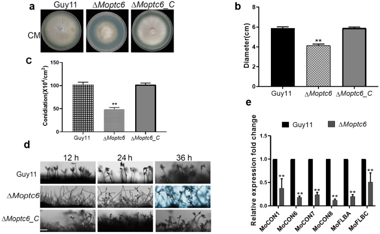
Rice blast caused by Magnaporthe oryzae pathotype is the worst disease that leads to serious food insecurity globally. Understanding rice blast disease pathogenesis is therefore essential for the development of a blast disease mitigation strategy. Reverse phosphorylation mediated by phosphatases performs a vital function in the activation of diverse biological mechanisms within eukaryotic. However, little has been reported on the roles of PP2Cs in the virulence of blast fungus. In this current work, we deployed functional genomics and biochemical approaches to characterize type 2C protein phosphatase MoPtc6 in blast fungus. Deletion of MoPTC6 led to a drastic reduction in conidiophore development, conidia production, hyphal growth, and stress tolerance. Western blotting assay demonstrated that the phosphorylation level of MoOsm1 was decreased while MoMps1 was increased in the MoPtc6 deletion mutant, and comparative transcriptome assay revealed a higher number of expressed genes between mutant and wild type. Localization assay confirmed that MoPtc6 is sub-localized in the cytoplasm of mycelia, spores, and in the appressoria of M. oryzae. Furthermore, disruption of MoPTC6 impaired appressoria turgor pressure and glycogen utilization; more findings revealed attenuation of hyphal penetration and virulence upon deletion of MoPTC6. Generally, present findings suggested the role of MoPtc6 in the growth and virulence of M. oryzae.
Keywords: Magnaporthe oryzae; phosphorylation; protein phosphatases; stress tolerance; virulence.
Conflict of interest statement
The authors declare no conflict of interest.
Figures


Similar articles
-
Protein Phosphatases MoPtc5, MoPtc1, and MoPtc2 Contribute to the Vegetative Growth, Stress Adaptation, and Virulence of Magnaporthe oryzae.J Fungi (Basel). 2025 Mar 18;11(3):231. doi: 10.3390/jof11030231. J Fungi (Basel). 2025. PMID: 40137268 Free PMC article.
-
The Calcium Chloride Responsive Type 2C Protein Phosphatases Play Synergistic Roles in Regulating MAPK Pathways in Magnaporthe oryzae.J Fungi (Basel). 2022 Dec 8;8(12):1287. doi: 10.3390/jof8121287. J Fungi (Basel). 2022. PMID: 36547620 Free PMC article.
-
Type 2C Protein Phosphatases MoPtc5 and MoPtc7 Are Crucial for Multiple Stress Tolerance, Conidiogenesis and Pathogenesis of Magnaporthe oryzae.J Fungi (Basel). 2022 Dec 20;9(1):1. doi: 10.3390/jof9010001. J Fungi (Basel). 2022. PMID: 36675822 Free PMC article.
-
The Devastating Rice Blast Airborne Pathogen Magnaporthe oryzae-A Review on Genes Studied with Mutant Analysis.Pathogens. 2023 Feb 26;12(3):379. doi: 10.3390/pathogens12030379. Pathogens. 2023. PMID: 36986301 Free PMC article. Review.
-
Effector-triggered susceptibility by the rice blast fungus Magnaporthe oryzae.New Phytol. 2024 Feb;241(3):1007-1020. doi: 10.1111/nph.19446. Epub 2023 Dec 10. New Phytol. 2024. PMID: 38073141 Review.
References
-
- Anjago W.M., Zhou T., Zhang H., Shi M., Yang T., Zheng H., Wang Z. Regulatory network of genes associated with stimuli sensing, signal transduction and physiological transformation of appressorium in Magnaporthe oryzae. Mycology. 2018;9:211–222. doi: 10.1080/21501203.2018.1492981. - DOI - PMC - PubMed
-
- Eseola A.B., Ryder L.S., Osés-Ruiz M., Findlay K., Yan X., Cruz-Mireles N., Molinari C., Garduño-Rosales M., Talbot N.J. Investigating the cell and developmental biology of plant infection by the rice blast fungus Magnaporthe oryzae. Fungal Genet. Biol. 2021;154:103562. doi: 10.1016/j.fgb.2021.103562. - DOI - PubMed
-
- Escalona-Montaño A.R., Zuñiga-Fabián M., Cabrera N., Mondragón-Flores R., Gómez-Sandoval J.N., Rojas-Bernabé A., González-Canto A., Gutierrez-Kobeh L., Perez-Montfort R., Becker I. Protein serine/threonine phosphatase type 2C of Leishmania mexicana. Front. Cell. Infect. Microbiol. 2021;11:641356. doi: 10.3389/fcimb.2021.641356. - DOI - PMC - PubMed
Grants and funding
LinkOut - more resources
Full Text Sources
Research Materials
