

AI-Powered Object Detection in Radiology: Current Models, Challenges, and Future Direction
- PMID: 40422999
- PMCID: PMC12112695
- DOI: 10.3390/jimaging11050141
AI-Powered Object Detection in Radiology: Current Models, Challenges, and Future Direction
Abstract
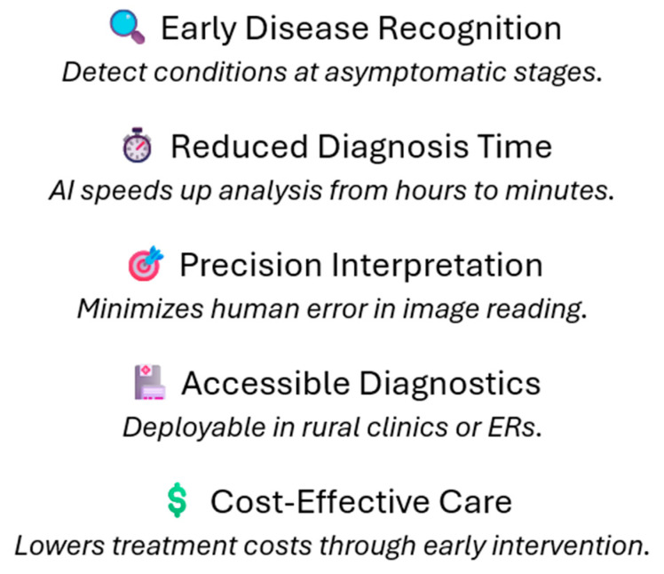
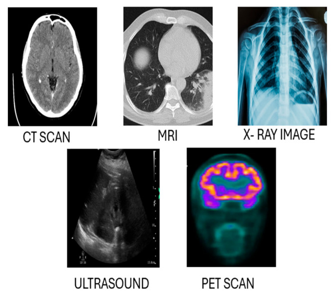
Artificial intelligence (AI)-based object detection in radiology can assist in clinical diagnosis and treatment planning. This article examines the AI-based object detection models currently used in many imaging modalities, including X-ray Magnetic Resonance Imaging (MRI), Computed Tomography (CT), and Ultrasound (US). The key models from the convolutional neural network (CNN) as well as the contemporary transformer and hybrid models are analyzed based on their ability to detect pathological features, such as tumors, lesions, and tissue abnormalities. In addition, this review offers a closer look at the strengths and weaknesses of these models in terms of accuracy, robustness, and speed in real clinical settings. The common issues related to these models, including limited data, annotation quality, and interpretability of AI decisions, are discussed in detail. Moreover, the need for strong applicable models across different populations and imaging modalities are addressed. The importance of privacy and ethics in general data use as well as safety and regulations for healthcare data are emphasized. The future potential of these models lies in their accessibility in low resource settings, usability in shared learning spaces while maintaining privacy, and improvement in diagnostic accuracy through multimodal learning. This review also highlights the importance of interdisciplinary collaboration among artificial intelligence researchers, radiologists, and policymakers. Such cooperation is essential to address current challenges and to fully realize the potential of AI-based object detection in radiology.
Keywords: artificial intelligence; convolutional neural network; diagnostic accuracy; object detection; radiology.
Conflict of interest statement
The authors declare no conflicts of interest.
Figures


References
-
- Elhanashi A., Lowe D., Saponara S., Moshfeghi Y. Proceedings of the Real-Time Image Processing and Deep Learning, Orlando, FL, USA, 3 April–13 June 2022. Proceedings Volume 12102. SPIE; Bellingham, WA, USA: 2022. Deep learning techniques to identify and classify COVID-19 abnormalities on chest X-ray images; p. 1210204. - DOI
Publication types
LinkOut - more resources
Full Text Sources
