

Primary Pleural Lymphoma in an Immune-Competent Patient: A Diagnostic and Therapeutic Challenge
- PMID: 40423034
- PMCID: PMC12113573
- DOI: 10.3390/jpm15050162
Primary Pleural Lymphoma in an Immune-Competent Patient: A Diagnostic and Therapeutic Challenge
Abstract
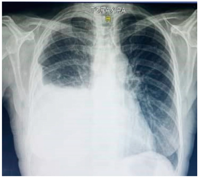
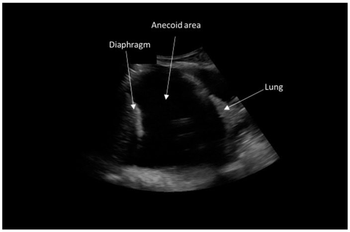
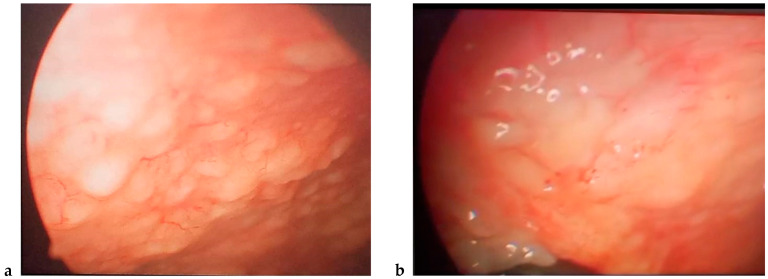
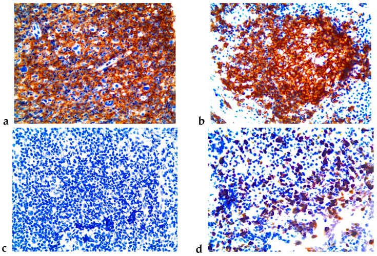
Background: Primary pleural lymphoma is a rare disease posing diagnostic and therapeutic challenges. Case presentation: We present a 65-year-old woman with dyspnoea, cough, and asthenia, with no significant past medical history. Chest X-ray and computed tomography showed extensive right pleural effusion. Video-assisted thoracoscopy demonstrated multiple pleural nodules, while pleural fluid analysis revealed a lymphocytic exudate, and finally, a primary pleural lymphoma diagnosis was confirmed by immunohistochemistry analysis in pleural nodules biopsy. Discussion: In this regard, eight cycles of chemotherapy with cyclophosphamide, doxorubicin, vincristine, dexamethasone, and rituximab were indicated, and after one year of follow-up, complete clinical and radiological remission was observed. Conlusions: We conclude that video-assisted thoracoscopy with an appropriate histopathological examination remains the gold standard for diagnosis, while R-CHOP chemotherapy plus rituximab may represent a highly effective therapeutic choice.
Keywords: R-CHOP chemotherapy; immunohistochemistry; pleura; primary pleural lymphoma; video-assisted thoracoscopy.
Conflict of interest statement
The authors declare no conflicts of interest.
Figures

Similar articles
-
[Primary pleural diffuse large B cell lymphoma:a case report and review of literature].Zhonghua Jie He He Hu Xi Za Zhi. 2014 Nov;37(11):835-9. Zhonghua Jie He He Hu Xi Za Zhi. 2014. PMID: 25604114 Review. Chinese.
-
[A case of malignant lymphoma diagnosed by thoracoscopy with local anesthesia].Nihon Kokyuki Gakkai Zasshi. 2005 Oct;43(10):622-5. Nihon Kokyuki Gakkai Zasshi. 2005. PMID: 16285597 Japanese.
-
Rituximab: a review of its use in non-Hodgkin's lymphoma and chronic lymphocytic leukaemia.Drugs. 2003;63(8):803-43. doi: 10.2165/00003495-200363080-00005. Drugs. 2003. PMID: 12662126 Review.
-
Rituximab-dose-dense chemotherapy with or without high-dose chemotherapy plus autologous stem-cell transplantation in high-risk diffuse large B-cell lymphoma (DLCL04): final results of a multicentre, open-label, randomised, controlled, phase 3 study.Lancet Oncol. 2017 Aug;18(8):1076-1088. doi: 10.1016/S1470-2045(17)30444-8. Epub 2017 Jun 28. Lancet Oncol. 2017. PMID: 28668386 Clinical Trial.
-
A case report on the effect of rituximab on pyothorax-associated lymphoma.Medicine (Baltimore). 2019 Dec;98(50):e18393. doi: 10.1097/MD.0000000000018393. Medicine (Baltimore). 2019. PMID: 31852157 Free PMC article.
References
-
- Pereira L.J., Mohrbacher S., Neves P.D.M.d.M., Zacchi F.F.S., Medeiros I.U.D., Sato V.A.H., Oliveira É.S., Pereira L.V.B., Cuvello-Neto A.L., Baiocchi O., et al. Primary Effusion Lymphoma: A Rare and Challenging Diagnosis for Recurrent Pleural Effusion. Diagnostics. 2023;13:370. doi: 10.3390/diagnostics13030370. - DOI - PMC - PubMed
-
- Farissi Y., Kouevidjin-Eppou G., Mazieres J., Abakarim O. Primary pleural lymphoma: A case report. PAMJ Clin. Med. 2024;14:45. doi: 10.11604/pamj-cm.2024.14.45.43145. - DOI
-
- Jiménez L.P., Martín A.R., Graves M.G., Mesa A.M., de Neumología S., de la Victoria H.U.V. Linfoma pleural primario asociado a piotórax. Rev. Esp. Patol. Torác. 2018;30:196–199.
Publication types
LinkOut - more resources
Full Text Sources
Research Materials
Miscellaneous
