. 2025 Jun 3;122(22):e2420704122.
doi: 10.1073/pnas.2420704122. Epub 2025 May 27.

Stimulus representations in visual cortex shaped by spatial attention and microsaccades

Affiliations

Stimulus representations in visual cortex shaped by spatial attention and microsaccades

Karthik Srinivasan et al. Proc Natl Acad Sci U S A. .

Abstract

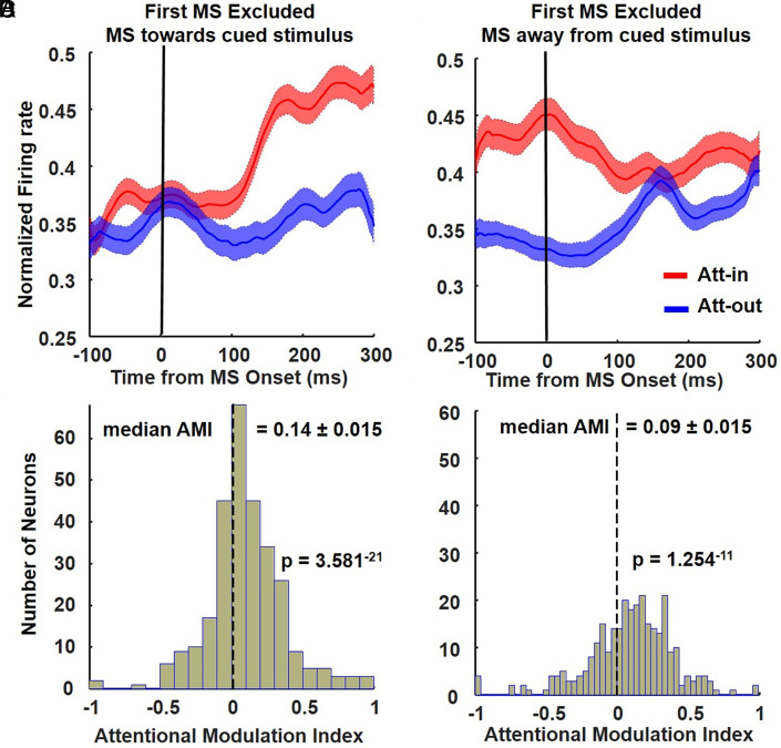
Microsaccades (MSs) are commonly associated with covert spatial attention, yet their impact on cortical processing of visual objects remains unclear. Rhesus macaques, randomly cued to attend to a target object amid distracters, were rewarded for detecting a color change in the target. While spatial attention does not affect the object tuning curves of V4 cells, the direction of MS significantly influenced object representations in V4 throughout the entire trial. Specifically, intervals following an MS toward the target exhibited superior stimulus decoding and sharper tuning curves compared to intervals following an MS away from the target. Furthermore, MSs directed toward the target enhanced neuronal responses to behaviorally relevant color changes, leading to faster reaction times. This sharpening effect stems from both a refreshing of the initial sensory response and an amplification of attention effects. The firing rate enhancement associated with spatial attention is delayed until the occurrence of the first MS directed toward the target. Subsequently, a positive effect of attention on firing rate, influenced by MS direction, was found throughout the trial across deep and superficial layers of V4, lateral pulvinar, and IT cortex. In summary, these findings underscore a crucial link between covert attention, object processing, and their coordination with MSs.

Keywords: attention; microsaccades; v4; vision.


Conflict of interest statement

Competing interests statement:The authors declare no competing interest.

Figures

Fig. 1.
Fig. 1.
The role of the first MS and subsequent MSs during the attention period in area V4. (A and B) Normalized population firing rates (n = 293) combined across monkeys and sessions (k = 34) locked to the onset of the first MS toward (A) or away from the target location in the RF (B) for both Att-in and Att-out conditions, respectively. Enhancement of firing rates with attention only occurred following the first MS toward but not away from the cued stimulus in the RF. (C and D). Histogram distribution of the Attentional Modulation Index (AMI) for the 300 ms following the first MS directed toward (C) or away (D) from the cued stimulus.
Fig. 2.
Fig. 2.
The effects of MS direction on the enhancement of V4 responses with attention depend on MS order. (A) If the first MS is directed toward the target stimulus in the RF, responses are enhanced by attention on the first and second MS. Top shows normalized firing rates across the population, and Bottom shows distribution of AMI values. (B) If the first MS is directed away from the target stimulus in RF, responses are not enhanced by attention until the next MS toward the target. Top shows normalized firing rates across the population, and Bottom shows distribution of AMI values.
Fig. 3.
Fig. 3.
(A and B) normalized population firing rates in V4 (n = 293) are locked to all the subsequent MSs excluding the first MS. (C and D) Histograms of AMI values for conditions A and B, respectively.
Fig. 4.
Fig. 4.
Mean AMI values of the V4 population in all layers combined versus superficial and deep layers separately. (A) First MS only. For neurons in all layers combined, as well as in superficial layers (supragranular) and deep layers (infragranular) there is a significant AMI for the MS toward intervals. There is a greater AMI in the superficial layers than in the deep layers. (B) Same as in B but with the first MS excluded. All AMI comparisons for statistical purposes are with respect to zero (no attention modulation of the population); * (black) of P < 0.05, and ** (blue) of P < 0.01 indicate AMIs significantly different from zero.
Fig. 5.
Fig. 5.
Object decoding of V4 neurons during the full stimulus time period and MS-triggered intervals. (A) Linear decoder performance for object categorization by V4 neurons (n = 132) from the cue first/stimulus second sessions including the transient stimulus onset period in the 1,000 ms following stimulus onset. (B) Linear decoder performance for object categorization by V4 neurons (n = 150) for 1,000 ms during the stimulus first/cue second condition, which does not include a stimulus transient. (C) Linear decoder performance for the 300 ms time periods for both Att-in and Att-out immediately after an MS is directed toward the cued stimulus (n = 282). Decoder performance was significant in intervals following MSs directed toward the target in the RFs of V4 (All the MS poststimulus and spatial cue presented were included). (D) Same as in (C), but when the MS is directed away from the cued stimulus. (AD) P-values are for the comparison of the attend-in conditions to chance.
Fig. 6.
Fig. 6.
Comparison of classification accuracy of the decoder across conditions.
Fig. 7.
Fig. 7.
Object selectivity of V4 neurons across the population for full stimulus vs MS-triggered intervals (All MS were included). (A) Mean normalized population firing rates of V4 neurons (n = 282) to the Att-in condition ranked from the most preferred to the least preferred objects, demonstrating sharper object tuning profiles for the neurons for both full stimulus transient and MS-triggered intervals, compared to full stimulus sustained (pink). (B) Same as in (A) but for the Att-out condition. See SI Appendix, Fig. S10 DE for the same analysis but excluding the first MS, SI Appendix, Fig. S11, for example, of differential object selectivity in exemplar V4 neurons depending on MS type, and SI Appendix, Fig. S15 for alternative OSI analyses.

Update of

References

    1. Posner M. I., Orienting of attention. Q. J. Exp. Psychol. 32, 3–25 (1980), 10.1080/00335558008248231. - DOI - PubMed
    1. Desimone R., Duncan J., Neural mechanisms of selective visual attention. Annu. Rev. Neurosci. 18, 193–222 (1995), 10.1146/annurev.ne.18.030195.001205. - DOI - PubMed
    1. Maunsell J. H. R., Neuronal mechanisms of visual attention. Annu. Rev. Vis. Sci. 1, 373–391 (2015), 10.1146/annurev-vision-082114-035431. - DOI - PMC - PubMed
    1. Moore T., Zirnsak M., Neural mechanisms of selective visual attention. Annu. Rev. Psychol. 68, 47–72 (2017), 10.1146/annurev-psych-122414-033400. - DOI - PubMed
    1. Rn Denison, Yuval-Greenberg S., Carrasco M., Directing voluntary temporal attention increases fixational stability. J. Neurosci. 39, 353–363 (2019), 10.1523/JNEUROSCI.1926-18.2018. - DOI - PMC - PubMed

LinkOut - more resources