. 2025 Jul;30(4):100085.
doi: 10.1016/j.cstres.2025.100085. Epub 2025 May 25.

Dynamics of heat shock protein 70 kDa in heat-shocked and hypoxic human endothelial cells

Affiliations

Dynamics of heat shock protein 70 kDa in heat-shocked and hypoxic human endothelial cells

Luiza B C T Coimbra et al. Cell Stress Chaperones. 2025 Jul.

Abstract

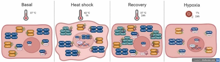
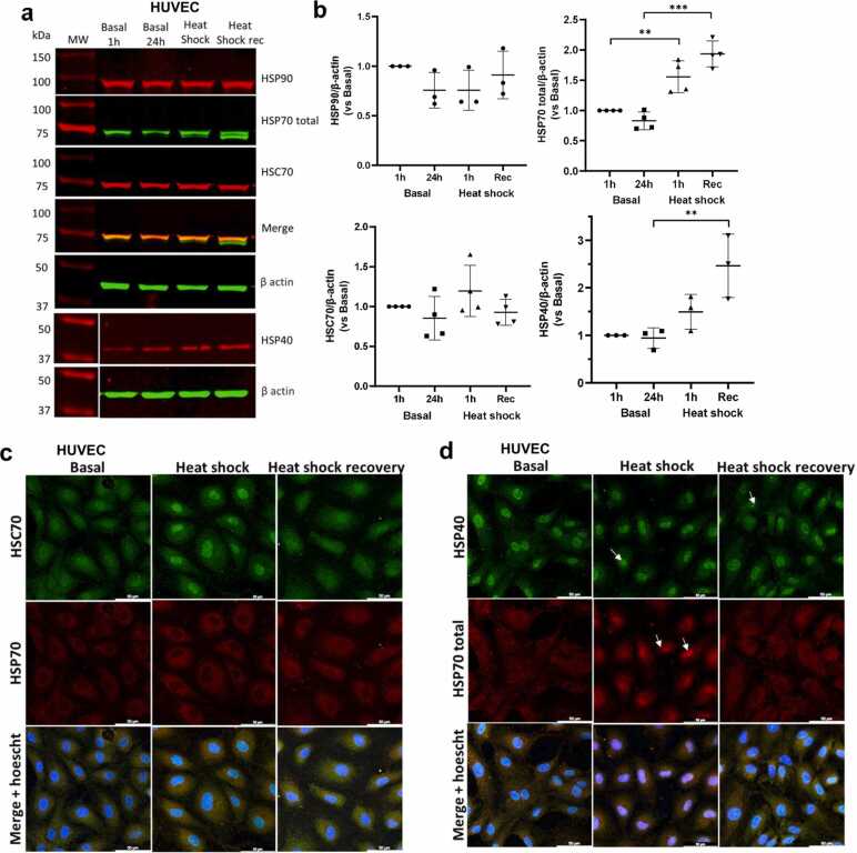
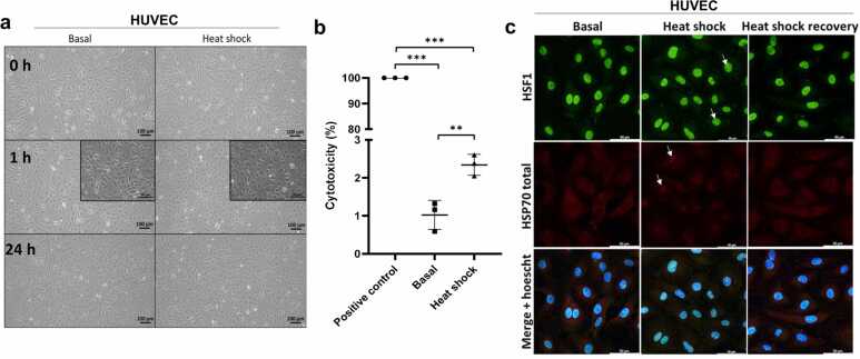
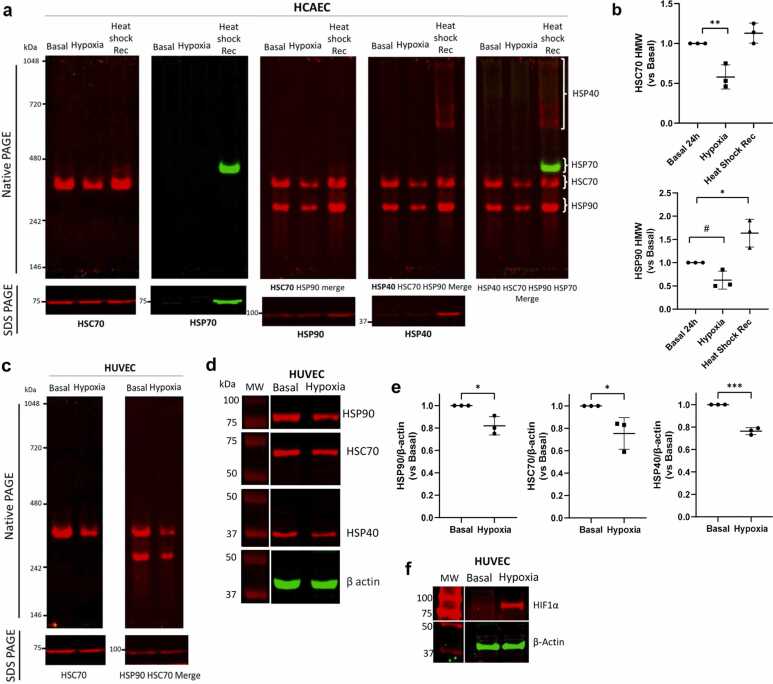
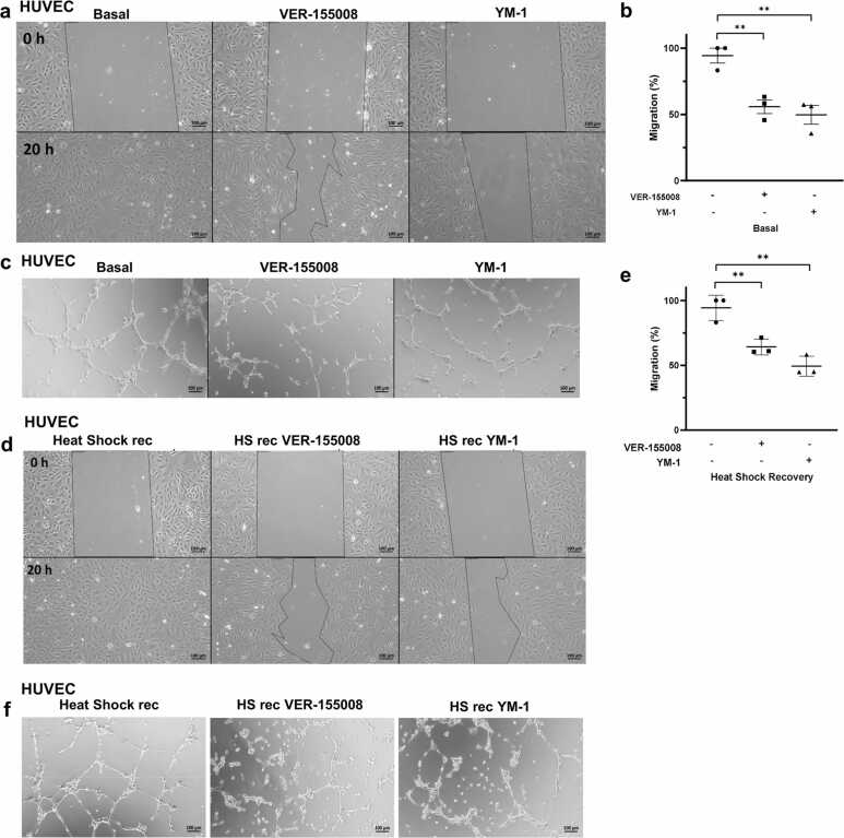
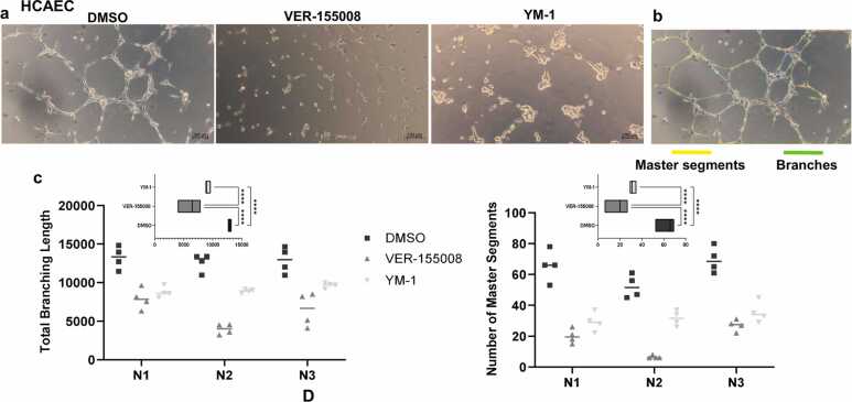
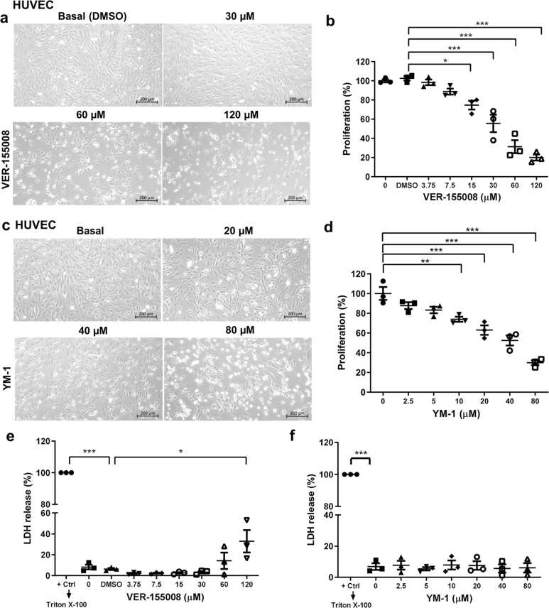
Heat shock proteins (HSPs) play crucial roles in human endothelial cell functions such as migration and angiogenesis. However, human HSP dynamics under stress conditions such as heat shock (HS) and hypoxia in human endothelial cells (ECs) are enigmatic, and the characteristics of HSPs in ECs after exposure to thermal stress and a low-oxygen environment are unknown. We hypothesized that ECs adapt to HS and hypoxia by modulating chaperome oligomerization and that HSP70 is a major determinant of the endothelial phenotype. HSP70 inhibition with VER-155008 or YM-1 in primary human endothelial cells decreases EC proliferation, migration, and angiogenesis at baseline and after heat shock recovery. We showed that vascular-independent HSC/P70 multimeric complexes in primary human veins (HUVEC) and coronary artery ECs (HCAEC) accumulate after HS and are decreased by hypoxia. HS recovery increases the number of HSP90 dimers, inducible HSP70, and HSP40 macromolecular complexes, whereas HSC70 returns to baseline. We demonstrated that the HS response and hypoxia regulate HSPs through a new layer of complexity, oligomerization, in addition to classical cochaperone/NEF interactions. The biphasic temporal oligomerization of molecular chaperones in the recovery phase provides a novel face of the heat shock response. In addition, shifts in the subcellular location and upregulation of HSP70 were also observed here. The decrease in HSP expression caused by hypoxia raises the possibility that decreased chaperone power contributes to the endothelial dysfunction found in atherosclerosis, thrombosis, and cancer. Together, these results show that HSP70 is pivotal to the healthy endothelial response in veins and coronary arteries, and we revealed human HSP dynamics in the vascular response to proteotoxic stress.

Keywords: Endothelial cell; HSP70; Heat-shock response; Hypoxia; Oligomerization.


Conflict of interest statement

Declarations of interest The authors declare that they have no known competing financial interests or personal relationships that could have appeared to influence the work reported in this paper.

Figures

ga1
Graphical abstract
Fig. 1
Fig. 1
Screening of the heat shock (HS) response in primary human endothelial cells. (a) Whole HUVEC lysates after HS for 1 h and recovery for 24 h or basal conditions were subjected to Western blot analysis for HSP40 (CST, 4868S), total HSP70 (Invitrogen, MA3-006), HSC70 (Invitrogen, PA5-27337), and HSP90 (CST, 4877S) expression. The data are representative of n = 3–4 independent experiments with cells from different lots (19TL127368, 19TL028325, 19TL023264, and 21TL195720). (b) Data normalized to the loading control β-actin are presented as the means ± SEMs; one-way ANOVA, with Tukey’s post hoc test; **P < 0.01, ***P < 0.001 versus the respective control; HSP40 and HSP90, n = 3; HSP70 total and HSC70, n = 4. (c) Immunofluorescence staining of HSC70 (green, Invitrogen PA5-27337) and HSP70 (red, Invitrogen MA3-009) and (d) HSP40 (green, CST 4356S) and total HSP70 (red, Invitrogen MA3-006) in HUVECs exposed to HS and recovery or basal conditions. Nuclei were stained with Hoechst (blue). Representative images of n = 3 independent experiments using cells from lots 19TL028324 and 19TL023264 (Lonza). The arrows indicate the nuclear focus of the proteins in ECs after HS. Scale bar: 50 µm. Abbreviation used: HUVEC, human vein.
Fig. 2
Fig. 2
Morphological characterization of thermal stress in human endothelial cells. (a) Primary HUVECs were exposed to heat shock (HS) at 42 °C for 1 h and allowed to recover at 37 °C for 24 h. Images were acquired using optical microscopy (ZEISS Primovert) before HS (0 h), immediately after HS (1 h), and after recovery (24 h). Scale bar: 100 µm. Inset scale bar: 50 µm. Representative images of n = 8 independent experiments using cells from different lots (19TL028325, 19TL023264, 19TL127368, and 21TL195720, Lonza). (b) Supernatants from these cells were collected, and HS cytotoxicity was measured by quantifying lactate dehydrogenase (LDH) activity. The data are shown as the means ± SEMs of 3 independent experiments (lot 19TL127368 and 19TL028325), ordinary one-way ANOVA, with Tukey’s multiple comparisons test, **P < 0.01, ***P < 0.001 versus positive control. HUVECs were treated with Triton X-100 to achieve maximum LDH release from the supernatant. (c) HSF1 (green, CST 4356S) and total HSP70 (red, Invitrogen MA3-006). Abbreviation used: HUVEC, human vein.
Fig. 3
Fig. 3
Temporal modulation of human HSP high-molecular-weight complexes by heat shock in primary ECs. (a) Invitrogen’s NativeMark™ was subjected to Native PAGE, stained with Coomassie blue, and used as a standard to identify high-molecular-weight complexes in native PAGE. (b) Whole-cell lysates from HUVECs exposed to heat shock, recovery, or basal conditions were subjected to native PAGE and sequentially stained for HSP40 (CST, 4868S), HSC70 (Invitrogen PA5-27337), HSP70 (Invitrogen MA3-009), and HSP90 (CST, 4877S). Below, sodium dodecyl sulfate-PAGE for each protein is displayed. The data are representative of n = 3–6 independent experiments with cells from different lots (19TL127368, 19TL028325, 19TL023264, and 21TL195720). (c) Merged image of all the stained samples shown in (b). The data are representative of n = 3–6 independent experiments in cells from different lots (19TL127368, 19TL028325, 19TL023264, and 21TL195720). (d) Quantification of the high-molecular-weight complex intensities from specific proteins from (b) versus Basal n. The data are presented as one-way ANOVA, with Tukey’s post hoc test; **P < 0.01, ***P < 0.001 versus the respective control; HSP40, n = 3; HSP70, n = 4; HSC70, n = 5; and HSP90, n = 6. (E) Schematic representation of the native PAGE procedure. Proteins in their native state from cell lysates were separated on a 6% polyacrylamide gel and transferred to a nitrocellulose membrane overnight. The membrane was blocked with nonfat milk, incubated with primary antibody overnight, and then incubated with secondary antibody. The membrane was scanned using the LI-COR Odyssey® 2-channel near-infrared fluorescence imaging system, and the fluorescence levels of the last HSP probed were quantified. The same membrane was then incubated overnight with a primary antibody against another chaperone, followed by secondary antibody incubation, scanning with the near-infrared fluorescence imager, and quantification. This process was repeated daily until all chaperones were stained on the same membrane, in this order: day 1 anti-HSP40; day 2 anti-HSC70; day 3 anti-HSP90; day 4 anti-HSP70. Abbreviations used: EC, endothelial cell; HSP, heat-shock protein; HUVEC, human vein.
Fig. 4
Fig. 4
Hypoxia induces decreases in multimeric HSPs in human coronary arteries (HCAECs) and vein endothelial cells (HUVECs). (a) Whole-cell lysates from HCAECs exposed to hypoxia, heat-shock recovery, or basal conditions were subjected to native PAGE and sequentially stained for HSC70 (Invitrogen PA5-27337), HSP70 (Invitrogen MA3-009), HSP90 (CST 4877S), and HSP40 (CST 4868S), as shown in the top panels. Below, sodium dodecyl sulfate-PAGE results for each protein are displayed. Data are representative of n = 3 independent experiments using two different lots of cells (20TL365545 and 20TL064651, Lonza). Both native PAGE and Western blot experiments were performed using two different lots of cells (20TL365545 and 20TL064651). (b) Quantification of HSC70, HSP70, and HSP90 high-molecular-weight complexes in (a). Data are presented as the means ± SEMs, one-way ANOVA, with Tukey’s post hoc n = 3, *P < 0.05, **P < 0.01 versus basal conditions. Unpaired t test (#P < 0.05) was used to compare only the HSP90 hypoxic and basal groups. (c) Whole-cell lysates from HUVECs exposed to hypoxia or basal conditions were subjected to native PAGE and sequentially stained for HSC70 (Invitrogen PA5-27337) and HSP90 (CST 4877S), as shown in the top panels. Below, sodium dodecyl sulfate-PAGE for each protein is displayed. The data are representative of n = 2 independent experiments using cells from lot 19TL028324. (d) Whole-cell lysates of HUVECs exposed to hypoxia or basal conditions were subjected to Western blot analysis for HSP40 (CST 4868S), total HSP70 (Invitrogen MA3-006), HSC70 (Invitrogen PA5-27337), and HSP90 (CST 4877S). The data are representative of n = 3 independent experiments using cells from lots 19TL028324 and 23TL086130. (e) Data from (d) are presented as the mean ± SEM, unpaired Student’s t test, n = 3 with *P < 0.05 and ***P < 0.001. β-actin, loading control. (f) HUVECs subjected to basal or hypoxic 1% O2 (94% N2, 5% CO2) for 24 h were subjected to Western blot analysis for HIF1α (CST, #48085) with β-actin (Sigma, A5441) loading control. The assay was performed three times using two different lots of cells (21TL195720 and 23TL086130).
Fig. 5
Fig. 5
HSP70 plays a central role in the primary human endothelial cell (EC) phenotype. (a) Migration was evaluated in a wound healing assay in HUVECs cultured for 1 h in EBM-2 under basal conditions or in the presence of 30 µM VER-155008 or 20 µM YM-1. After scratching, the medium was changed to EGM-2 with the respective inhibitors, and the cells were allowed to migrate for 20 h. Representative images of n = 3 independent experiments using two different lots of cells (21TL195720 and 19TL028324) are shown. Scale bar: 100 µm. (b) The area of each scratch at 0 and 20 h was measured, and the percentage of migration was calculated as follows: migration = (original wound area − area after healing)/original wound area × 100. The data from (a) are presented as the means ± SEMs; ordinary one-way ANOVA with Tukey’s multiple comparisons test, **P < 0.01 versus the control; n = 3. (c) HUVECs were cultured under basal conditions and then plated in Matrigel in the presence of the inhibitors VER-155008 (50 µM) or YM-1 (40 µM). After 5 h of incubation, images were acquired. Representative images of n = 3 independent experiments using cells from lot 21TL195720. Scale bar: 100 µm. (d) HUVEC migration after heat-shock recovery, followed by 30 µM VER-155008 or 20 µM YM-1 treatment, was evaluated in a wound healing assay. Representative images of three independent experiments using two different cell lots (21TL195720 and 19TL028324) are shown. Scale bar: 100 µm. (e) The area of each scratch at 0 and 20 h was measured from (a). Data are presented as the mean ± SEM; one-way ANOVA with Tukey’s post hoc test, n = 3; **P < 0.01 versus conditions. (f) HUVECs exposed to heat shock for 1 h and allowed to recover for 24 h, as in (d), were treated with 50 µM VER-155008 or 40 µM YM-1 and subjected to 5 h of tube formation. Representative images of three independent experiments using cells from lot 21TL195720. Scale bar: 100 µm. Abbreviation used: HUVEC, human vein.
Fig. 6
Fig. 6
HSP70 loss of function impairs coronary artery angiogenesis in vitro (a) HCAECs were cultured under basal conditions and then plated in Matrigel in the presence of the inhibitors VER-155008 (50 µM) or YM-1 (40 µM). After 5 h of incubation, images were acquired. Representative images of triplicate independent experiments are shown. (b) Representative image of angiogenesis analysis with a map of selected parameters. (c) Quantification of the total branching length (left) and number of master segments (right). Each point represents repeated measurements taken in three independent experiments (N) for each treatment (symbol). The insets show the results of two-way ANOVA with Tukey’s post hoc test; ****P < 0.0001 indicates significant differences among means. Abbreviations used: HCAEC, human coronary artery endothelial cell; HUVEC, human vein.
Fig. 7
Fig. 7
Characterization of VER-155008 and YM-1 in primary human endothelial cells. (a) Phase-contrast images (ZEISS Primovert) of primary HUVECs treated with 30, 60, or 120 µM VER-155008 for 24 h and (c) 2.5, 5, 10, 20, 40, or 80 µM YM-1. The volume of DMSO used corresponded to the highest concentration of VER-155008 (120 µM). Scale bar: 200 µm. (b) Cell proliferation was measured by counting the number of cells after treatment with 3.75, 7.5, 15, 30, 60, or 120 µM VER-155008 for 24 h and (d) 2.5, 5, 10, 20, 40, or 80 µM YM-1. (e) and (f) A cytotoxicity assay was performed by quantifying the activity of lactate dehydrogenase in the supernatants. The data are presented as the means ± SEMs. Statistical significance was determined by ordinary one-way ANOVA with Tukey’s multiple comparisons test; *P < 0.05, **P < 0.01, ***P < 0.001 versus the respective control. All experiments were independently performed three times using two different lots of cells (21TL195720 and 19TL028324). Abbreviation used: HUVEC, human vein.

Similar articles

References

    1. Balchin D., Hayer-Hartl M., Hartl F.U. In vivo aspects of protein folding and quality control. Science. 2016;353 doi: 10.1126/science.aac4354. - DOI - PubMed
    1. Araujo T.L.S., Venturini G., Moretti A.I.S., et al. Cell-surface HSP70 associates with thrombomodulin in endothelial cells. Cell Stress Chaperones. 2019;24:273–282. doi: 10.1007/s12192-018-00964-y. - DOI - PMC - PubMed
    1. Benjamin I.J., McMillan D.R. Stress (heat shock) proteins: molecular chaperones in cardiovascular biology and disease. Circ Res. 1998;83:117–132. doi: 10.1161/01.RES.83.2.117. - DOI - PubMed
    1. Finka A., Goloubinoff P. Proteomic data from human cell cultures refine mechanisms of chaperone-mediated protein homeostasis. Cell Stress Chaperones. 2013;18:591–605. doi: 10.1007/s12192-013-0413-3. - DOI - PMC - PubMed
    1. García-Cardeña G., Fan R., Shah V., et al. Dynamic activation of endothelial nitric oxide synthase by Hsp90. Nature. 1998;392:821–824. doi: 10.1038/33934. - DOI - PubMed

MeSH terms

LinkOut - more resources