. 2025 May 27;16(1):4899.
doi: 10.1038/s41467-025-59713-2.

Multiplex base editing to protect from CD33 directed drugs for immune and gene therapy

Affiliations

Multiplex base editing to protect from CD33 directed drugs for immune and gene therapy

Florence Borot et al. Nat Commun. .

Abstract

The selection of genetically engineered immune or hematopoietic cells in vivo after gene editing remains a clinical problem and requires a method to spare on-target toxicity to normal cells. Here, we develop a base editing approach exploiting a naturally occurring CD33 single nucleotide polymorphism leading to removal of full-length CD33 surface expression on edited cells. CD33 editing in human and nonhuman primate hematopoietic stem and progenitor cells protects myeloid progeny from CD33-targeted therapeutics without affecting normal hematopoiesis in vivo, thus demonstrating potential for improved immunotherapies with reduced off-leukemia toxicity. For broader application to gene therapies, we demonstrate highly efficient (>70%) multiplexed adenine base editing of the CD33 and gamma globin genes, resulting in long-term persistence of dual gene-edited cells with HbF reactivation in nonhuman primates. Using the CD33 antibody-drug conjugate Gemtuzumab Ozogamicin, we show resistance of engrafted, multiplex edited human cells in vivo, and a 2-fold enrichment for edited cells in vitro. Together, our results highlight the potential of adenine base editors for improved immune and gene therapies.


Conflict of interest statement

Competing interests: This study was funded by a grant from Vor Biopharma and 1R21CA256461 at Columbia University and by grants from NIH/NHLBI (R01 HL136135_2) and NIH/NCI (R01 CA266556) at Fred Hutchinson Cancer Center. Columbia University has licensed technology that is the subject of this study to Vor Biopharma. F.B., A.M.A., and S.M. are coinventors on issued and pending patent applications licensed to Vor Biopharma. S.M. has equity ownership and is on the Scientific Advisory Board of Vor Biopharma. R.B.W. received laboratory research grants and/or clinical trial support from Aptevo, Celgene/Bristol Myers Squibb, ImmunoGen/AbbVie, Janssen, Jazz, Kite, Kura, Pfizer, and Vor Biopharma, and has been a consultant to Wugen. HPK is or was a consultant to and has or had ownership interests with Rocket Pharmaceuticals, Homology Medicines, Vor Biopharma and Ensoma Inc. H-P.K. has also been a consultant to CSL Behring and Magenta Therapeutics. D.R.L. is a consultant for Prime Medicine, Beam Therapeutics, Pairwise Plants, and Chroma Medicine, and Nvelop Therapeutics, companies that use or deliver genome editing or genome engineering agents and owns equity in these companies. The remaining authors declare no competing interests.

Figures

Fig. 1
Fig. 1. ABE8e introduces A > G conversion at targeted nucleotide with up to 95% efficiency and negligible indels.
a Top Possible splicing outcomes at CD33 exon 1–3, ag: Splicing acceptor site (SA). gt: Splicing donor site. (Ab icon was created in BioRender. Du, X. (2025) https://BioRender.com/y35d204). Bottom Intron 1 (lower case)/exon2 (upper case) junction DNA sequence with highlight of exon 2 SA (red) and Exon Splicing Enhancer site (ESE in yellow). b CD33Δ2 lacks exon 2 due to the polymorphism rs12459419 that results in an altered ESE site. c Sequences of the protospacers designed to either edit the ESE with BE4max (sgCB1 and CB2) or the SA with ABE8e (sgABE). Protospacer adjacent motif (PAM) is in red. d Sanger sequencing profiles of edited CD34+ cells compared to the wild-type sequence (top) 7 days post electroporation. Edited nucleotides are indicated by arrows. e Editing efficiency (HTS analysis) at the targeted nucleotides and bystanders, as well as indels. f FACS analysis of the edited CD34+ cells 7 days post electroporation with antibody clone P67.6 which recognizes an epitope located in exon 2. g PCR on cDNA with sets of primers, specific to CD33Δ2 (spanning exon junction 1–3), or common to all isoforms (in exons 1, 5 and 7), (L: Ladder in bp). Sanger sequencing of PCR products confirm the absence of exon 2 in edited cells while all other exons are intact. 1 independent experiment. h ML-1 cells were mock electroporated (ML-1mock), or electroporated with ABE8e and CD33 monoclonal antibody (mAb) staining compared to parental ML-1 cells or ML-1 cells in which both alleles of CD33 had been disrupted via CRISPR targeting of exon 1 (ML-1CD33KO). P67 mAb (i), 9G2 (ii) binds to the C2-set domain of CD33, whether V-set is present or not, and 11D5 (iii and iv) binds to the C2-set domain of CD33 when the V-set is absent, e.g., CD33 lacking exon 2. Specificity of 11D5 to CD33Δ2 is shown (iv.) using mouse 3T3 cells that lack human CD33 expression are shown (m3T3) with forced expression of either CD33Δ2 (m3T3+CD33Δ2) or full length CD33 (m3T3+CD33FL). Source data are provided as a Source Data file.
Fig. 2
Fig. 2. ABE8e edited HSPCs are resistant to CD33-targeted therapy in vitro and in vitro differentiated CD33Δ2-edited myeloid cells display intact phagocytic capacity.
a In vitro differentiated unedited (UE) or CD33Δ2-edited monocytes show comparable phagocytosis capacity, as measured by E.coli bioparticles internalization. Left, Representative FACs plots of E. coli bioparticles internalization. Treatment with actin polymerization inhibitor, cytochalasin D, abrogates phagocytosis. Right, Graph of phagocytosis quantification. Unpaired two-tailed t-test. b CD33Δ2-edited CD34+ cells resist Gemtuzumab Ozogamicin (GO) cytotoxicity in vitro. Cells were incubated 48 h with GO and cytotoxicity analyzed by FACS using Sytox Blue or 7AAD as a viability dye. CD33 Δ2-edited CD34+ show same resistance to GO cytotoxicity than a donor with homozygous rs12459419 A14V SNP (TT genotype). Error bars show ±SEM. (2 independent experiments, 2 donors, run in triplicates) c, ML1 CD33 WT or KO cells and Unedited, or ABE8e-edited mPB CD34+ cells were assessed for resistance to the CD33/CD3 bispecific T-cell engager (BiAb, generated from published sequences and described in Correnti et al.). Target cells were incubated with healthy donor T cells for 2 days and absolute cell number and viability were detected by flow cytometry analysis following staining with 4’,6-diamidino-2-phenylindole (DAPI). Results were normalized to reactions that were not treated with the drug (1 experiment run in triplicates, 1 donor). Source data are provided as a Source Data file.
Fig. 3
Fig. 3. CD33Δ2-edited CD34+ engraft, recapitulate a complete hematopoietic system and display resistance to GO CD33-targeted therapy in vivo.
a Schematic of the experiment was created in BioRender. Du, X. (2025) https://BioRender.com/y35d204. b, c Measure of engraftment by percentage of human CD45+ cells and of hematopoietic repopulation by frequency of progenitors myeloid (CD123) and lymphoid (CD10), as well as mature myeloid (CD14) and lymphoid (CD19), and T cells (CD3) within the human CD45 population in peripheral blood at 8 weeks post-transplantation and in the BM at 16 weeks post-transplantation. (Unedited: n = 5, Edited: n = 9, 2 independent experiments, 2 donors, unpaired two-tailed t-test). d On-target editing (HTS analysis) in edited cells kept in vitro (pre-Transplant) or harvested in the BM 16 weeks post-Transplantation. (2 independent experiments, 2 donors, one-way ANOVA, Tukey’s multiple comparisons test) e Left, frequency of CD14+ myeloid cells in BM of 12 weeks post-transplanted mice. Right, frequency of CD14+CD33 cells in the BM of 12 weeks post-transplanted mice before and one week after Gemtuzumab Ozogamicin (GO) treatment (0.5 ug per mouse). UE, unedited cells. (n = 6, 2 independent experiments, 2 donors, one-way ANOVA, Tukey’s multiple comparisons test). All error bars in this figure show ±SEM. Source data are provided as a Source Data file.
Fig. 4
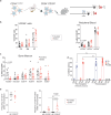
Fig. 4. Multiplex CD33/HBG ABE8e editing of human CD34+ cells and GO selection for multiplex edits in vitro.
a Schematic of HBG −175 ABE8e target site. Adenines in the editing window are marked in bold red and PAM in light red. b HBG ABE8e editing efficiency measured in human mPB CD34+ HSPCs after treatment by mock electroporation (unedited, UE) or by HBG ABE8e mRNA. Results are from one representative donor. c HBG editing efficiency in human mPB CD34+ HSPCs treated by mock electroporation (UE), by HBG ABE8e mRNA (single) or by CD33/HBG ABE8e mRNA (multiplex). Results are from 4 different donors, 4 independent experiments. d CD33 editing efficiency in human mPB CD34+ HSPCs treated by mock EP (UE) or by CD33/HBG ABE8e mRNA (multiplex). Results are from 5 different donors, 5 independent experiments. e Colony-forming potential of mPB CD34+ cells treated by multiplex ABE8e editing. Results are from 1 representative donor, 3 technical replicates. M=macrophages, G=granulocytes, GM=granulocyte/macrophage, GEMM=granulocyte/erythrocyte/macrophage/monocyte BFU-E=erythroid. f Frequency of colony-forming cells displaying unedited (UE), edits at each gene target, or edits at both gene targets in the same colony. Results are from 2 different human donors, 2 independent experiments. g Frequency of colony-forming cells with unedited (UE), monoallelic (mono) or biallelic (bi) CD33 edits, and accompanying edits (0 to 4) at the HBG target site in two different donors. Number of colonies analyzed is shown on top. N/A=Not Applicable. h Unedited (UE) or multiplex edited ML1 cells treated for 6 consecutive days with 10 pg/mL Gemtuzumab Ozogamicin (GO) starting at 5 days post-electroporation. Viable count was determined by trypan blue staining. Results are from 1 representative experiment. i, Editing efficiency measured at the CD33 (left) or HBG (right) gene targets in multiplex edited ML1 cells from h treated or not with GO at 8 days post GO removal (14 days post-electroporation). All error bars in this figure show ±SEM. Source data are provided as a Source Data file.
Fig. 5
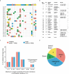
Fig. 5. Off-target ABE8e editing with single or multiplex guides in CD34+ HPSCs.
a Visualization of on-target (CD33) and top 23 off-target sites associated with ABE8e editing of the CD33 exon 2 splice acceptor site in multiplex-edited (CD33 + HBG) and single-edited (CD33 or HBG) CD34+ HPSCs. These off-target (OT) sites include eighteen loci (OT1-OT18) nominated by CIRCLE-seq using ABE8e-SpCas9-NG for the CD33 target as well as five loci that showed statistically significant editing from prior off-target analyses in engrafted cells, which had been nominated by CIRCLE-seq using ABE8e (wild type Cas9) (OT19-OT23). Alignment of each site to the sgABE protospacer is shown. b Table summarizing the top 23 nominated off-target loci. Loci with an asterisk (*) indicate that statistically significant editing was observed at this site at one or more nucleotides (A5, A7, or both). Quantification of editing at each site is shown in Fig.S9 and S10. c Histogram demonstrating the mean percent of reads with A > G editing at any site within the sgABE protospacer window for validated off-target sites, classified by multiplex (CD33 + HBG) or single-edited (CD33 or HBG) status. Among the top off-target sites analyzed, no substantial difference in off-target editing was detected among multiplex vs. single-edited CD34+ HPSCs. d Pie chart indicating genomic context of top nominated off-target sites that showed statistically significant editing. The majority of off-target sites were located in intergenic or intronic regions. Results shown are from 3 human donors, 3 independent experiments. Source data are provided as a Source Data file.
Fig. 6
Fig. 6. Engraftment of multiplex ABE8e-edited human mPB CD34+ cells in NSG mice.
a Measure of engraftment by percentage of human CD45+ cells in different tissues at 14 to 16 weeks post-transplantation. Results are from 2 different human donors, 2 independent experiments, n = 11 for PB and n = 6 for BM/spleen for the unedited (UE) group; n = 12 for PB and n = 4 for BM/spleen for multiplex editing. b Hematopoietic repopulation by frequency of mature myeloid (CD14), lymphoid (CD19), and T cells (CD3) within the human CD45 population in peripheral blood. CD33 expression within the CD14+ subset is also shown. Results are from 2 different human donors, 2 independent experiments, n = 11 for UE and n = 12 animals for multiplex editing. c Representative flow plots showing gating strategy of human HSC (CD34+CD38low) in BM of multiplex edited mouse at necropsy. d Engraftment of multiplex edited vs. unedited mPB CD34+ cells in BM as measured by human CD45 expression and of hematopoietic repopulation by frequency of mature myeloid (CD14) and B and T lymphoid (CD19 and CD3) within the human CD45 population in peripheral blood at 14 to 16 weeks post-transplantation. CD33 expression in total BM cells and HSC content (CD34+CD38low) is also shown. Results are from 2 different human donors, 2 independent experiments, n = 6 for unedited and n = 4 animals for multiplex editing. e Representative flow plots of CD33 expression in BM HSC (CD34+CD38low) from engrafted mice at necropsy. All statistical analyses in this figure were done with an unpaired two-tailed t-test. All error bars in this figure show ±SEM. Source data are provided as a Source Data file.
Fig. 7
Fig. 7. In vivo protection of ABE8e multiplex edited cells from GO treatment.
a Schematic and timeline of mouse engraftment experiment with three consecutive administrations of Mylotarg (0.05 mg/kg). The mouse cartoon was created in BioRender. Du, X. (2025) https://BioRender.com/g56e371. b Human cell engraftment in BM of mice engrafted with unedited (UE) or multiplex ABE8e edited CD34+ cells at 14 weeks post-transplant. c, Frequency of human CD45+/CD33+ cells measured from the same animal as b. Statistical analysis shows unpaired two tailed t-test. d Editing efficiency measured at CD33 (left) and HBG (right) targets measured in mouse BM engrafted with multiplex edited cells at 14 weeks post-transplant. e Frequency of human monocytes (% HuCD45+CD14+) in BM of mice engrafted with UE or multiplex ABE8e edited CD34+ cells pre- (wk14) and post-Mylotarg (wk18) treatment. f Frequency of human HSCs (% HuCD45+CD34+CD38low) in BM of engrafted mice pre- and post-Mylotarg treatment. 1 experiment, 1 human donor, n = 3 for the unedited group and n = 5 for the multiplex edited group. All statistical analyses were done with one-way ANOVA, Tukey’s multiple comparisons test. All error bars in this figure show ±SEM. Source data are provided as a Source Data file.
Fig. 8
Fig. 8. Long term engraftment of ABE8e multiplex edited CD90+ HSPCs in rhesus macaques.
ABE8e editing efficiency at the CD33 (a) and HBG (b) targets measured in the infusion product of each transplanted animal at 5 days post multiplex editing or post mock electroporation (unedited, UE). c, Frequency of unedited, edits at each gene target, or at both targets in colony-forming cells (CFCs) obtained from infusion products from a and b. n = 71 for A18031 and n = 37 for A18038. d Frequency of colony-forming cells with unedited (UE), monoallelic (mono) or biallelic (bi) CD33 edits, and accompanying edits (0 to 4) at the HBG target site in the two infusion products. Number of colonies analyzed is shown on top. e Tracking of CD33 and HBG editing in peripheral blood of both transplanted animals. For simplicity, only editing efficiency at the relevant adenines are shown (A7 for CD33 and A5 for HBG). f CD33 expression in CD11b+CD14 peripheral blood granulocytes in both transplanted animals as compared to an untransplanted control animal (A16227). g, Fetal hemoglobin (HbF) expression as measured by flow cytometry staining for F-cells in peripheral blood of both transplanted animals as compared to the untransplanted control. h Colony forming cells (CFCs) derived from BM aspirates taken from both transplanted animals at 8- or 6-months post-transplant for A18031 and A18038, respectively. i, Frequency of unedited, edits at each gene target, or at both targets in CFCs from g n = 57 for A18031 and n = 45 for A18038. All error bars in this figure show ±SEM. Source data are provided as a Source Data file.

Update of

References

    1. Borot, F. et al. Gene-edited stem cells enable CD33-directed immune therapy for myeloid malignancies. Proc. Natl. Acad. Sci. USA116, 11978–11987 (2019). - PMC - PubMed
    1. Kim, M. Y. et al. Genetic Inactivation of CD33 in Hematopoietic Stem Cells to Enable CAR T Cell Immunotherapy for Acute Myeloid Leukemia. Cell173, 1439–1453.e1419 (2018). - PMC - PubMed
    1. Humbert, O. et al. Engineering resistance to CD33-targeted immunotherapy in normal hematopoiesis by CRISPR/Cas9-deletion of CD33 exon 2. Leukemia33, 762–808 (2019). - PubMed
    1. Genovese, P. et al. Targeted genome editing in human repopulating haematopoietic stem cells. Nature510, 235–240 (2014). - PMC - PubMed
    1. Schiroli, G. et al. Precise Gene Editing Preserves Hematopoietic Stem Cell Function following Transient p53-Mediated DNA Damage Response. Cell Stem Cell24, 551–565.e558 (2019). - PMC - PubMed

MeSH terms

LinkOut - more resources