

ESUR consensus MRI for endometriosis: indications, reporting, and classifications
- PMID: 40425757
- PMCID: PMC12559033
- DOI: 10.1007/s00330-025-11579-0
ESUR consensus MRI for endometriosis: indications, reporting, and classifications
Abstract
Objective: To propose an update of ESUR endometriosis guidelines to reflect advances in MRI indications, reporting, and classifications.
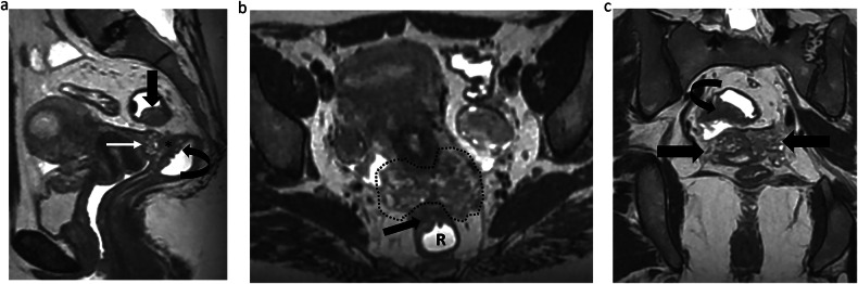
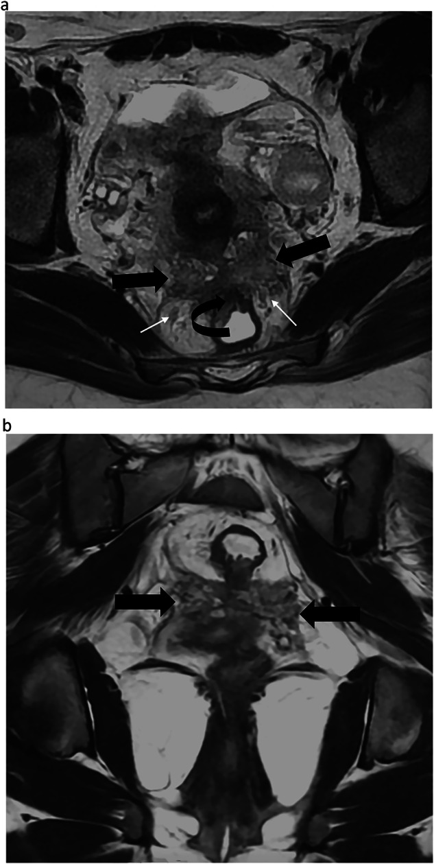
Methods: The ESUR Research Committee appointed two chairs (I.T.N., L.M.) to supervise the development of the updated guidelines. Following literature research, a survey was delivered to 20 experts in gynecological imaging from 10 countries. Two rounds of surveys were conducted to obtain a consensus according to a Delphi process method. In this article, the results regarding MR indication, the use of standardized reports, and classifications are presented RESULTS: Magnetic resonance imaging (MRI) is recommended when transvaginal ultrasonography is inconclusive in diagnosing endometriosis or negative, in a symptomatic patient, before surgery or interventional procedure, or after surgical treatment if symptoms persist. ESUR panelists consider the roles of an MR classification: to improve communication between radiologist and surgeon (100%, 20/20) and between the radiologist and the patient (45%, 9/20), to predict operating time if surgery is planned (70%, 14/20), to predict the length of hospital stay after surgery (40%, 8/20), and to predict postoperative complications (70%, 14/20). ESUR panelists strongly agree that using an MR classification is useful (19/20, 95%), especially the radiological score, deep-pelvic endometriosis index (dPEI). Among the ESUR expert group, 9/20 experts (45%) used or agreed to use drawings in their report to improve communication with patients.
Conclusion: Standardized MR reporting is crucial and should include the use of MR classification. Drawings are considered an option, knowing that communication with the patient and surgeon is of paramount importance.
Key points: Question ESUR's endometriosis guidelines were last published in 2017; an update is provided to reflect advances in MRI indications, reporting, and classifications. Findings MRI is advised for inconclusive/negative transvaginal ultrasound in symptomatic patients, before surgery, or post-treatment if symptoms persist. A structured report enhances communication with surgeons and patients. Clinical relevance A standardized report based on a compartmental analysis of the location of endometriotic nodules, with optional drawings, is essential for comprehensive mapping and optimal communication with both patient and surgeon.
Keywords: Consensus; Endometriosis, Pelvis; Magnetic resonance imaging.
© 2025. The Author(s).
Conflict of interest statement
Compliance with ethical standards. Guarantor: The scientific guarantor of this publication is Isabelle Thomassin-Naggara. Conflict of interest: The authors of this manuscript declare relationships with the following companies: I.T.N. discloses the following: Speakers bureaus: European Society of Breast Imaging (active), Société d’imagerie de la femme (active), American College of Radiology O-RADS (active), Bayer (ended), Siemens Healthineers (ended), Guerbet (ended), Bard (ended). Ponctual remunerated lectures: GE, Siemens, Guerbet, Hologic, Canon, Guebet, Bracco, GSD, Samsung, Fujifilm, Incepto, ICAD. Research Grants: ASCORDIA: ADNEX MR Scoring System: Impact of an MR scoring system on therapeutic strategy of pelvic adnexal masses PHRC ID RCB 2015-A01593-46. P.R. discloses the following: Consultancies: ZIWIG and EDAP TMS France. Luciana P.Chamie discloses the following: Ponctual remunerated lectures: Cleveland Clinic Imaging Institute. The remaining authors declare no conflicts of interest. Statistics and biometry: No complex statistical methods were necessary for this paper. Informed consent: Not applicable. Ethical approval: Institutional Review Board approval was not required because this paper is a recommendation paper. Study subjects or cohorts overlap: None. Methodology: Consensus paper based on the DELPHI process
Figures


References
Publication types
MeSH terms
LinkOut - more resources
Full Text Sources
Medical
