. 2025 Dec;14(1):2508050.
doi: 10.1080/2162402X.2025.2508050. Epub 2025 May 27.

Increasing the odds: antibody-mediated delivery of two distinct immunogenic T-cell epitopes with one antibody

Affiliations

Increasing the odds: antibody-mediated delivery of two distinct immunogenic T-cell epitopes with one antibody

Willemijn van der Wulp et al. Oncoimmunology. 2025 Dec.

Abstract

Antibody-epitope conjugates (AECs) proved to be a promising new therapeutic strategy to redirect virus-specific CD8+ T cells toward cancer cells by delivering T-cell epitopes. To be able to redirect a larger fraction of the virus-specific T-cell population, it is beneficial to deliver a broader selection of T-cell epitopes. We investigated two different methods to generate AECs with two distinct virus-specific T-cell epitopes fused to one antibody. Epitopes were either placed in a tandem-like fashion at the C-terminus of the AEC (t-AEC) or bispecific-AECs (bs-AECs) were generated via controlled Fab-arm exchange to generate bs-AECs with two identical antigen binding domains, but two distinct epitopes on each Fab-arm. Our study revealed that maintaining a free epitope terminus was required for efficient delivery of the virus-specific T-cell epitopes. Consequently, viral-epitope delivery using t-AECs was suboptimal as the concatenated epitopes were less effectively delivered to the target cells. However, well-defined bs-AECs containing both CMV and EBV epitopes were successfully generated and both in vitro and in vivo efficacy was evaluated. Our results demonstrate that bispecific-AECs can efficiently deliver EBV and CMV epitopes simultaneously to multiple cancer cell lines from different origins, thereby redirecting and activating two distinct populations of virus-specific T cells. Furthermore, our in vivo findings indicate that when both virus-specific T-cell populations are present and tumor cells express the proteases required for efficient epitope delivery, bs-AECs exhibit similar efficacy in reducing tumor burden compared to AECs. To conclude, our study demonstrates the feasibility of redirecting two groups of virus-specific T cells using a single antibody and highlights the potential of bs-AECs both in vitro and in vivo.

Keywords: Antibody-epitope conjugates (AECs); bispecific-AECs; immunotherapy; redirecting T-cells; tandem-AECs; virus-specific T-cells.


Conflict of interest statement

BB, JS, and LG are (former) employees at Genmab BV and have ownership interests (including stocks, warrants, patents, etc.).

Figures
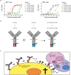
Figure 1.
Figure 1.
Epitopes in an enclosed position are not delivered properly (a) HeLa-A2 cells were exposed for 1 h to titrated concentrations of the different peptides. The sequences of the peptides used within the experiment are listed in Table 1. The exposed HeLa-A2 cells were cocultured for 18 hrs with either the EBV- or CMV-specific T cells as indicated, and T-cell activation was analyzed by measuring IFN-γ production of the T cells within the supernatant. Plotted values are the means of duplicates (SEM) and each graph shows a representative figure of three independently performed experiments. (b) An overview of the bs-AECs with two distinct epitopes attached to one antibody (CTX-EBV and CTX-CMV or TRS-EBV and TRS-CMV) where DuoBody technology was used to generate bispecific-AECs (bs-CTX-EBVxCTX-CMV or bs-TRS-EBVxTRS-CMV) without changing the specificity of the binding domain. (c) Schematic overview of the hypothesized mechanism of the bs-AECs, in which the bs-AEC first recognized the antibody target, followed by proteolytic release of the epitope by proteases secreted by the tumor cells. After release the epitopes eventually gets presented on the HLA molecules present on the tumor cells, which can be recognized by the two different virus specific CD8+ T cells.
Figure 2.
Figure 2.
Both the EBV- and CMV-epitope of bs-TRS-EBVxTRS-CMV are delivered and induce T cell activation and target cell killing. (a–b) to determine whether both AECs were able to deliver their epitopes, HeLa-A2 tHer2 cells were exposed to TRS-EBV and -CMV and cocultured with either (a) EBV- or (b) CMV-specific T cells. (c–d) HeLa-A2 tHer2 cells were exposed to the wildtype (WT) of TRS, TRS-EBV, -CMV and different bs-AECs, and subsequently cocultured with (c) EBV-specific T cells or (d) CMV-specific T cells. (e) HeLa-A2 tHer2 cells were exposed to bs-TRS-EBVxTRS-CMV and cocultured with either EBV-, CMV- or a 50%/50% mixture of EBV- and CMV-specific T cells with a constant E:T ratio for all three T cell combinations (a-e). For all coculture assays, T cell activation was analyzed by measuring the IFN-γ production of the T cells within the supernatant after 18 hrs of coculture. (f) To measure specific cell killing HeLa-A2 tHer2 cells were exposed to TRS-EBV, -CMV, or bs-TRS-EBVxTRS-CMV, followed by a 72 hrs coculture with a 50%/50% mixture of EBV- and CMV-specific T cells. Tumor cell killing was measured with an AlamarBlue assay. (a-f) Plotted values are the means of duplicates (SEM) and each graph shows a representative figure of three independently performed experiments.
Figure 3.
Figure 3.
Tumor cells treated with bs-AECs efficiently activate virus-specific T cells. (a) SKOV3-A2, (c) H292-A2 and (d) MDA-MB231 were exposed for 1 h to wildtype CTX (CTX-WT), CTX-EBV, -CMV, or bs-CTX-EBVxCTX-CMV, and subsequently cocultured for 72 hrs with a 50%/50% mixture of EBV- and CMV-specific T cells. (b) SKOV3-A2 were exposed to TRS (WT), TRS-EBV, -CMV, or bs-TRS-EBVxTRS-CMV followed by a coculture with a 1:1 mixture of EBV- and CMV-specific T cells. (a-d) Supernatant was harvested after 18 hrs of coculture to determine IFN-у production as a measure of T-cell activation. The tumor cell killing was measured after 72 hrs of coculture using the Alamar blue assay. Plotted values are the means of duplicates (SEM) and each graph shows a representative figure of three independently performed experiments.
Figure 4.
Figure 4.
Bs-AECs induce additive T-cell activation when mixtures of EBV- and CMV-specific T cells are present. (a) SKOV3-A2 and (d) H292-A2 cells were exposed to 16 nM bs-CTX-EBVxCTX-CMV and (b) SKOV3-A2 and (c) HeLa-A2 tHer2 were exposed to 16 nM bs-TRS-EBVxTRS-CMV, and (a-d) subsequently cocultured with titrated amounts of EBV-specific T cells (EBV/(-)) or CMV-specific T cells ((-)/CMV) or the mixture of these titrated EBV- and CMV-specific T cells (EBV/CMV). The highest number of T cells in the EBV- and CMV-specific T-cell titration was 4.000 T cells per well and per titration step the number of T cells per well was reduced by 570. In the combination of titrated EBV- and CMV-specific T cells a total of 4.000 T-cells was present. The T-cell activation was analyzed by measuring the IFN-γ production of the T cells within the supernatant after 18 hrs of coculture. Plotted values are the means of duplicates (SEM) and each graph shows a representative figure of three independently performed experiments.
Figure 5.
Figure 5.
One EBV-epitope of bsAECs can still reduce tumor outgrowth in an U266 xenograft model. (a) U266-tEGFR cells were exposed to 16 nM of CTX-WT, -EBV, -CMV or bs-CTX-EBVxCTX-CMV and cocultured with either EBV- or CMV-specific T cells. Plotted values are the means of duplicates (SEM) of three independent experiments. (b) U266-tEGFR were exposed to 16 nM bs-CTX-EBVxCTX-CMV and subsequently cocultured with titrated amounts of EBV-specific T cells (EBV/(-)) or CMV-specific T cells ((-)/CMV) or the mixture of these titrated EBV- and CMV-specific T cells (EBV/CMV). The highest number of T cells in the EBV- and CMV-specific T-cell titration was 4.000 T cells per well and per titration step the number of T cells per well was reduced by 570. (c) Overview of the experimental set-up of the in vivo experiment. (d) NSG mice engrafted with 2 × 106 luciferase positive U266-tEGFR cells were i.V. injected with 2.5 × 106 EBV and 2.5 106 CMV TCR-transduced CD8+ T cells at day 14. On day 15 and 18 after tumor injection mice were i.P. injected with 100 μg CTX-EBV, -CMV or bs-CTX-EBVxCTX-CMV and 100 μg pembrolizumab. Tumor outgrowth of the U266-tEGFR2 was visualized by bioluminescence imaging 1–2 times per week of the ventral side. Significance was determined with a two-way ANOVA, with a Tukey`s multiple comparisons on log-transformed data and only visualized within the graph day 36. (e) The survival analysis was performed with the Kaplan–Meier method and significance was assessed with the mantel cox method and corrected using the Bonferroni method. Treatment groups were compared to the combined control groups. The control group (Ab only) received bs-TRS-EBVxTRS-CMV and pembrolizumab, but no T cells, to demonstrate that binding of the antibody and delivery of the epitopes does not influence tumor outgrowth. Control group (T cells only) received the mixture of virus specific T cells and pembrolizumab. The endpoint was determined when the tumor outgrowth reached a bioluminescent signal of 1 × 107, which is considered the endpoint.
Figure 6.
Figure 6.
Both the EBV- and CMV-epitope of bs-AECs can reduce tumor outgrowth in an SKOV3-A2 xenograft model. (a) Overview of the experimental set-up of the in vivo experiment. (b) NSG mice engrafted with 6 × 106 luciferase positive SKOV3-A2 cells were subsequently i.V. injected with 2.5 × 106 EBV and 2.5 106 CMV TCR-transduced CD8+ T cells at day 14. On day 15, 18 and 21 after tumor injection mice were i.P. injected with 100 μg TRS-EBV, -CMV or bs-TRS-EBVxTRS-CMV and 100 μg pembrolizumab. Tumor outgrowth was determined by means of caliper measurement. The control group (Ab only) received bs-TRS-EBVxTRS-CMV and pembrolizumab, but no T cells, to demonstrate that binding of the antibody and delivery of the epitopes does not influence tumor outgrowth. Control group (T cells only) received the mixture of virus specific T cells and pembrolizumab. (c) The survival analysis was performed with the Kaplan–Meier method and significance was assessed with the mantel cox method and corrected using the Bonferroni method. Treatment groups were compared to the combined control groups. The control group (Ab only) received bs-TRS-EBVxTRS-CMV and pembrolizumab, but no T cells, to demonstrate that binding of the antibody and delivery of the epitopes does not influence tumor outgrowth. The endpoint was defined at a tumor outgrowth of 1000 mm3.

References

    1. Jung K, Son MJ, Lee SY, Kim JA, Ko DH, Yoo S, Kim C-H, Kim Y-S.. Antibody-mediated delivery of a viral Mhc-I epitope into the cytosol of target tumor cells repurposes virus-specific Cd8(+) T cells for cancer immunotherapy. Mol Cancer. 2022;21(1):102. doi: 10.1186/s12943-022-01574-0. Epub 2022/04/24. - DOI - PMC - PubMed
    1. Sefrin JP, Hillringhaus L, Mundigl O, Mann K, Ziegler-Landesberger D, Seul H, Tabares G, Knoblauch D, Leinenbach A, Friligou I, et al. Sensitization of tumors for attack by virus-specific Cd8+ T-Cells through antibody-mediated delivery of immunogenic T-Cell Epitopes. Front Immunol. 2019;10:1962. doi: 10.3389/fimmu.2019.01962. Epub 2019/09/27. - DOI - PMC - PubMed
    1. van der Wulp W, Gram AM, Bleijlevens B, Hagedoorn RS, Araman C, Kim RQ, Drijfhout JW, Parren PWHI, Hibbert RG, Hoeben RC, et al. Comparison of methods generating antibody-epitope conjugates for targeting cancer with virus-specific T cells. Front Immunol. 2023;14:1183914. doi: 10.3389/fimmu.2023.1183914. Epub 2023/06/01. - DOI - PMC - PubMed
    1. Millar DG, Ramjiawan RR, Kawaguchi K, Gupta N, Chen J, Zhang S, Nojiri T, Ho WW, Aoki S, Jung K, et al. Antibody-mediated delivery of viral epitopes to tumors harnesses Cmv-specific T cells for cancer therapy. Nat Biotechnol. 2020;38(4):420–13. doi: 10.1038/s41587-019-0404-8. Epub 2020/02/12. - DOI - PMC - PubMed
    1. Robert C. A decade of immune-checkpoint inhibitors in cancer therapy. Nat Commun. 2020;11(1):3801. doi: 10.1038/s41467-020-17670-y. Epub 2020/08/01. - DOI - PMC - PubMed

Substances

LinkOut - more resources