

Recent advances in osteonecrosis of the femoral head: a focus on mesenchymal stem cells and adipocytes
- PMID: 40426076
- PMCID: PMC12108002
- DOI: 10.1186/s12967-025-06564-6
Recent advances in osteonecrosis of the femoral head: a focus on mesenchymal stem cells and adipocytes
Abstract
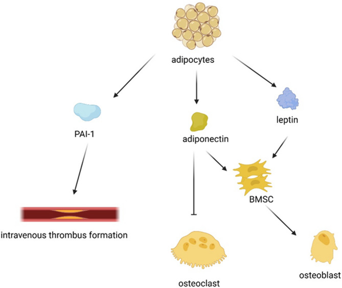
Osteonecrosis of the femoral head (ONFH) is a debilitating orthopedic disease characterized by femoral head collapse and destruction of bone and articular cartilage, resulting in severe joint pain and loss of hip mobility. Bone marrow mesenchymal stem cells (BMSCs) exhibit multilineage differentiation potential, including osteoblasts, adipocytes, fibroblasts, chondrocytes, and neurocytes. The imbalance between osteogenesis and adipogenesis in BMSCs plays a critical role in ONFH pathogenesis. Factors such as alcohol consumption and glucocorticoid exposure promote adipogenic differentiation while inhibiting osteogenic differentiation, leading to excessive adipocyte accumulation, reduced bone formation, and vascular impairment. We highlight the molecular mechanisms underlying ONFH with a particular focus on the role of BMSCs and further discuss the involvement of adipocytes. Moreover, we suggest that the use of adipose-derived mesenchymal stem cells (ADMSCs) is a viable approach for stem cell therapy and may have immense potential in ONFH. Several signaling pathways, including the Wnt, TGFβ/BMP, and PI3K/AKT pathways, along with various RNAs and other regulators, govern the osteogenesis and adipogenesis of BMSCs. These signaling pathways target essential transcription factors, such as Runx2 for osteogenesis and PPARγ and C/EBPs for adipogenesis. Adipocytes and their secreted adipokines, including leptin and adiponectin, strongly influence ONFH progression. Emerging therapies involving ADMSCs show potential for promoting bone regeneration and neovascularization. Our review provides a comprehensive overview of the current understanding of ONFH mechanisms by focusing on mesenchymal stem cells and adipocytes and suggests future research directions for therapeutic interventions.
Keywords: Adipocyte; Adipokine; Mesenchymal stem cell; Osteonecrosis of the femoral head; Signaling pathways.
© 2025. The Author(s).
Conflict of interest statement
Declarations. Ethics approval and consent to participate: Not applicable. Consent for publication: Not applicable. Competing interests: The authors declare no competing interests.
Figures


Similar articles
-
Increased COUP-TFII Expression Mediates the Differentiation Imbalance of Bone Marrow-Derived Mesenchymal Stem Cells in Femoral Head Osteonecrosis.Biomed Res Int. 2019 Dec 8;2019:9262430. doi: 10.1155/2019/9262430. eCollection 2019. Biomed Res Int. 2019. PMID: 31886265 Free PMC article.
-
Chrysophanic acid shifts the differentiation tendency of BMSCs to prevent alcohol-induced osteonecrosis of the femoral head.Cell Prolif. 2020 Aug;53(8):e12871. doi: 10.1111/cpr.12871. Epub 2020 Jun 29. Cell Prolif. 2020. PMID: 32597546 Free PMC article.
-
Icariin Protects against Glucocorticoid-Induced Osteonecrosis of the Femoral Head in Rats.Cell Physiol Biochem. 2018;47(2):694-706. doi: 10.1159/000490023. Epub 2018 May 22. Cell Physiol Biochem. 2018. PMID: 29794448
-
The shift in the balance between osteoblastogenesis and adipogenesis of mesenchymal stem cells mediated by glucocorticoid receptor.Stem Cell Res Ther. 2019 Dec 5;10(1):377. doi: 10.1186/s13287-019-1498-0. Stem Cell Res Ther. 2019. PMID: 31805987 Free PMC article. Review.
-
Application of biomaterials in treating early osteonecrosis of the femoral head: Research progress and future perspectives.Acta Biomater. 2023 Jul 1;164:15-73. doi: 10.1016/j.actbio.2023.04.005. Epub 2023 Apr 18. Acta Biomater. 2023. PMID: 37080444 Review.
Cited by
-
Exercise-Induced Muscle-Fat Crosstalk: Molecular Mediators and Their Pharmacological Modulation for the Maintenance of Metabolic Flexibility in Aging.Pharmaceuticals (Basel). 2025 Aug 19;18(8):1222. doi: 10.3390/ph18081222. Pharmaceuticals (Basel). 2025. PMID: 40872612 Free PMC article. Review.
-
microRNA-576-5p ameliorates dexamethasone-induced BMSC injury by suppressing ANXA2.Sci Rep. 2025 Aug 20;15(1):30612. doi: 10.1038/s41598-025-16883-9. Sci Rep. 2025. PMID: 40836105 Free PMC article.
References
-
- Xu HH, Li SM, Fang L, Xia CJ, Zhang P, Xu R, et al. Platelet-rich plasma promotes bone formation, restrains adipogenesis and accelerates vascularization to relieve steroids-induced osteonecrosis of the femoral head. Platelets. 2021;32:950–9. - PubMed
-
- Li J, Wang Y, Li Y, Sun J, Zhao G. The effect of combined regulation of the expression of peroxisome proliferator-activated receptor-gamma and calcitonin gene-related peptide on alcohol-induced adipogenic differentiation of bone marrow mesenchymal stem cells. Mol Cell Biochem. 2014;392:39–48. - PubMed
Publication types
MeSH terms
Grants and funding
LinkOut - more resources
Full Text Sources
