. 2025 May 28;23(1):388.
doi: 10.1186/s12951-025-03419-6.

Pulmonary hazards of nanoplastic particles: a study using polystyrene in in vitro models of the alveolar and bronchial epithelium

Affiliations

Pulmonary hazards of nanoplastic particles: a study using polystyrene in in vitro models of the alveolar and bronchial epithelium

Sara Michelini et al. J Nanobiotechnology. .

Abstract

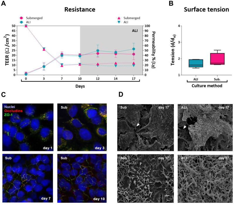
Background: Nanoplastics (NPs) are released into the environment through the degradation of plastic objects, leading to human exposure. Due to their small size, concerns have been raised about the potential hazards to the respiratory tract, as ultrafine and nanoparticles are known to penetrate till the alveolar regions of the lungs, potentially impairing their functions. Thus, in the present study, we used model polystyrene nanoparticles doped with the fluorescent metal europium (PS-Eu) to enhance the understanding of NPs hazard and investigate adverse outcomes associated with exposure in human lungs using alveolar (A549) and bronchial (Calu-3) cell models grown in 2D and 3D submerged conditions or quasi air-liquid interface (ALI) conditions (3D).

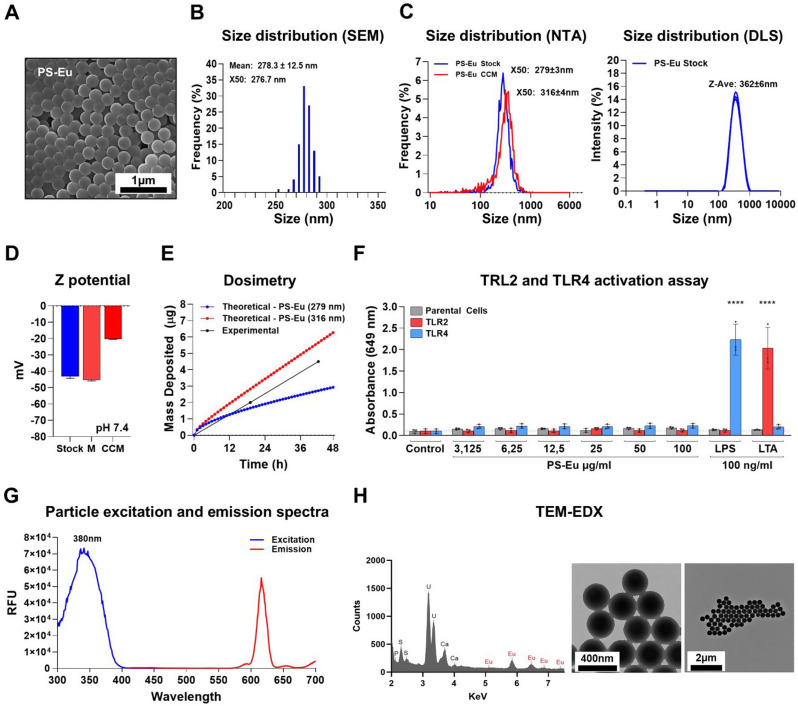
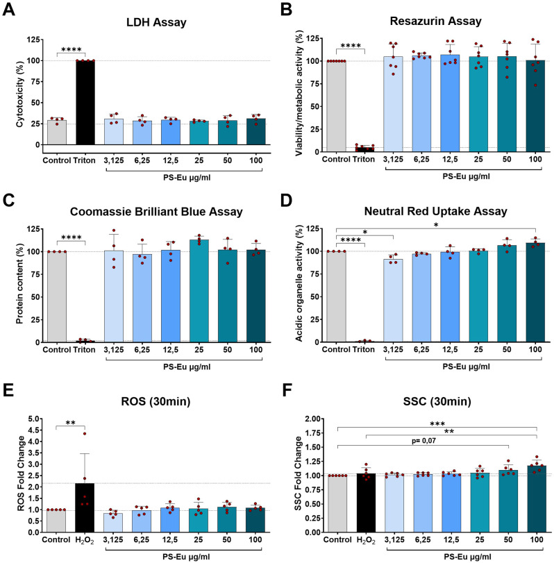
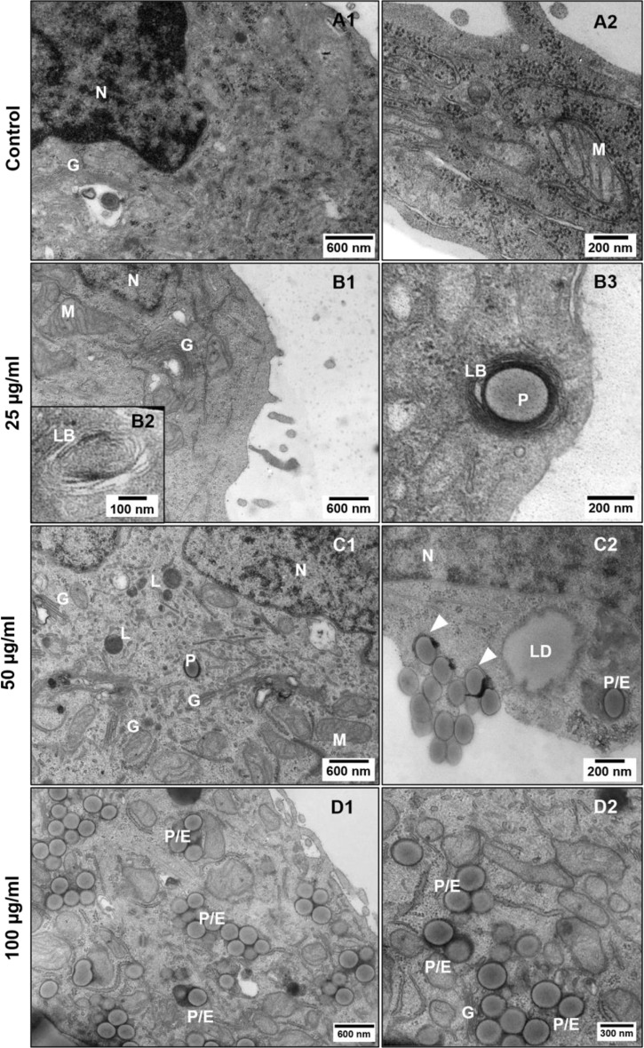
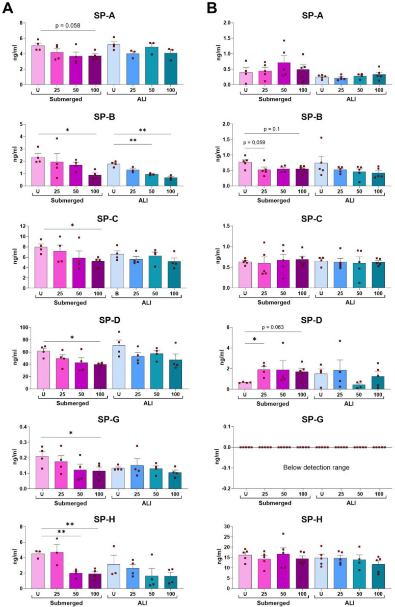
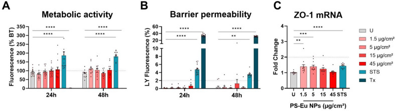
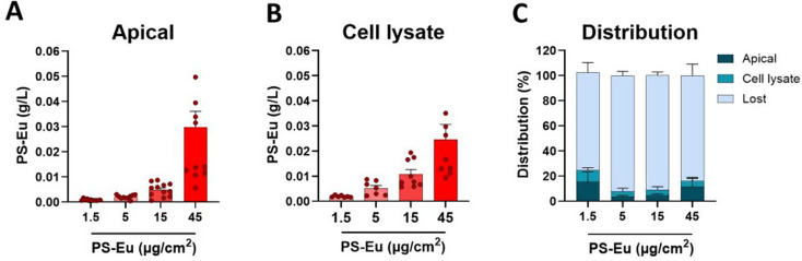
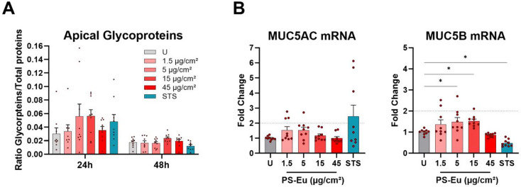
Results: Briefly, after in-dept physicochemical characterization of the particles, we assessed their impact on ROS production, cell viability (AlamarBlue and lactate dehydrogenase assays) and barrier integrity (lucifer yellow assay and TEER measurement), finding no negative effects in either model. However, in alveolar cells, particles increased acidic organelle activity. Transmission electron microscopy and Raman microscopy showed, in both models, a dose- and cell-dependent particle uptake with PS-Eu accumulating in numerous and large endo-lysosomes, which, in transwells-grown A549 cells, often contained also lamellar bodies (LBs), organelles involved in surfactants storage and secretion. After extensively quantifying surfactant proteins (SP) in the pellet and supernatant fractions of treated A549 cells, we observed a significant reduction in several members of this family, including surfactant protein B, which is crucial for lamellar body formation and surface tension regulation in the lungs. In quasi-ALI Calu-3 cultures instead, PS-Eu significantly upregulated interleukin 6 (IL-6) and increased transforming growth factor beta β (TGF-β), zonula occludens 1 (ZO-1), and mucin (MUC) 5B mRNA expressions causing a moderate proinflammatory response.

Conclusion: Our results show that PS-Eu exposure does not induce acute cytotoxicity in these models, but affects cell-specific functions like surfactant, mucin, and cytokine production. This underscores the limitations of relying solely on standard cytotoxicity tests for particle hazard assessment and highlights the importance of investigating cell function-specific signaling pathways. To support researchers in hazard assessment, we propose specific classes of biomarkers to test in in vitro lung models.


Conflict of interest statement

Declarations. Ethics approval and consent to participate: Not applicable. Consent for publication: Not applicable. Competing interests: The authors declare no competing interests.

Figures

Fig. 1
Fig. 1
Particle characterization. (A) a representative SEM image of PS-Eu stock suspension and correspondent size distribution calculated using ImageJ on the same images (B); (C) Left: size distribution of stock suspension (blue) or PS-Eu incubated for 48 h in CCM (red) calculated via NTA. Right: PS-Eu stock suspension size distribution calculated via DLS. (D) Z potential of PS-Eu stock suspension or PS-Eu incubated for 48 h in FBS-free medium (M) or in CCM. (E) PS-Eu deposited mass over 48 h calculated theoretically and experimentally combining the ISDD model and NTA. (F) Graph showing the level of TLR2 and TLR4 ligand contamination of PS-Eu stock suspension measured using the HEK293 reporter cell assay. 100 ng/ml of LPS and LTA were used as positive controls for TLR2 and TLR4 activation, respectively. Grey: parental cells. Red/Blue: cells expressing the human TLR2 or the TLR4 gene. (G) PS-Eu excitation and emission spectra in CCM. (H) TEM-EDX spectrum of PS-Eu stock showing the presence of the Eu element. Abbreviations: PS-Eu: europium doped polystyrene nanoparticles; SEM; Scanning electron microscopy; TEM: Transmission electron microscopy; CCM: Complete culture medium; NTA: nanoparticle tracking analysis; DLS: dynamic light scattering; FBS: fetal bovine serum; ISDD: in vitro sedimentation, diffusion and dosimetry; HEK293: Human embryonic kidney 293 cells; LPS: lipopolysaccharide; LTA: lipoteichoic acid; TLR: Toll-like receptor; RFU: relative fluorescence units; TEM-EDX: Transmission electron microscopy–energy dispersive X-ray
Fig. 2
Fig. 2
Impact of PS-Eu on A549 cells grown in submerged 2D conditions. Graphs represent the results obtained with (A) lactate dehydrogenase (LDH) assay (cytotoxicity), (B) Resazurin assay (cell viability/mitochondrial activity), (C) Coomassie brilliant blue (CBB) assay (protein content), (D) Neutral red uptake (NRU) assay (acidic organelles activity) of cells incubated for 48 h with increasing concentrations of particles. Additionally in (E) and (F) are shown reactive oxygen species (ROS) production, and cell inner granularity measurement (SSC), measured 30 min post treatment with flow cytometry. Statistical analysis was done using One-way ANOVA vs. untreated control. In SSC measurement, analysis was also performed vs. H2O2 control. All graphs show mean ± standard deviation and every dot represents an independent experiment. Abbreviations: A549: adenocarcinoma human alveolar basal epithelial cells; PS-Eu: europium doped polystyrene nanoparticles
Fig. 3
Fig. 3
Particle internalization assessment in 2D grown A549 cells treated with increasing concentrations of nanoplastics. Control cells (A1-2), cells treated for 48 h with 25 µg/ml (B1-3), 50 µg/ml (C1-2) and 100 µg/ml (D1-2) PS-Eu. Abbreviations and legend: A549: adenocarcinoma human alveolar basal epithelial cells; LB, lamellar body; LD, lipid droplet; Arrowheads, plasma membrane foldings during particle internalization; G, Golgi apparatus; N, nucleus; P, PS-Eu particles; E, endosomes; L, lysosomes; M, mitochondria
Fig. 4
Fig. 4
3D model characterization. (A) graph depicting over time formation of the lung alveolar barrier, in terms of transepithelial electrical resistance (TEER, solid line), and permeability measured with lucifer yellow assay (Ly) (dashed line) in quasi-air-liquid interface (ALI, blue) and submerged (pink) cultures; (B) graph depicting the surface tension of the epithelial barrier estimated with the drop-spreading method; (C) Confocal microscopy of adenocarcinoma human alveolar basal epithelial cells (A549) grown for different periods in the transwells in submerged condition. Blue: DAPI (nuclei), red: occludins, green: zonula oclludens-1 (ZO-1); magnification 1000x. (D) Scanning electron microscopy images of cells grown for 17 days in the transwells, (left) cells grown in submerged conditions, (right) cells grown in quasi-ALI conditions. Magnification top figures: x2500, bottom figures: x10000. Arrowheads indicate the membrane pores. Sub: submerged; d: diameter
Fig. 5
Fig. 5
Impact of PS-Eu on A549 cells grown on transwells (3D). After incubating cells with increasing concentration of PS-Eu (25, 50 and 100 µg/ml) the following parameters were measured. (A – B) Impact on epithelial barrier formation in terms of resistance (transepithelial electrical resistance (TEER)) and epithelial permeability (quantified by lucifer yellow (Ly) assay) measured in cells grown either in submerged (pink/purple) or quasi-air-liquid interface (ALI) conditions (blue). Data are presented as fold changes from the values measured prior treatment. (C-D) Particle translocation measured by detecting Eu fluorescence in the bottom compartment (D) of the transwell. The fluorescence in the top compartment (C) was measured to confirm particle presence after treatment. B, negative control, cell-free transwells. B 100, positive control for translocation, a cell-free transwells treated with 100 µg/ml PS-Eu. (E-F) Effect of PS-Eu particles on cell viability/metabolic activity (measured by Resazurin assay) and their cytotoxic potential (measured by LDH assay). Data are presented as fold change vs. the positive controls (U, for Resazurin assay, and Triton, for LDH assay). Significance was calculated using One-way ANOVA vs. the respective untreated controls (#). Significance vs. Triton is not shown. (G) PS-Eu particle impact on the surface tension of the epithelial barrier. Statistical analysis was performed either using One-way ANOVA on particle-treated samples vs. the U controls (#), or with a Two-way ANOVA vs. all samples (*). The numbers enclosed in the bars represent the fold change vs. the correspondent U control. All graphs in Fig. 4 show mean ± standard deviation and every dot represents an independent experiment. (H) representative images obtained during the drop-spreading experiment. Ly: lucifer yellow; D: diameter; U: untreated; LDH: lactate dehydrogenase
Fig. 6
Fig. 6
Ultrastructural changes in quasi-ALI A549 cultures treated with increasing particle concentrations. Transmission electron microscopy images showing untreated control cells (A1-5) and cells treated with 25 (B1-3), 50 (C1-3) and 100 µg/ml (D1-9) of PS-Eu. Abbreviations and legend: ALI, quasi-air-liquid interface; A549: adenocarcinoma human alveolar basal epithelial cells; LB, lamellar body; LD, lipid droplet; E, endosomes at different stages of maturation; Arrowhead, aggregate; Arrows, mitochondria associated ER membranes (MAMs); Black arrow, dilated ER cisternae; N, nucleus; P, PS-Eu particles; M, mitochondrion; V, microvilli; S, surfactant; G, Golgi apparatus; White asterisk, secretory lysosome; Red asterisk, tight junctions
Fig. 7
Fig. 7
Ultrastructural changes in submerged-grown A549 cells treated with increasing particle concentrations. Transmission electron microscopy images showing untreated control cells (A1-3), and cells treated with 25 (B1-3), 50 (C1-3) and 100 µg/ml (D1-3) of PS-Eu. Abbreviations and legend: A549: adenocarcinoma human alveolar basal epithelial cells; LB, lamellar body; LD, lipid droplet; L, lysosome; E, endosomes at different stages of maturation; G, Golgi apparatus; arrowhead, aggregate; arrows, mitochondria associated ER membranes (MAMs); star, membrane tightly surrounding a nanoparticle; N, nucleus; P, PS-Eu particles; M, mitochondrion; V, microvilli
Fig. 8
Fig. 8
Impact of PS-Eu particles (25, 50 and 100 µg/ml) on surfactant protein (SP) production. Surfactant proteins were quantified via ELISA performed on (A) the cell pellet (intracellular fraction) or (B) cell supernatant (secreted fraction) of 48 h-treated adenocarcinoma human alveolar basal epithelial cells (A549) grown either in quasi-air-liquid interface (ALI) or 3D submerged conditions. Statistical analysis was performed using One-way ANOVA on particle-treated samples vs. the untreated (U) controls. Graphs show mean ± standard deviation and each dot represents an independent experiment
Fig. 9
Fig. 9
Cytotoxicity of PS-Eu on quasi-ALI Calu-3 cultures. Cultured human airway epithelial cells (Calu-3) were grown in transwells in quasi-air-liquid interface (ALI) conditions for 7 days before 24 h and 48 h of treatment with droplets of europium doped polystyrene nanoparticles (PS-Eu) (from 1.5 to 45 µg/cm2). Staurosporine (STS) at 0.75 µM and Triton X-100 (Tx) at 0.01% were used as positive controls. The metabolic activity of the cultures (A) was evaluated by adding Alamar Blue (AB) to the basal compartment and quantifying its metabolization after 2 h by spectrofluorimetry. Results are expressed as % of the metabolic activity measured before treatment (BT) for each culture (dotted line). The barrier integrity (B) was evaluated by adding lucifer yellow (Ly) to the apical compartment, and the % of translocation to the basal compartment was quantified after 1 h by spectrofluorimetry. Gene expression analysis of zonula occludens-1 (ZO-1) mRNA (C) performed using ubiquitin C (UBC) and hypoxanthine phosphoribosyl transferase (HPRT) as reference genes. Results are expressed as fold change of untreated control (U). 3 independent experiments with 4 (A, B) or 3 (C) technical replicates were performed and reported as mean ± standard error of mean. Statistical analysis was performed with two-way ANOVA in A and B, and one-way Anova in C
Fig. 10
Fig. 10
Particle localization in 2D grown Calu-3 cells. Cultured human airway epithelial cells (Calu-3) were exposed to 45 µg/cm2 of europium doped polystyrene nanoparticles (PS-Eu) for 24 h and then images via transmission electron microscopy. (A) A cell fully loaded with PS-Eu particles. (B-D) Three successive stages of particle internalization. (E) Numerous PS-Eu individually distributed in the apical cytoplasm with membrane adherent to the particle surface. (F-G) Large particle-containing vesicles in the interior of the cell. (H) Autophagosome with PS-Eu particles, LBs and glycogen-like granules. (I) Granules adhering on particle surface and PS-Eu particles interacting with membrane debris in the extracellular space. Legend: white arrow: PS-Eu particles; N: Nucleus; black arrow: dilated cisterna of endoplasmic reticulum; white star: lamellar body (LB); red arrowhead: glycogen resembling-granules
Fig. 11
Fig. 11
Particle localization in quasi-ALI Calu-3 cultures. (A) Transmission electron microscopy images of cells exposed to 45 µg/cm2 of europium doped polystyrene (PS) nanoparticles (PS-Eu) for 6 h, 24 h and 48 h under quasi-air-liquid interface conditions (ALI). White arrow, PS-Eu; blue arrowhead, mucus vesicles. (B-C) Detection of PS-Eu in the epithelium by Raman microscopy. (B) Combined Raman image of cultured human airway epithelial cells (Calu-3) exposed to PS-Eu for 24 h. The Raman image of cells (green) and PS-Eu (red) was taken at z = 7 μm above the transwell. (C) Raman spectrum of PS-Eu measured inside the epithelium. The Raman bands specific to PS (1001 cm-1), Eu (2440 and 2504 cm-1), and proteins and lipids (1441, 2850, 2930 cm-1) are indicated
Fig. 12
Fig. 12
Quantification of PS-Eu in quasi-ALI Calu-3 cultures by fluorescence spectroscopy. Cells were exposed to 1.5–45 µg/cm² of PS-Eu particles for 48 h and then PS-Eu fluorescence was quantified in the compartment apical (A) and basal (not shown) as well as in the cell lysate (B) by fluorescence spectroscopy at λEx = 264 nm. Distribution percentages were calculated relatively to the quantity of PS-Eu used for exposure (C). Cells were lysed using milliQ water. Data are reported as mean ± standard error of mean. Abbreviations: quasi-air-liquid interface conditions (ALI); cultured human airway epithelial cells (Calu-3)
Fig. 13
Fig. 13
Cytokine, chemokine and antioxidant gene expression in quasi-ALI Calu-3 cultures after treatment with PS-Eu. Calu-3 cells were cultured in quasi-ALI conditions for 7 days before 24 h treatment with droplets (18 µl/cm2) of PS-Eu. Particles were applied from 1.5 to 45 µg/cm2 and 0.75 µM staurosporine (STS) was used as positive control. After exposure, RT-qPCR was performed using UBC and HPRT as reference genes. Soluble mediators (IL-8, IL-6, TNF-α, MCP-1, CCL5 and TGF-β) (A), and oxidative stress markers (SOD2, HO-1 and NQO1) (B) mRNA expression is shown as fold change of control. 3 independent experiments with 3 technical replicates were performed and reported as mean ± standard error of mean. Statistical analysis was performed with one-way ANOVA. Abbreviations: mRNA: messenger ribonucleic acid; ALI: quasi-air-liquid interface conditions; Calu-3: cultured human airway epithelial cells; PS-Eu, europium doped polystyrene nanoparticles; IL-8: interleukin 8; IL-6: interleukin 6; MCP-1: monocyte chemoattractant protein-1; CCL5: chemokine (C-C motif) ligand 5; TNF-α: tumor necrosis factor-α; TGF-β: transforming growth factor-β; SOD2: superoxide dismutase 2, HO-1: heme-oxygenase 1 (HO-1); NQO-1: NAD(P)H quinone dehydrogenase 1; HPRT: hypoxanthine guanine phosphoribosyltransferase; UBC: ubiquitin C; U, untreated control
Fig. 14
Fig. 14
Glycoprotein production and mucin gene expression of quasi-ALI-grown Calu-3 cultures treated with PS-Eu. Calu-3 cells were cultured in quasi-air-liquid interface conditions (ALI) for 7 days before 24 h treatment with droplets (18 µl/cm2) of PS-Eu. Particles were applied from 1.5 to 45 µg/cm² and 0.75 µM staurosporine (STS) was used as positive control. After exposure, glycoprotein content of the apical secretome was quantified by ELLA (A) and reported to the total protein content of each transwell measured by bicinchoninic acid assay (BCA). RT-qPCR analysis was performed using UBC and HPRT as reference genes. Mucin mRNA expressions (B) is shown as fold change of control. 3 independent experiments with 3 technical replicates were performed and reported as mean ± standard error of mean. Statistical analysis was performed with one-way ANOVA. Abbreviations: Calu-3: cultured human airway epithelial cells; PS-Eu, europium doped polystyrene nanoparticles; ELLA: enzyme-linked lectin assay; HPRT: hypoxanthine guanine phosphoribosyl transferase; UBC: ubiquitin C; MUC: mucin; mRNA: messenger ribonucleic acid; U, untreated control
Fig. 15
Fig. 15
graphical depiction of the proposed classes of biomarkers

References

    1. Geyer R, Jambeck JR, Law KL. Production, use, and fate of all plastics ever made. Sci Adv [Internet]. 2017;3(7):e1700782. Available from: https://www.science.org/doi/abs/10.1126/sciadv.1700782 - PMC - PubMed
    1. Siddiqui SA, Singh S, Bahmid NA, Shyu DJH, Domínguez R, Lorenzo JM, et al. Polystyrene microplastic particles in the food chain: characteristics and toxicity. A review. Science of the Total Environment. Volume 892. Elsevier B.V.; 2023. - PubMed
    1. Barnes DKA, Galgani F, Thompson RC, Barlaz M. Accumulation and fragmentation of plastic debris in global environments. Philosophical Trans Royal Soc B: Biol Sci. 2009;364(1526):1985–98. - PMC - PubMed
    1. Kik K, Bukowska B, Sicińska P. Polystyrene nanoparticles: Sources, occurrence in the environment, distribution in tissues, accumulation and toxicity to various organisms. Environmental Pollution [Internet]. 2020;262:114297. Available from: https://www.sciencedirect.com/science/article/pii/S026974911933595X - PubMed
    1. Hwang J, Choi D, Han S, Jung SY, Choi J, Hong J. Potential toxicity of polystyrene microplastic particles. Sci Rep [Internet]. 2020;10(1):7391. Available from: 10.1038/s41598-020-64464-9 - PMC - PubMed

LinkOut - more resources