Review
. 2025 May 27;20(1):256.
doi: 10.1186/s13023-025-03740-1.

mTOR pathway diseases: challenges and opportunities from bench to bedside and the mTOR node

Affiliations
Review

mTOR pathway diseases: challenges and opportunities from bench to bedside and the mTOR node

Laura Mantoan Ritter et al. Orphanet J Rare Dis. .

Abstract

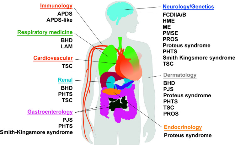
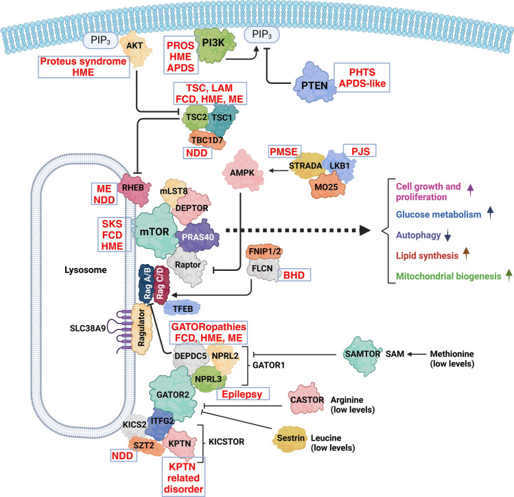
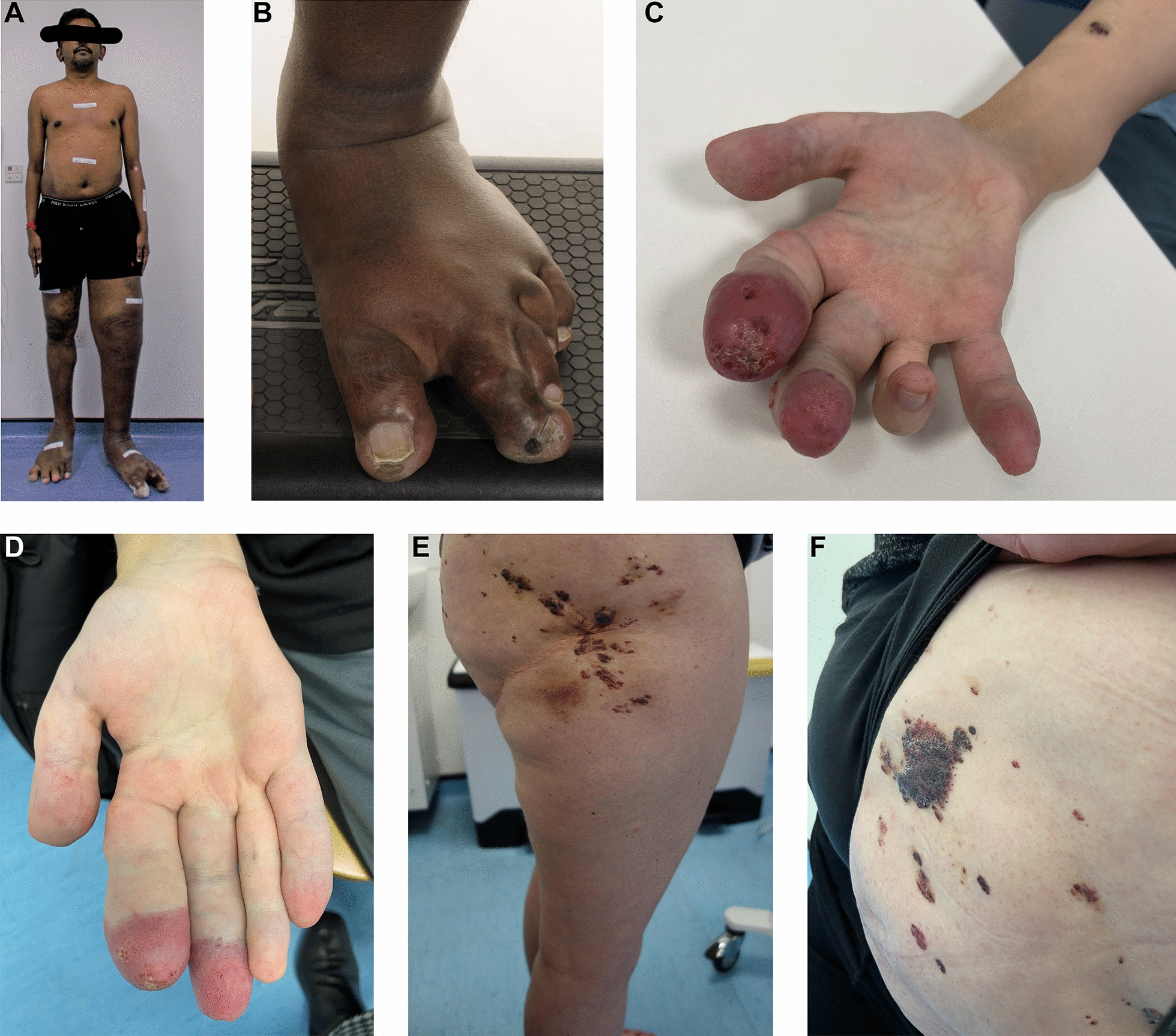
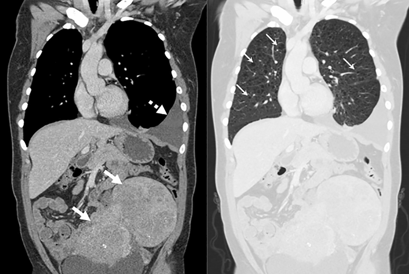
Mechanistic target of rapamycin (mTOR) is a highly conserved serine/threonine kinase that regulates key cellular processes including cell growth, autophagy and metabolism. Hyperactivation of the mTOR pathway causes a group of rare and ultrarare genetic diseases. mTOR pathway diseases have diverse clinical manifestations that are managed by distinct medical disciplines but share a common underlying molecular basis. There is a now a deep understanding of the molecular underpinning that regulates the mTOR pathway but effective treatments for most mTOR pathway diseases are lacking. Translating scientific knowledge into clinical applications to benefit the unmet clinical needs of patients is a major challenge common to many rare diseases. In this article we expound how mTOR pathway diseases provide an opportunity to coordinate basic and translational disease research across the group, together with industry, medical research foundations, charities and patient groups, by pooling expertise and driving progress to benefit patients. We outline the germline and somatic mutations in the mTOR pathway that cause rare diseases and summarise the prevalence, genetic basis, clinical manifestations, pathophysiology and current treatments for each disease in this group. We describe the challenges and opportunities for progress in elucidating the underlying mechanisms, improving diagnosis and prognosis, as well as the development and approval of new therapies for mTOR pathway diseases. We illustrate the crucial role of patient public involvement and engagement in rare disease and mTOR pathway disease research. Finally, we explain how the mTOR Pathway Diseases node, part of the Research Disease Research UK Platform, will address these challenges to improve the understanding, diagnosis and treatment of mTOR pathway diseases.

Keywords: AKT; Birt-Hogg-Dubé; Everolimus; PI3K; PTEN; Peutz-Jeghers; Rapamycin; Rare diseases; Tuberous sclerosis complex; mTOR.


Conflict of interest statement

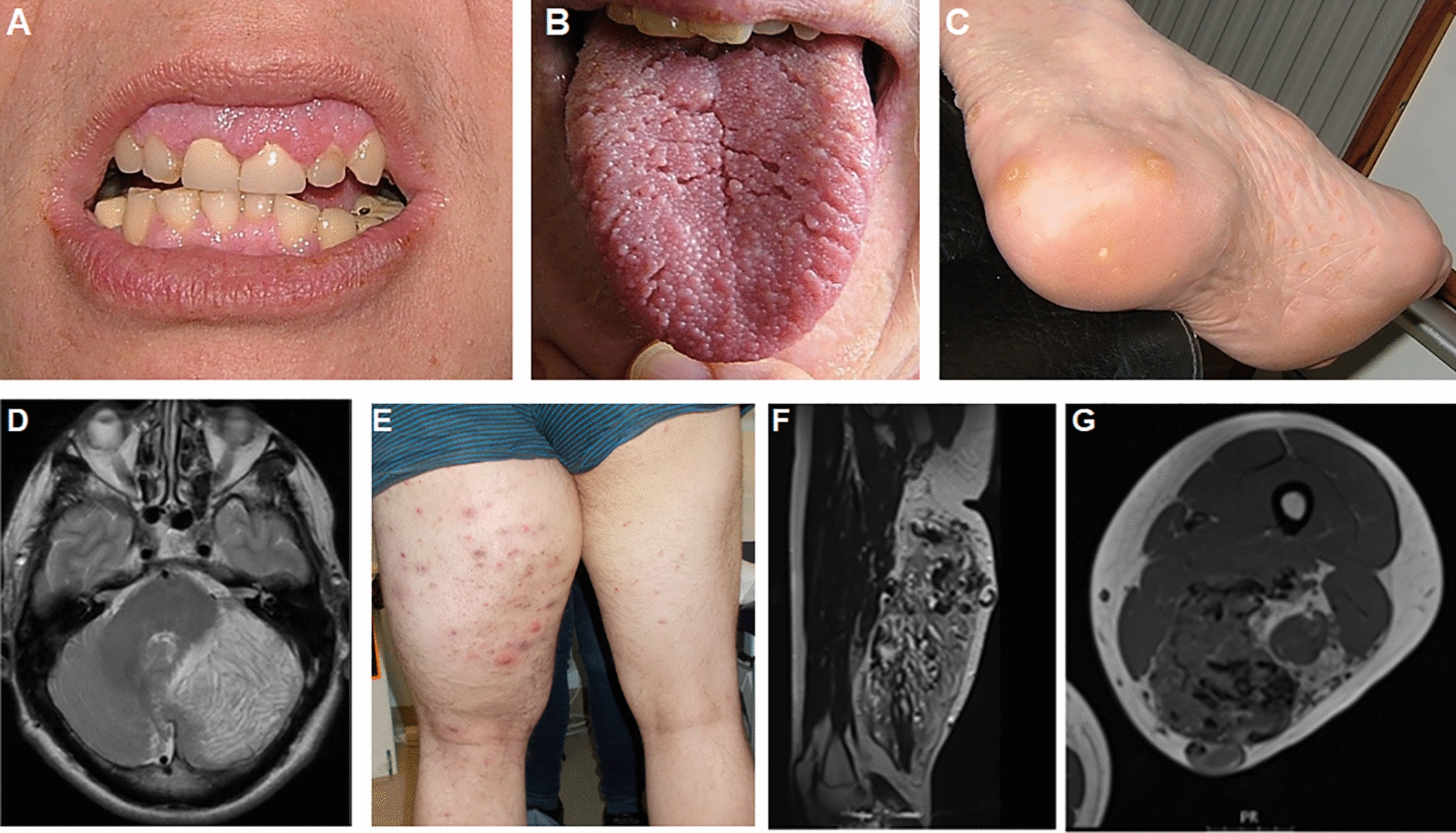
Declarations. Ethics approval and consent to participate: Patient data were recorded as part of routine clinical practice and not as part of a research study so do not require ethical approval, apart from data in Fig. 9 which was part of a study approved by the Wales REC committee REC number 22/WA/0326. Consent for publication: Consent for publication was given for all patient related data. Competing interests: SD is a UCB employee and SD receives stock or stock options from their employment. SH has received consultancy fees and research funding from Pharming in the past. DP and JMB are members of the scientific advisory board for CoSyne.

Figures

Fig. 1
Fig. 1
The different medical specialities that manage patients with mTOR pathway diseases. APDS: activated PI3K delta syndrome, BHD: Birt-Hogg-Dubé, FCDIIA/B: focal cortical dysplasia type IIA/B, HME: hemimegalencephaly, LAM: lymphangioleiomyomatosis, ME: megalencephaly, PMSE: polyhydramnios, megalencephaly and symptomatic epilepsy syndrome, PHTS: PTEN hamartoma tumour syndrome, PJS: Peutz-Jeghers syndrome, PROS: PIK3CA-related overgrowth spectrum, TSC: tuberous sclerosis complex
Fig. 2
Fig. 2
The mTOR pathway with rare diseases (shown in red) caused by mutations in specific proteins. APDS: activated PI3K delta syndrome, BHD: Birt-Hogg-Dubé, FCD: focal cortical dysplasia type IIA/B, HME: hemimegalencephaly, LAM: lymphangioleiomyomatosis, ME: megalencephaly, NDD: neurodevelopmental disease, PMSE: polyhydramnios, megalencephaly and symptomatic epilepsy syndrome, PHTS; PTEN hamartoma tumour syndrome, PJS: Peutz-Jeghers syndrome, PROS: PIK3CA-related overgrowth spectrum, SKS: Smith Kingsmore syndrome, TSC: tuberous sclerosis complex
Fig. 3
Fig. 3
PHTS clinical manifestations. A Oral mucosal features. Gingival papillomatosis and neuromas may encroach on the dentition and cause difficulty with dental hygiene. B Papillomatosis of the tongue. C Plantar and palmar keratoses. Some are hyperkeratotic and may resemble viral verrucous lesions. D Axial T2 MRI image showing abnormal tissue in the left cerebellar hemisphere with the characteristic tigroid appearance and apparently preserved cerebellar folia of Lhermitte-Duclos Disease. E Segmental overgrowth of left thigh. This presented in adolescence, with no evidence of the lesion in early childhood. F, G MRI images demonstrating vascular malformation with fast and slow flow elements infiltrating muscle and fat
Fig. 4
Fig. 4
Peutz-Jeghers syndrome polyps. A 42-year-old female PJS patient being treated with ischaemic polypectomy
Fig. 5
Fig. 5
Neurological manifestations of TSC. 34-year-old female TSC patient with a germline TSC1 null mutation (Pro266ArgfsTer5). T2w (A) and postcontrast T1w (B) MRI images showing a large enhancing mass (SEGA) at the foramen of Monro with obstructive hydrocephalus and surrounding oedema. Background multiple cortical tubers were present, one of which is seen in the right frontal lobe (white arrow). C Histology of subependymal giant cell astrocytoma showing of spindle and epithelioid cells with abundant eosinophilic cytoplasm. D Immunohistochemistry for thyroid transcription factor 1 (TTF-1) displays expression in the nuclei. Bar: 50 mm
Fig. 6
Fig. 6
Clinical manifestations of KPTN-related disorder. A Craniofacial appearance of a 19-year-old male with KPTN-related disorder, a macrocephalic neurodevelopmental disorder with subtle facial dysmorphology, including frontal bossing, a prominent chin, small downslanting palpebral fissures and a broad nasal tip. Reproduced from [184]. B Occipitofrontal circumference in centimeters for a male individual with KPTN-related disorder. OFC increased from below the 50th centile (mean) at birth to over two standard deviations (SD) above the mean by the age of two years (blue line). Centiles given in brackets. Reproduced from [190]
Fig. 7
Fig. 7
Hemimegalencephaly MRI. MRI from a female at age 2 months. A, B T2w and T2w images showing enlargement of the right occipital and adjacent posterior temporal/parietal lobes with diffuse thickening of the cortex, abnormal shallow overlying sulcation and underlying white matter signal abnormality. Findings are consistent with posterior quadrantic dysplasia (localised or hemi hemimegalencephaly)
Fig. 8
Fig. 8
Hemimegalencephaly histology. A, B Complex histological abnormalities in posterior quadrantic dysplasia (localised or hemi hemimegalencephaly) (Luxol fast blue-Nissl staining). The cortical lamination is abnormal with myelinated fibre layer in the middle of the cortex. The neurons are almost exclusively pyramidal cells with absence of granular cell layers. C The lamina 1 contains increased number of cells, occasionally pyramidal cells, and in areas parallel running myelinated fibres are seen. D In places the grey and white matter demarcation is blurred due to large neurons splaying into the subcortical white matter, immunohistochemistry for neuronal nuclear protein N—NeuN). E Immunohistochemistry for NeuN. F Occasional the heterotopic nodules are also noted. (Bars A, B, D, E: 1 mm; Bars C, F: 100 µm)
Fig. 9
Fig. 9
Focal cortical dysplasia (FCD) type IIb. A Slicing of the lesion after formalin fixation reveals blurred demarcation between cortex and white matter, confirmed by Luxol fast blue-Nissl staining (LFB-N) (B), also showing neurons splaying into the white matter. C Dyslamination of the cortex with presence of large (dysmorphic) neurons by immunohistochemistry for neuronal nuclear protein N (NeuN). D The dysmorphic neurons have abnormal orientation and frequently show chromatolysis (haematoxylin & eosin staining). E The ballooned cells are most common in the subcortical white matter (LFB-N). F Immunohistochemistry for CD34 may label glial cells with bushy ramified processes around some of the balloon cells. Bar A: 1 cm, Bars B, C: 1 mm; Bars D-F: 100 mm
Fig. 10
Fig. 10
Clinical manifestations of PROS. A, B A 44-year-old male with CLOVES—congenital lipomatous malformation of the trunk, segmental overgrowth of both legs, extensive vascular and venous malformations, and scoliosis. There is macrodactyly of the toes on both feet with typical appearance (wide sandal gap and relatively short hallux). Skin biopsy of affected tissue identified a pathogenic variant in PIK3CA (c.1039-1041AAA. P.Val346_347ins Lys) in 50% of cells. C, D Extensive venous malformations of the right hand with leaking lymphangiectactic blisters at the tips of the index and middle finger and macrodactyly. Skin biopsy of the affected tissue identified a gain-of-function pathogenic variant in PIK3CA (c.1633G > A. P.Glu545Lys) in 13% of cells. E, F A 40-year-old female with a lympho-veno-vascular malformation of the right thigh and buttock and segmental overgrowth of the right leg (diagnosed as Klippel–Trénaunay syndrome). Current problems with pain and leakage from lymphangiectatic blisters (seen in the images). Skin biopsy identified a pathogenic variant in PIK3CA c.3140A > G. p.His1047arg in 4% of cells
Fig. 11
Fig. 11
Clinical manifestations of LAM. Coronal CT scan of the chest and abdomen of a patient with LAM. Soft tissue windows (left) show a large, complex abdominal lymphangioleiomyoma (white arrows) and a left sided chylous pleural effusion (dashed arrow). Lung windows (right) show multiple air-filled lung cysts distributed throughout the lung parenchyma (some highlighted by small arrows)

Similar articles

References

    1. O’Connor DJ, Gabaldo M, Aartsma-Rus A, Hechtelt JA. Defining rare conditions in the era of personalized medicine. Nat Rev Drug Discov. 2023;22(11):857–8. - PubMed
    1. Nguengang Wakap S, Lambert DM, Olry A, Rodwell C, Gueydan C, Lanneau V, et al. Estimating cumulative point prevalence of rare diseases: analysis of the Orphanet database. Eur J Hum Genet. 2020;28(2):165–73. - PMC - PubMed
    1. Fermaglich LJ, Miller KL. A comprehensive study of the rare diseases and conditions targeted by orphan drug designations and approvals over the forty years of the Orphan Drug Act. Orphanet J Rare Dis. 2023;18(1):163. - PMC - PubMed
    1. UK Rare Diseases Framework [Available from: https://www.gov.uk/government/publications/uk-rare-diseases-framework. Accessed on 30th Sept 2024
    1. mTOR Pathway Diseases [Available from: https://rd-research.org.uk/node/mtor-pathway-diseases/. Accessed on 30th Sept 2024

MeSH terms

Substances