. 2025 May 27;23(1):384.
doi: 10.1186/s12951-025-03458-z.

DNA binding effects of LDH nanozyme for aseptic osteolysis mitigation through STING pathway modulation

Affiliations

DNA binding effects of LDH nanozyme for aseptic osteolysis mitigation through STING pathway modulation

Zi Fu et al. J Nanobiotechnology. .

Abstract

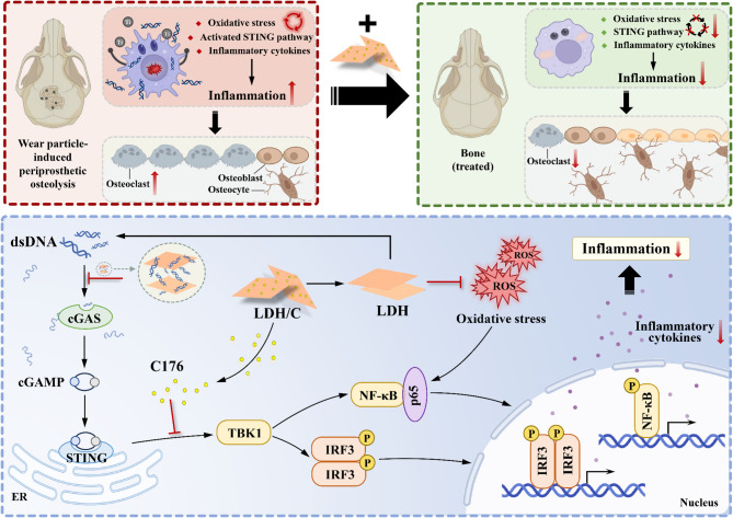
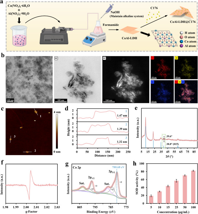
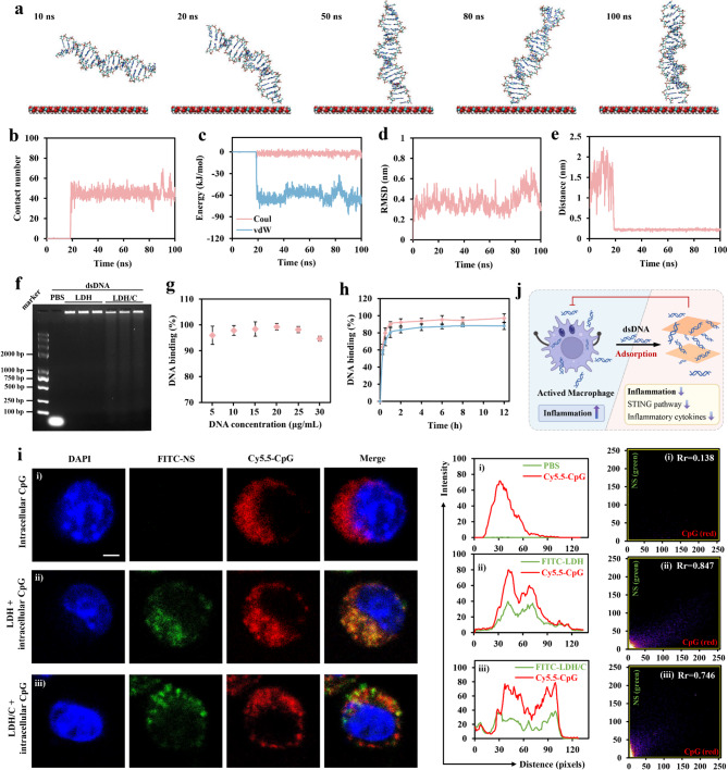
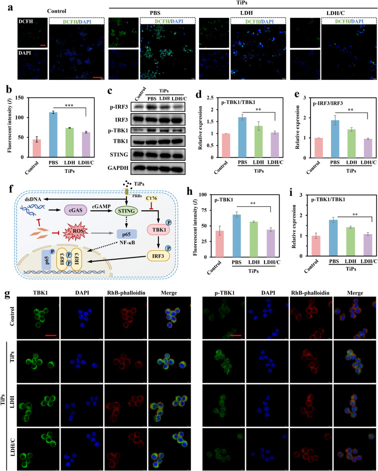
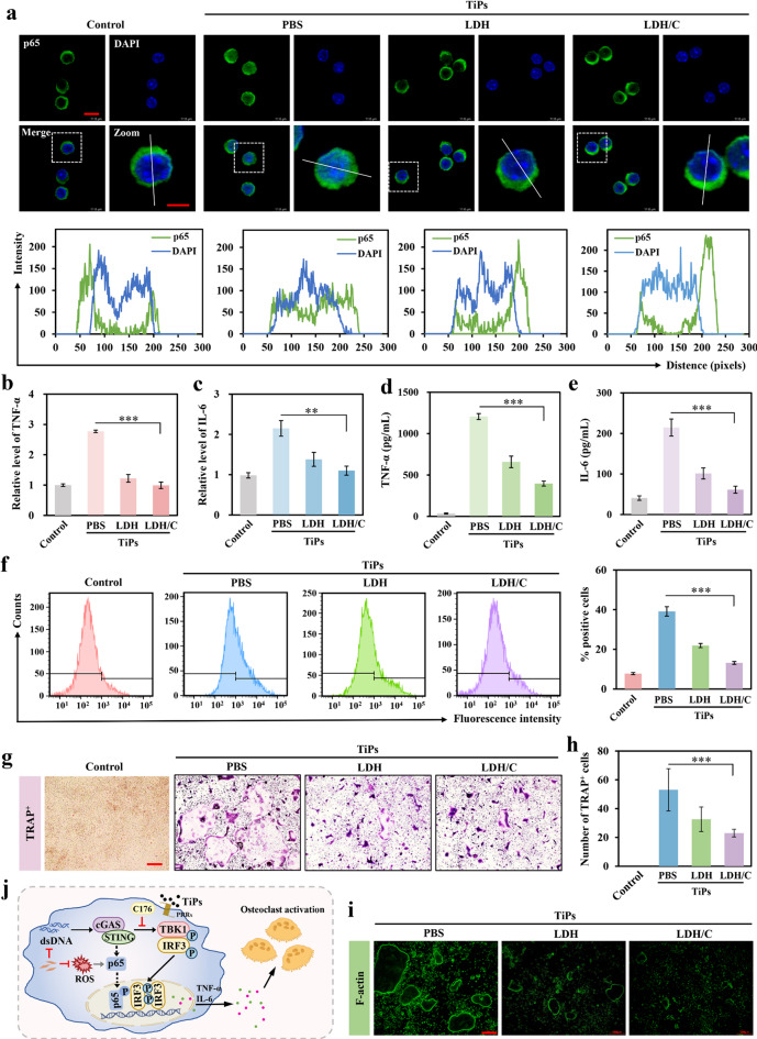
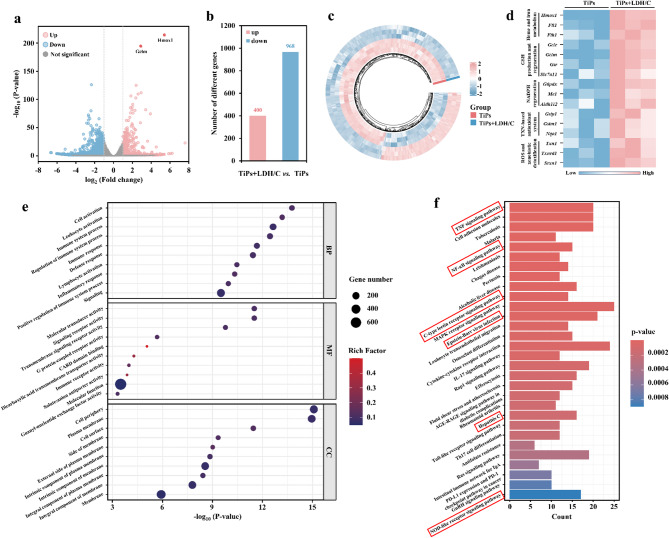
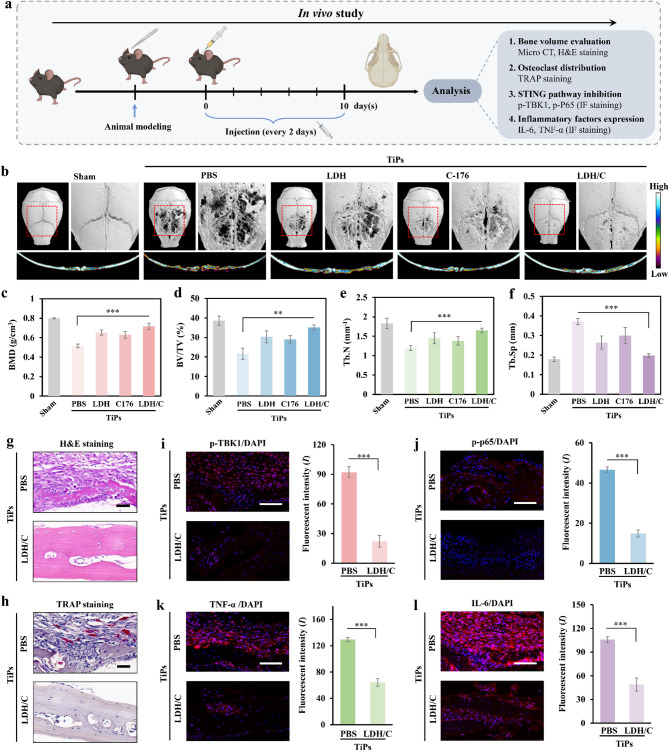
Persistent and intense inflammation is recognized as the primary cause of wear-particle-induced aseptic osteolysis, which ultimately resulting in aseptic prosthesis loosening. Reducing inflammation plays a significant role in mitigating osteolysis, and the STING pathway has emerged as a promising therapeutic target for its prevention. Specifically, damaged periprosthetic cells of aseptic osteolysis release double-stranded DNA (dsDNA) into the osteolytic microenvironment, serving as a specific stimulus for the STING pathway. Herein, we found that layered double hydroxide (LDH) nanozyme exhibited a robust DNA-binding capacity primarily mediated by van der Waals interactions, which showed superior performance in inhibiting dsDNA-induced inflammation of aseptic osteolysis. Importantly, such binding capability enabled effective co-loading LDH with STING inhibitor C176, thus facilitating inhibition of the STING pathway. Such synergistic actions contributed to ameliorate the inflammatory milieu and remodel the osteolysis microenvironment successfully to reduce cranial bone damage, which was confirmed on animal model of osteolysis. Collectively, this strategy demonstrated an effective approach by utilizing synergistic effects to establish a positive feedback loop in the treatment of osteolysis, thereby alleviating TiPs-induced periprosthetic osteolysis and preventing postoperative complications.

Keywords: Inflammation; Nanozyme; Osteolysis; Oxidative stress; cGAS-STING pathway.


Conflict of interest statement

Declarations. Ethics and consent to publish: All animal experiments were authorized by the Ethics Committee of the Shanghai Jiao Tong University (A2023128-001). Competing interests: The authors declare no competing interests.

Figures

Scheme 1
Scheme 1
The synergistic mechanism for the mitigation of aseptic osteolysis with LDH/C-based therapeutics
Fig. 1
Fig. 1
Characterization of LDH and LDH/C. (a) The preparation schematic of LDH and LDH/C. (b) TEM image, bright-field image, dark-field image, and corresponding element mapping of LDH nanosheets. (c) AFM image, and (d) corresponding thickness of LDH. (e) XRD patterns and (f) EPR spectra of LDH nanosheets. (g) A high-resolution XPS spectrum of Co 2p in LDH nanosheets. (h) The SODase activity of LDH nanosheets with different concentration. The quantitative data in h reported as means ± S.D. from three independent replicates, and statistical significance was ascertained by a Student’s t-test, with signifiers for P-values at *P < 0.05, **P < 0.01, and ***P < 0.001)
Fig. 2
Fig. 2
LDH/C exhibits strong dsDNA-binding capabilities and effectively eliminates free dsDNA. (a) Representative snapshots for the binding process of dsDNA to LDH nanosheets at different key time points. (b) The time evolution of the contact number for the dsDNA adsorption process to the LDH nanosheets. (c) The van der Waals (vdW) and coulombic (Coul) energies between the dsDNA and the LDH nanosheets. (d) The root-mean-square deviation (RMSD) of dsDNA presented as a function of simulation time. (e) Time evolution of the minimum distance of the base pair from the LDH surface. (f) Agarose gel electrophoresis of free dsDNA, dsDNA + LDH, and dsDNA + LDH/C on 1.5% agarose gel. (g) The dsDNA binding percentages of LDH with various DNA concentration. (h) The dsDNA binding percentages of LDH or LDH/C as a function of incubation time. (i) Representative images, relative fluorescence intensity spectra, and Pearson’s coefficient (Rr) of Raw 264.7 cells incubation with dsDNA (red), LDH (green), and DAPI staining (blue) for intracellular colocalization measured (scale bars: 2.5 μm). (j) The scheme of dsDNA absorption by LDH/C. The quantitative data in g and h reported as means ± S.D. from three independent replicates, and statistical significance was ascertained by a Student’s t-test, with signifiers for P-values at *P < 0.05, **P < 0.01, and ***P < 0.001
Fig. 3
Fig. 3
LDH/C inhibited over-activation of the STING pathway in vitro. (a) Representative images of Raw 264.7 cells for ROS detection with DCFH-DA (green) and DAPI staining (blue) after different treatments, scale bar: 100 μm. (b) Relative fluorescence intensity quantitative analysis of Raw 264.7 cells for ROS detection after different treatments. (c) Representative immunoblotting of p-IRF3, IRF3, p-TBK1, TBK1, and STING in BMDMs after indicated treatments, and GAPDH was employed as a control. (d-e) The relative protein expression quantitative analysis of (d) p-TBK1/TBK1, and (e) p-IRF3/IRF3. (f) Diagrammatic representation of the mechanism by which LDH/C inhibits the STING pathway. (g) Representative images of Raw 264.7 cells for TBK1 and p-TBK1 (green), RhB-phalloidin (red), and DAPI (blue) staining after different treatments (scale bar: 20 μm). (h-i) The relative fluorescence intensity quantitative analysis of (h) p-TBK1, and (i) p-TBK1/TBK1 in Raw 264.7 cells after various treatments. The quantitative data in b, d-e, and h-i reported as means ± S.D. from three independent replicates, and statistical significance was ascertained by a Student’s t-test, with signifiers for P-values at *P < 0.05, **P < 0.01, and ***P < 0.001
Fig. 4
Fig. 4
LDH/C alleviates inflammation and inhibits osteoclast activation. (a) Representative images and intracellular colocalization fluorescence intensity spectra of Raw 264.7 cells incubation with p65 (green) and DAPI staining (blue) (scale bars: 10 μm and 5 μm). (b-c) The relative expressions level of (b) TNF-α and (c) IL-6 assessed by qPCR. ELISA of (d) TNF-α and (e) IL-6. (f) The M1 macrophages assessed by flow cytometry and the quantification of the positive rate of M1 macrophages with treatment of separate experimental groups. (g) Representative TRAP staining images of osteoclasts (scale bar = 500 μm). (h) Quantitation of number of TRAP-positive multinucleated cells with varying treatments. (i) Representative images for podosome belts with green fluorescent F-actin (scale bar = 500 μm). (j) A description of the mechanism of LDH/C-induced inhibition of osteoclast activation. The quantitative data in b-f and h reported as means ± S.D. from three independent replicates, and statistical significance was ascertained by a Student’s t-test, with signifiers for P-values at *P < 0.05, **P < 0.01, and ***P < 0.001
Fig. 5
Fig. 5
Differentially expressed genes (DEG) and involved pathway of Raw 264.7 cells treated with TiPs followed by LDH/C treatment. (a) Volcano plot showing the distribution and expression changes of differential genes in the TiPs and TiPs + LDH/C groups. Genes highlighted in blue (indicating downregulated genes) and red (indicating upregulated genes) with absolute fold change > 2 and P value < 0.05, respectively. (b) The number of different genes after treated by TiPs and TiPs + LDH/C. (c) Heatmap of significantly differentially expressed genes from TiPs + LDH/C vs. TiPs (adjusted P-value cut-off < 0.05). (d) Heatmap of significantly differentially expressed genes with inflammation from TiPs + LDH/C vs. TiPs. (e, f) The (e) GO, and (f) KEGG enrichment of downregulated DEG in the TiPs and TiPs + LDH/C groups
Fig. 6
Fig. 6
LDH/C mitigates the pathological manifestations associated with TiPs-induced mice inflammation osteolysis. (a) The experimental timeline for conducting the mice inflammation osteolysis experiment in vivo. (b) Micro-CT scanner reconstructions and transverse sections (pseudocolor) of the calvarial anatomy from each experimental cohort. Quantitatively analyzed parameters of (c) bone mineral density (BMD), (d) bone volume percentage (BV/TV), (e) trabecular number (Tb.N.), and (f) trabecular separation (Tb.Sp). (g) Calvaria sections from each mouse group subjected to H&E staining (scale bar = 50 μm). (h) Calvaria sections from each mouse group subjected to TRAP staining (scale bar = 50 μm). Immunohistochemical assays and quantitative analysis for (i) p-TBK1, (j) p-p65, (k) TNF-α, and (l) IL-6 (all marked in red while DAPI marked in blue) (scale bars at 200 μm and zoomed in at 100 μm for detailed images). The quantitative data in c-f and i-l reported as means ± S.D. from five independent replicates, and statistical significance was ascertained by a Student’s t-test, with signifiers for P-values at *P < 0.05, **P < 0.01, and ***P < 0.001

Similar articles

References

    1. Ormsby RT, Solomon LB, Yang D, Crotti TN, Haynes DR, Findlay DM, Atkins GJ. Osteocytes respond to particles of clinically-relevant conventional and cross-linked polyethylene and metal alloys by up-regulation of resorptive and inflammatory pathways. Acta Biomater. 2019;87:296–306. - PubMed
    1. Wu Y, He F, Zhang C, Zhang Q, Su X, Zhu X, Liu A, Shi W, Lin W, Jin Z, Yang H, Lin J. Melatonin alleviates titanium nanoparticles induced osteolysis via activation of butyrate/GPR109A signaling pathway. J Nanobiotechnol. 2021;19(1):170. - PMC - PubMed
    1. Springer BD, Sotile WM. The psychology of total joint arthroplasty. J GROMACS Arthroplasty. 2020;35(6S):S46–9. - PubMed
    1. Tian Y, Terkawi MA, Onodera T, Alhasan H, Matsumae G, Takahashi D, Hamasaki M, Ebata T, Aly MK, Kida H, Shimizu T, Uetsuki K, Kadoya K, Iwasaki N. Blockade of XCL1/Lymphotactin ameliorates severity of periprosthetic osteolysis triggered by Polyethylene-Particles. Front Immunol. 2020;11:1720. - PMC - PubMed
    1. Fritz EA, Glant TT, Vermes C, Jacobs JJ, Roebuck KA. Chemokine gene activation in human bone marrow-derived osteoblasts following exposure to particulate wear debris. J Biomed Mater Res A. 2006;77(1):192–201. - PubMed

LinkOut - more resources