Review
. 2025 Apr 22;14(5):422.
doi: 10.3390/antibiotics14050422.

The Role of Flexibility in the Bioactivity of Short α-Helical Antimicrobial Peptides

Affiliations
Review

The Role of Flexibility in the Bioactivity of Short α-Helical Antimicrobial Peptides

Daniel Balleza. Antibiotics (Basel). .

Abstract

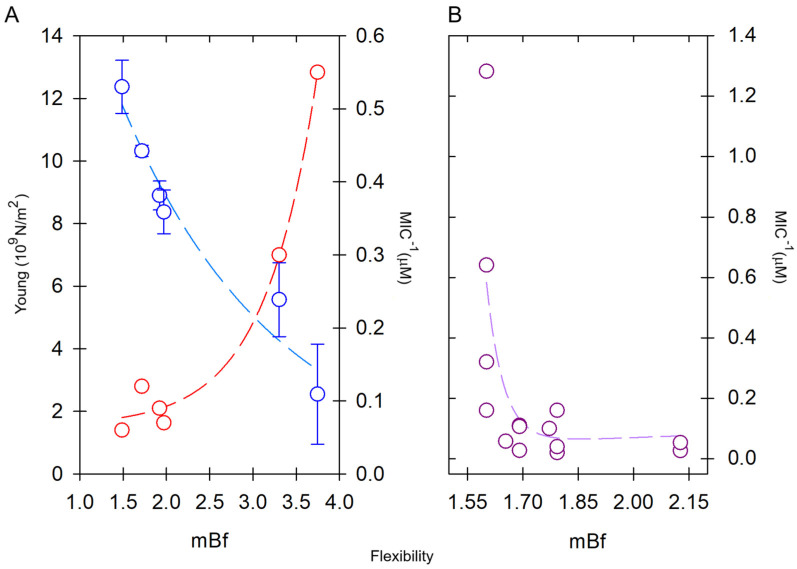
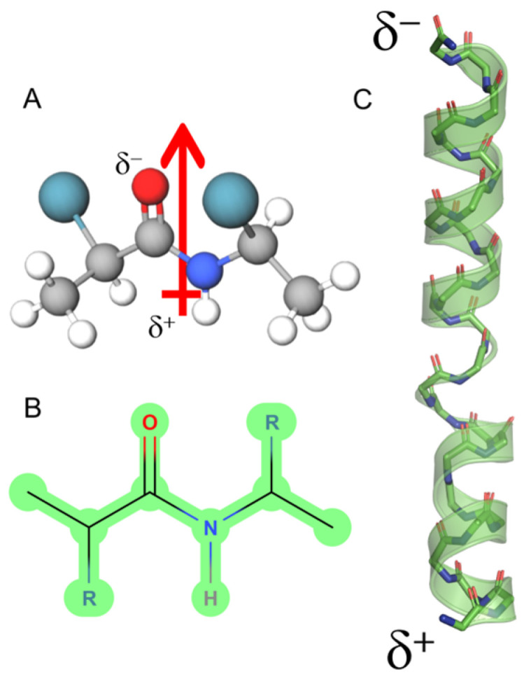
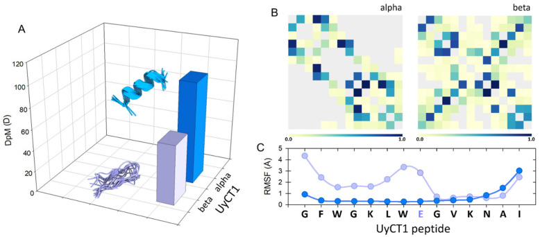
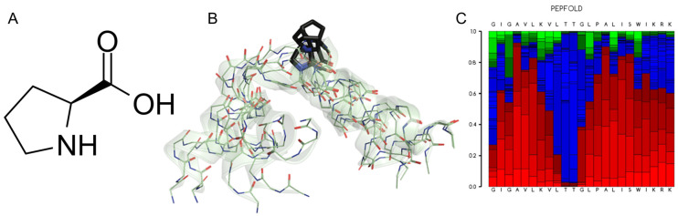
The formation of aqueous pores through the interaction of amphipathic peptides is a process facilitated by the conformational dynamics typical of these biomolecules. Prior to their insertion with the membrane, these peptides go through several conformational states until they finally reach a stable α-helical structure. The conformational dynamics of these pore-forming peptides, α-PFP, is, thus, encoded in their amino acid sequence, which also predetermines their intrinsic flexibility. However, although the role of flexibility is widely recognized as fundamental in their bioactivity, it is still unclear whether this parameter is indeed decisive, as there are reports favoring the view of highly disruptive flexible peptides and others where relative rigidity also predetermines high rates of permeability across membranes. In this review we discuss in depth all those aspects linked to the conformational dynamics of these small biomolecules and which depend on the composition, sequence and dynamic performance both in aqueous phase and in close interaction with phospholipids. In addition, evidence is provided for the contribution of the known carboxyamidation in some well-studied α-PFPs, which are preferentially associated with sequences intrinsically more rigid than those not amidated and generally more flexible than the former. Taken together, this information is of great relevance for the optimization of new antibiotic peptides.

Keywords: dipolar moment; intrinsic flexibility; short antimicrobial peptides.


Conflict of interest statement

The author declares no conflict of interest.

Figures

Figure 6
Figure 6
Effect of the intrinsic flexibility on pore-forming peptides with α-helical structure (α-PFPs). (A) Young’s modulus (red) is high in rigid peptides but decreases in flexible peptides, which are more active in terms of the MIC (blue). The flexibilities of the peptides are compared in increasing order: Maculatin-1.1 > Brevinin-1 > Pseudin-2 > ranateurin-1 >> HP(2-20) > CAP18. Young’s modulus data were taken from Liu et al. [129]. (B) Comparison of the bioactivity of six short scorpion peptides in increasing order of flexibility: VmCT1 > TsAP2 > Stigmurin > Ctriporin > Uy234 > Pin2. In this case, the most active peptides were those with low mean flexibility index (mBf), i.e., rigid. MIC data were reviewed for different bacteria in ref. [107].
Figure 1
Figure 1
Dipole moment of an α-helix peptide. (A) A peptide unit showing the orientation of the dipole vector (arrow). (B) The peptide bond in Fisher projection. (C) Schematic view of the macrodipole. The total assembly of the α-helix produces a macrodipole where the N-end exhibits a positive partial charge (δ+), whereas the C-end exposes a negative partial charge (δ). The solved structure for melittin is depicted (PDB ID:2MLT).
Figure 2
Figure 2
(A) Conceptual representation of the magnitude of the dipole moment (DpM) of the UyCT1 peptide as a typical α-helix (blue) or a β-sheet (light violet), where the DpM is significantly reduced from μ = 96.8 ± 20 D to μ = 56.8 ± 10.3 D. (B) Intramolecular contacts in alpha (left) and beta (right) configurations. (C) Atomic fluctuations (RMSF) mainly in side chains; in this peptide, the presence of a glutamate residue near the center of the structure, a residue of high intrinsic flexibility, favors a higher degree of atomic fluctuations towards the N-end of this peptide.
Figure 3
Figure 3
(A) L-proline, (2S)-pyrrolidine-2-carboxylic acid. (B) Five superimposed structures for Melittin generated by the PepFold server (https://bioserv.rpbs.univ-paris-diderot.fr, accessed on 12 December 2024) to reveal the Pro-kink motif. The role of the Pro-14 residue (black) stands out, which determines a high degree of potential mobility for the N-terminus of this peptide. (C) Graphical representation of the probabilities of potential substructures using the next code: helical (red), extended (green) and coil (blue).
Figure 4
Figure 4
Two different topologies for the configuration of aqueous pores through lipid membranes. (A) Toroidal pore, a typical interpretation for the mechanism of action for melittin and magainins. (B) Barrel-stave pore, a typical configuration found in voltage-dependent ion channels. In the schematic representation of the phospholipids, the head groups are shown in blue and the peptides are shown fully helical for melittin (green) and alamethicin (orange).
Figure 5
Figure 5
Types of curvature and self-assembly of lipids in nanostructures. (A) Inverted micelles, where C0 < 0 leads to inverted phases. (B) Planar lipid bilayer, C0 ≈ 0. (C) Micelles, C0 > 0. See text for details.
Figure 7
Figure 7
(A) Map of intrapeptide contacts for C-amidated melittin (PDB: 2mlt, left) and (B) the T10S C-carboxylated variant (MLT-S, right). Contact types are color-coded on the scale shown on each bar. The MLT-S variant shows some middle-distance contacts, which are less frequent in the wild-type peptide, in agreement with its lower intrinsic flexibility.

Similar articles

References

    1. Vorov O.K., Livesay D.R., Jacobs D.J. Nonadditivity in conformational entropy upon molecular rigidification reveals a universal mechanism affecting folding cooperativity. Biophys. J. 2011;100:1129–1138. doi: 10.1016/j.bpj.2011.01.027. - DOI - PMC - PubMed
    1. Uversky V.N. Protein intrinsic disorder and structure-function continuum. Prog. Mol. Biol. Transl. Sci. 2019;166:1–17. doi: 10.1016/bs.pmbts.2019.05.003. - DOI - PubMed
    1. Sun Z., Liu Q., Qu G., Feng Y., Reetz M.T. Utility of B-Factors in Protein Science: Interpreting Rigidity, Flexibility, and Internal Motion and Engineering Thermostability. Chem. Rev. 2019;119:1626–1665. doi: 10.1021/acs.chemrev.8b00290. - DOI - PubMed
    1. de Sá Ribeiro F., Lima L.M.T.R. Linking B-factor and temperature-induced conformational transition. Biophys. Chem. 2023;298:107027. doi: 10.1016/j.bpc.2023.107027. - DOI - PubMed
    1. Khatua P., Sinha S.K., Bandyopadhyay S. Size-Dependent Conformational Features of Aβ17-42 Protofilaments from Molecular Simulation Studies. J. Chem. Inf. Model. 2017;57:2378–2392. doi: 10.1021/acs.jcim.7b00407. - DOI - PubMed

LinkOut - more resources