. 2025 May 9;14(5):484.
doi: 10.3390/antibiotics14050484.

Heterogeneity of Biofilm Formation Among Staphylococcus aureus and Coagulase-Negative Staphylococcus Species in Clinically Relevant Intravenous Fat Emulsions

Affiliations

Heterogeneity of Biofilm Formation Among Staphylococcus aureus and Coagulase-Negative Staphylococcus Species in Clinically Relevant Intravenous Fat Emulsions

Gustavo R Alvira-Arill et al. Antibiotics (Basel). .

Abstract

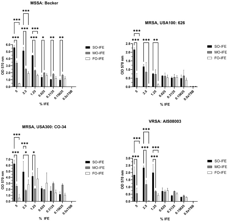
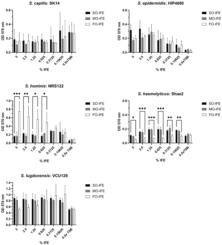
Background: Compared to soybean oil intravenous fat emulsion (SO-IFE), use of mixed-oil IFE (MO-IFE) is associated with reduced rates of catheter-related bloodstream infections caused by coagulase-negative Staphylococcus species (CoNS) in pediatric patients receiving parenteral nutrition. Methods: Using an in vitro biofilm model, this study aimed to assess the impact of IFEs on biofilm formation among Staphylococcus species. S. aureus, S. capitis, S. epidermidis, S. haemolyticus, S. hominis, and S. lugdunensis were cultivated as biofilms in media supplemented with SO-IFE, MO-IFE, or fish oil IFE (IFE). Biomass was quantified by the crystal violet method, and follow-up planktonic growth assays assessed antimicrobial effects of IFEs. Results: Compared to SO-IFE, MO-IFE and FO-IFE significantly inhibited biofilm formation of S. aureus but did not impact planktonic growth. Contrary to clinical data, CoNS biofilm formation was not impacted by any of the IFEs tested. S. aureus biofilm inhibition in IFEs was further investigated by comparing differences following growth in SO-IFE supplemented with capric acid, docosahexaenoic acid (DHA), or eicosapenaenoic acid (EPA) to concentrations matching those of MO-IFE. Capric acid supplementation was associated with significant reduction in biofilm formation compared to SO-IFE alone. However, this was attributed to a bactericidal effect based on follow-up planktonic growth assays. Conclusions: These results suggest that biofilm formation in S. aureus is variably impacted by fatty acid composition in clinically relevant IFEs, with capric acid exhibiting bactericidal activity against tested isolates.

Keywords: Staphylococcus; biofilm; catheter-related infections; fat emulsions; intravenous; parenteral nutrition.


Conflict of interest statement

J.S.S. reports grant funding from Merck Sharp and Dohme for research outside of this work. The other authors declare no conflicts of interest.

Figures

Figure 1
Figure 1
Biofilm formation of select Staphylococcus aureus isolates in varying concentrations of SO-IFE, MO-IFE, and FO-IFE. Representative isolates of four categories of S. aureus: methicillin-susceptible S. aureus (MSSA), methicillin-resistant S. aureus (MRSA) clade USA 100, MRSA clade USA 300, and vancomycin-resistant S. aureus (VRSA) were cultivated as biofilms for 24 h in 0.5× tryptic soy broth (TSB) supplemented with serial dilutions of soybean oil intravenous fat emulsion (SO-IFE), mixed-oil intravenous fat emulsion (MO-IFE), or fish oil intravenous fat emulsion (FO-IFE). Data expressed as mean ± standard deviation. N = 2 replicates. *, p < 0.05; **, p < 0.01; ***, p < 0.001 using two-way ANOVA and Dunnett’s multiple comparison post-test to SO-IFE at each concentration.
Figure 2
Figure 2
Biofilm formation of an expanded panel of Staphylococcus aureus isolates in clinically relevant concentrations of SO-IFE, MO-IFE, and FO-IFE. Laboratory or clinical isolates of S. aureus categorized as methicillin-susceptible S. aureus (MSSA), methicillin-resistant S. aureus (MRSA) clade USA 100, MRSA clade USA 300, and vancomycin-resistant S. aureus (VRSA) were cultivated as biofilms for 24 h in 0.5× tryptic soy broth supplemented with 5% soybean oil intravenous fat emulsion (SO-IFE), mixed-oil intravenous fat emulsion (MO-IFE), or fish oil intravenous fat emulsion (FO-IFE). Data expressed as mean ± standard deviation. N = 2 replicates. *, p < 0.05; **, p < 0.01; ***, p < 0.001 using two-way ANOVA and Dunnett’s multiple comparison post-test.
Figure 3
Figure 3
Impact of IFEs on Staphylococcus aureus planktonic growth. Planktonic cultures of laboratory or clinical isolates of S. aureus categorized as methicillin-susceptible S. aureus (MSSA), methicillin-resistant S. aureus (MRSA) clade USA 100, MRSA clade USA 300, and vancomycin-resistant S. aureus (VRSA) were grown in 0.5× tryptic soy broth (TSB) supplemented with 10% SO-IFE (soybean oil IFE), MO-IFE (mixed-oil IFE), or 5% FO-IFE (fish oil IFE) and subsequently enumerated by drop plate method. Data expressed as mean and standard deviation. N = 2 replicates. *, p < 0.05; **, p < 0.01; ***, p < 0.001 using a two-way ANOVA and Dunnett’s multiple comparison post-test.
Figure 4
Figure 4
Supplementation of SO-IFE with capric acid significantly reduces biofilm formation in select Staphylococcus aureus isolates. Representative isolates of S. aureus were cultivated as biofilms for 24 h in 0.5× tryptic soy broth (TSB) and 10% SO-IFE (soybean oil IFE) supplemented with serial dilutions of capric acid, DHA, and EPA at starting concentrations of 58.05 mM, 6.85 mM, and 7.44 mM, respectively. Data expressed as mean ± standard deviation. N = 3 replicates. *, p < 0.05; **, p < 0.01; ***, p < 0.001 using two-way ANOVA and Dunnett’s multiple comparison post-test.
Figure 5
Figure 5
Capric acid supplementation of SO-IFE significantly impacts growth of select Staphylococcus aureus isolates. Planktonic cultures of representative isolates of S. aureus were grown in 0.5× tryptic soy broth (TSB) and 10% SO-IFE (soybean oil IFE) supplemented with serial dilutions of capric acid, DHA, and EPA at concentrations of 58.05 mM, 6.85 mM, and 7.44 mM, respectively. Data expressed as mean ± standard deviation. N = 3 replicates. **, p < 0.01; ***, p < 0.001 using two-way ANOVA and Dunnett’s multiple comparison post-test. nd, not detected for SO-IFE + Capric acid.
Figure 6
Figure 6
Biofilm formation of select CoNS isolates in varying concentrations of SO-IFE, MO-IFE, and FO-IFE. Representative isolates of five CoNS species: Staphylococcus capitis, Staphylococcus epidermidis, Staphylococcus hominis, Staphylococcus haemolyticus, and Staphylococcus lugdunensis were cultivated as biofilms for 24 h in 0.5× tryptic soy broth (TSB) supplemented with serial dilutions of SO-IFE (soybean oil IFE, MO-IFE (mixed-oil IFE), or FO-IFE (fish oil IFE). Biomass quantified by crystal violet staining. Data expressed as mean ± standard deviation. N = 2 replicates. *, p < 0.05; **, p < 0.01; ***, p < 0.001 using two-way ANOVA and Dunnett’s multiple comparison post-test to SO-IFE at each concentration.
Figure 7
Figure 7
Biofilm formation of CoNS isolates in clinically relevant concentrations of SO-IFE, MO-IFE, and FO-IFE. Laboratory or clinical isolates of CoNS categorized as Staphylococcus capitis, Staphylococcus epidermidis, Staphylococcus haemolyticus, Staphylococcus hominis, and Staphylococcus lugdunensis were cultivated as biofilms for 24 h in 0.5× tryptic soy broth (TSB) supplemented with 5% SO-IFE (soybean oil IFE), MO-IFE (mixed-oil IFE), or FO-IFE (fish oil IFE) and biomass quantified by crystal violet staining. Data expressed as mean ± standard deviation. N = 2 replicates. *, p < 0.05; **, p < 0.01; ***, p < 0.001 using a two-way ANOVA and Dunnett’s multiple comparison post-test.

References

    1. American Society for Parenteral and Enteral Nutrition (A.S.P.E.N.) Board of Directors Clinical Guidelines for the Use of Parenteral and Enteral Nutrition in Adult and Pediatric Patients, 2009. JPEN J. Parenter. Enteral Nutr. 2009;33:255–259. doi: 10.1177/0148607109333115. - DOI - PubMed
    1. Mehta N.M., Skillman H.E., Irving S.Y., Coss-Bu J.A., Vermilyea S., Farrington E.A., McKeever L., Hall A.M., Goday P.S., Braunschweig C. Guidelines for the Provision and Assessment of Nutrition Support Therapy in the Pediatric Critically Ill Patient: Society of Critical Care Medicine and American Society for Parenteral and Enteral Nutrition. JPEN J. Parenter. Enteral Nutr. 2017;41:706–742. doi: 10.1177/0148607117711387. - DOI - PubMed
    1. Advani S., Reich N.G., Sengupta A., Gosey L., Milstone A.M. Central Line-Associated Bloodstream Infection in Hospitalized Children with Peripherally Inserted Central Venous Catheters: Extending Risk Analyses Outside the Intensive Care Unit. Clin. Infect. Dis. 2011;52:1108–1115. doi: 10.1093/cid/cir145. - DOI - PMC - PubMed
    1. Fonseca G., Burgermaster M., Larson E., Seres D.S. The Relationship Between Parenteral Nutrition and Central Line-Associated Bloodstream Infections: 2009–2014. JPEN J. Parenter. Enteral Nutr. 2018;42:171–175. doi: 10.1177/0148607116688437. - DOI - PMC - PubMed
    1. Netto R., Mondini M., Pezzella C., Romani L., Lucignano B., Pansani L., D’Argenio P., Cogo P. Parenteral nutrition is one of the most significant risk factors for nosocomial infections in a pediatric cardiac intensive care unit. JPEN J. Parenter. Enteral Nutr. 2017;41:612–618. doi: 10.1177/0148607115619416. - DOI - PubMed

LinkOut - more resources